What 6 organs are part of the digestive system?
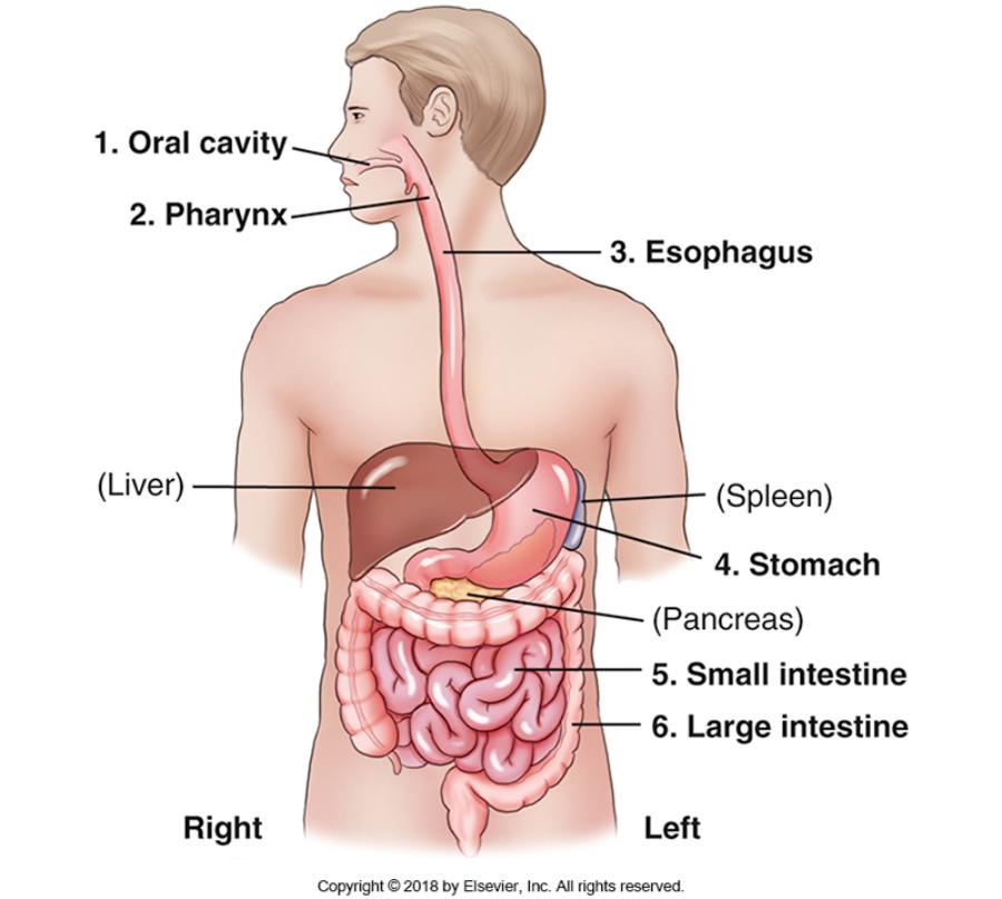
OPES Si Li = Oral Cavity, Pharynx, Esophagus, Stomach, Small intestine, Large intestinte
What is KUB
Kidney, ureters, bladder
What else does KUB refer to?
Abdomen supine. When shooting an abdomen supine x-ray we can demonstrate the borders of the psoas major muscles.
What are 3 abdominal muscles?
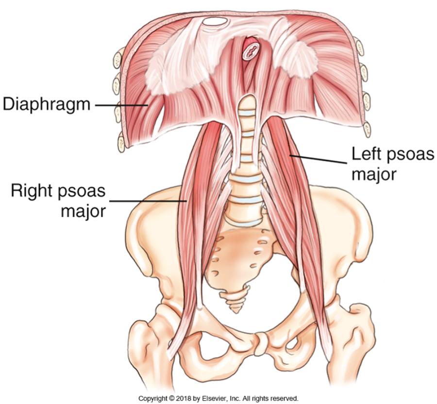
Diaphragm, R psoas major, L psoas major
Where does the psoas major muscle originate?
at the transverse process of L1
Where does the psoas major muscle insert?
at the lesser trochanter of the femur.
Psoas major muscle
Is one of the major flexor muscles of the trunk. It allows us to bend waist down.
Diaphragm
an inspiration muscle which separates the thoracic cavity from the abdominal cavity
Which organs are a part of the digestive tract?
Oral cavity, Pharynx, Esophagus, Stomach, Small intestine, and Large intestine
What are the accessory organs?
Liver, Gallbladder, and Pancreas
cholelithiasis
is the presence of gallstones in the gallbladder
What are the functions of the accessory organs?
They are involved in the digestion and break down of food particles.
Spleen
is a part of the lymphatic system located in the abdomen
stomach
first organ of the digestive system located in the abdominal cavity. Size and shape depends on the body habitus and contents of the stomach.
What connects the stomach to the duodenum?
Pylorus
How long is the small intestine?
15 to 18 feet
What parts are in the small intestine?
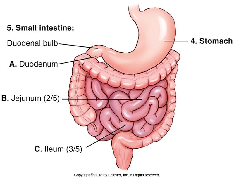
Duodenum (10 inches or 25cm in length), Jejunum (2/5), Ileum (3/5)
Where does the ileum end?
RLQ
Where is the ileocecal valve located?
at the conjunction where the ileum meets the cecum
Which organ is the last organ of the digestion system
large intestine
How long is the large intestine?
about 1.5 meters
What parts are in the large intestine?
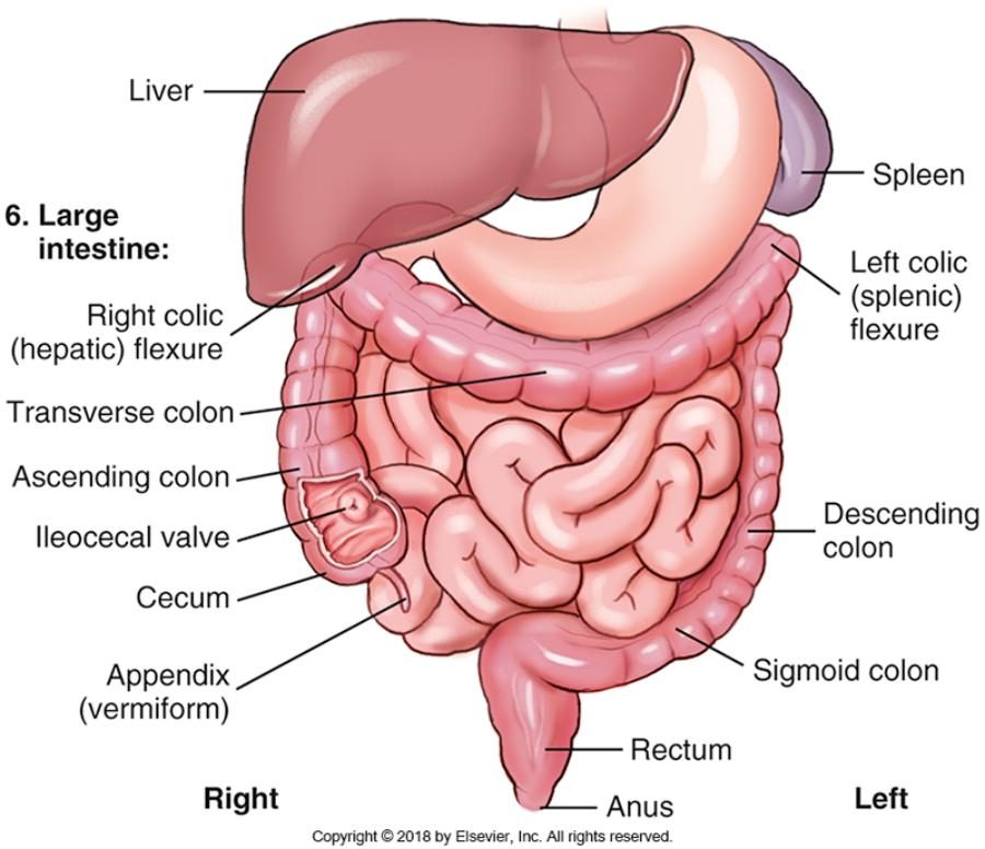
Ileocecal valve, Appendix (Vermiform), Cecum, Ascending Colon,Right colic (hepatic) flexure, Transverse colon, Left colic (splenic) flexure, Descending colon, Sigmoid colon, Rectum, Anus
What is the main function of the Ileocecal valve?
Prevents back flow of bowel movements.
What are the 9 abdominal regions?
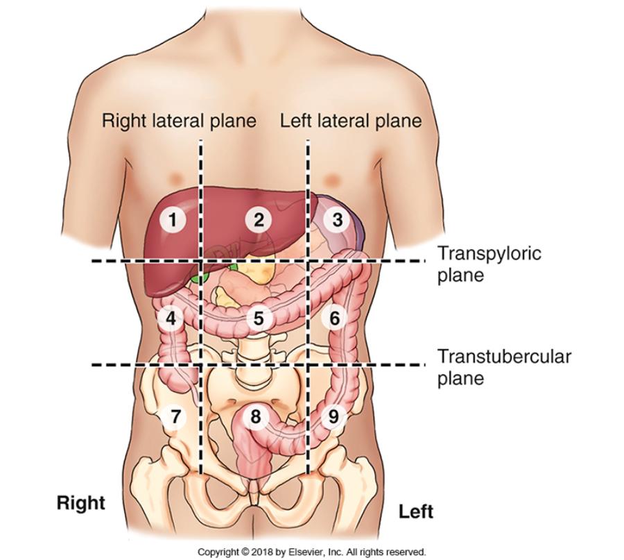
- Right hypochondriac
- Epigastric
- Left hypochondriac
- Right lateral (lumbar)
- Umbilical
- Left lateral (lumbar)
- Right inguinal (iliac)
- Pubic (hypogastric)
- Left inguinal (iliac)
What are considerations about Gonadal shielding for abdomen imaging?
Males always shield. Physician must decide if female may be shielded.
What are the 7 landmarks? Which 3 are most commonly used?
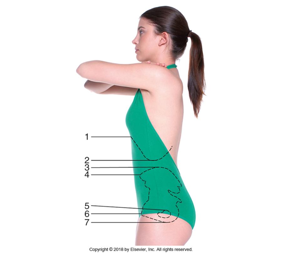
- Xiphoid process (T9-T10)
- Inferior costal margin (L2-L3)
- Iliac crest (L4-L5)
- ASIS
- Greater trochanter
- Symphysis pubis
- Ischial tuberosity
Most used
1. Iliac crest
2. ASIS
3. Greater trochanter
What are abdominal positioning considerations?
- Exposure on expiration - for diaphragm can go up
- IR markers - Lower hip bone area
- Collimation - always collimate tightly unless imaging an obese patient
What are abdominal exposure factors to consider?
- Analog kV (70-80)/Digital kV (80-90)
may change -/+ depending on body habitus - Short exposure time
- Adequate mAs
What does Pneumoperitonium refer to?
Free air or gas in the peritoneal cavity.
What is Intussusception?
When the distal small intestine, loops over itself and creates an obstruction. Needs to be treated within 48 hours before necrosis (tissue death). Most common in kids.
The largest solid organ in the abdomen
Liver
True or false? The stomach is located in the left upper quadrant of the abdomen?
True
The membrane covering the abdominal organs is termed
Visceral peritoneum
In which region or compartment of the abdomen is the pancreas located?
Retroperitoneal
The spleen is located in which quadrant?
LUQ
In which abdominal region is the majority of the transverse colon located in a person with a sthenic body type?
Umbilical region
Which segment of the large intestine is located in the right lower quadrant of the abdomen?
Cecum
In which region or compartment of the abdomen is the urinary bladder located?
Infraperitoneal
Which topographic landmark corresponds to the level of L2/L3?
Inferior costal margin
What is the greater trochanter is part of?
Proximal femur
A properly exposed radiograph of the abdomen should demonstrate
the:
1. Renal outline
2. Psoas muscles
3. Urinary bladder
1 and 2 only
What is the best method of reducing involuntary motion during an abdomen projection?
Short exposure time
Neoplasms of the abdominal organs can be evaluated with:
1.
Computed tomography
2. Magnetic resonance imaging
3. Sonography
1, 2 and 3
An abnormal collection of free air or gas within the peritoneal cavity is termed:
Pneumoperitoneum
Which pathologic condition is most common in young children?
Intussusception
Which one of the following structures must be demonstrated on a KUB projection?
- Symphysis pubis
- Obturator foramen
- Xiphoid process
- Ischial tuberosities
Symphysis pubis
Two AP projections in the supine position with the IR oriented crosswise may be needed for patients who have a body type that is:
- Asthenic
- Hypersthenic
- Hyposthenic
- Sthenic
Hypersthenic
The AP projection of the abdomen is a better choice than a PA projection when the kidneys are of primary interest due to:
- Less motion
- Increased patient comfort
- Less magnification of the kidneys
- Decreased gonadal dose for female patients
Less magnification of the kidneys
Which projection or position of the abdomen will best demonstrate an umbilical hernia?
- Left lateral decubitus position
- Dorsal decubitus position
- Right lateral decubitus position
- AP erect projection
Dorsal decubitus position
Where is the CR directed for a left lateral decubitus position of the abdomen?
- At level of iliac crest
- At level of xiphoid process
- At level of inferior costal margin
- 2 inches (5 cm) above iliac crests
2 inches (5 cm) above iliac crests
What is the optimal amount of time a patient should lie on his side prior to a left lateral decubitus projection?
- 1 to 2 minutes
- 5 to 10 minutes
- 10 to 20 minutes
- 20 to 40 minutes
10 to 20 min
An acute abdominal series includes AP projections in the:
1.
Supine position
2. Erect position
3. Dorsal decubitus position
1 and 2 only
Which projection is performed to demonstrate free air or gas in the abdominal cavity when the patient is unable to stand?
- AP supine abdomen
- Left lateral decubitus
- Right lateral decubitus
- PA supine abdomen
Left lateral decubitus
Clinical indications for an acute abdominal series include:
1.
Ascites
2. Ileus
3. Urinary calculi
1 and 2 only
Which positioning error is classified as a repeatable error for an AP supine-KUB projection?
- Four-side collimation not evident on radiograph
- Slight tilt of the spine
- Symphysis pubis not included on radiograph
- Ischial tuberosities not demonstrated on radiograph
Symphysis pubis not included on radiograph
Which structure is evaluated to determine rotation on an AP radiograph of the abdomen?
- Psoas major muscles
- Kidney shadows
- Iliac wings
- Symphysis pubis
Iliac wings
What is the specific positioning error if the right iliac wing is wider in appearance as compared to the left as seen on an AP supine abdomen radiograph?
- Rotation of the right side of the body toward the IR (right rotation)
- Rotation of the left side of the body toward the IR (left rotation)
- Tilt of the spine to the right
- Tilt of the spine to the left
Rotation of the right side of the body toward the IR (right rotation)
The position demonstrated in the abdomen image below is the:
- Dorsal decubitus
- Erect lateral
- Lateral decubitus
- Ventral decubitus
Dorsal decubitus
The abdomen image below could be improved by:
1. Directing the
CR to the level of the iliac crest
2. Decreasing mAs
3.
Decreasing kV
- 1 only
- 3 only
- 1 and 2 only
- 2 and 3 only
1 only
The abdomen image below could be improved by:
1. Aligning the
midsagittal plane to the center of the IR
2. Collimating to skin
surface
3. Decreasing mAs
- 2 only
- 3 only
- 1 and 2 only
- 1, 2, and 3
1, 2, and 3