

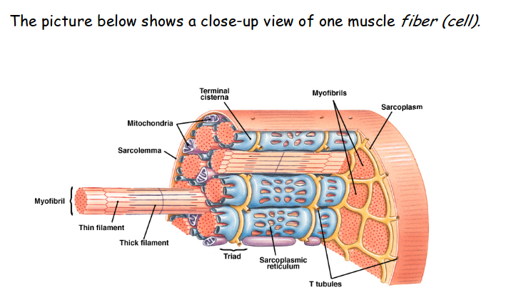
Myofibrils
1. Each muscle cell is made of 1,000's of protein cylinders called myofibrils. Each myofibril contains two types of protein strands called filaments:
i.Actin Filament (Thin Filament)
ii. Myosin filament (Thin filament)
2. Where Actin and myosin filaments overlap,each THICK filament is surrounded by 6 THIN filaments.
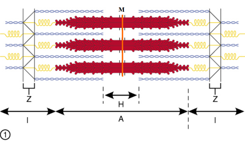
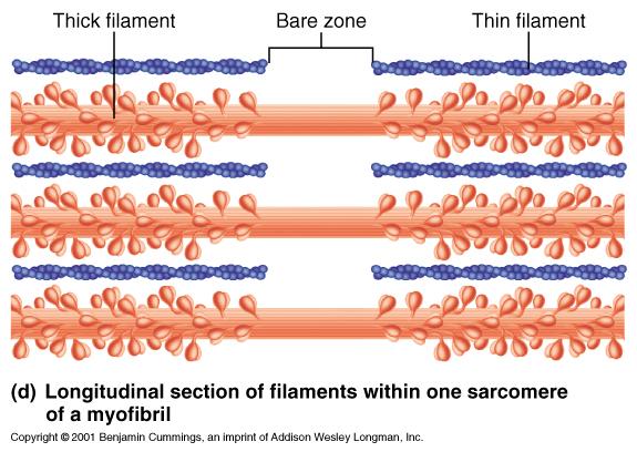
Sarcomere
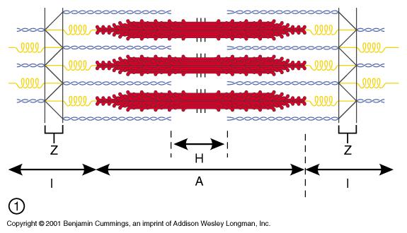
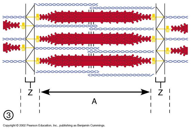
The thin and thick filaments form a repeating pattern of fibers.
1. Z Lines
2. A Band
3. H Zone
4. I Band

Z Lines
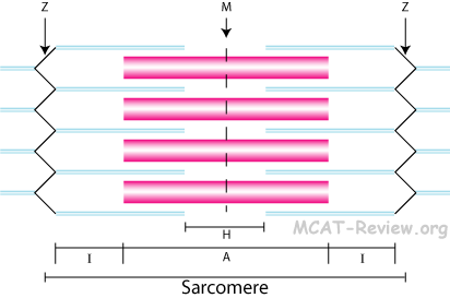
The boundaries of the sarcomere where ACTIN filaments are JOINED together, forming a ZIGZAG line.

A Band
The anisotropic band that extends form the start of the myosin filament to its end. Can also be described as the region of overlapping myosin and actin filaments.

H Zone
Region in center of A band where ONLY myosin is present.
I Band
The isotropic band, an area of actin filaments next to the Z line.
*shows up as a light stripe
Troponin
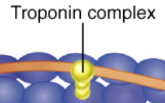
A protein that is attached to tropomyosin.
When calcium is present, troponin acts like a hand and pulls tropomyosin off of the active sites of the actin. This allows the muscle contraction to start.
Tropomysoin
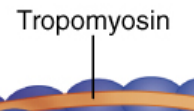
Thread-like proten that winds aroun the actin chain so that it covers the active sites (when muscle is relaxed).
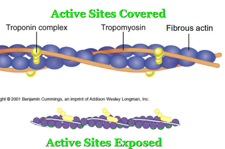
Actin Filament in Detail
Made up of three proteins:
1. Actin--Bead-like proteins which form a chain that is twisted back on itself.
*Each actin bead contains an active site--An area where the myosin head attaches to during muscle contraction.
2. Tropomysoin--Thread-like proten that winds aroun the actin chain so that it cobers the active sites (when muscle is relaxed).
3. Troponin--A protein that is attached to tropomyosin.
*Each contains a calcium-binding area/site--Calcium must bind to troponin before muscle contraction can take place. When calcium is present, troponin acts like a hand and pulls tropomyosin off of the active sites of the actin. This allows the muscle contraction to start.
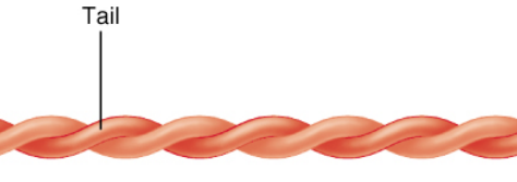
Myosin Rod
Elongated portion of "golf club" found in the H zone.
Myosin Head--*Most important part
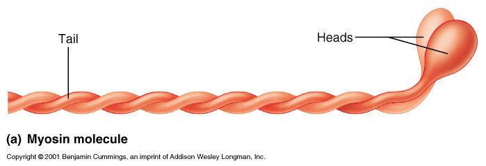
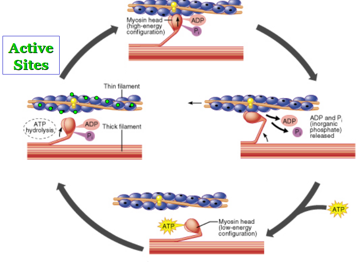
a. Hinge region--A bendable neck region that attaches the head to the rod. Bending of the hinge region moves the head and is directly responsible for muscle contraction!-allows swinging
b. ATP-binding area/site--ATP or ENERGY must bind to the myosin head to allow it to move.
Myosin Filament in Detail
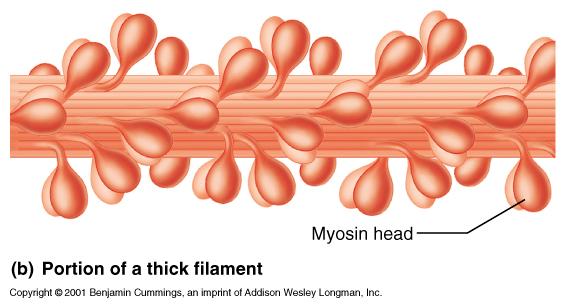
Each myosin filament is made up of 100's of golf-club shaped myosin proteins. Each myosin has two important parts:
1. Myosin Rod--Elongated portion of "golf club" found in the H zone.
2. Myosin Head--*Most important part
a. Hinge region--A bendable neck region that attaches the head to the rod. Bending of the hinge region moves the head and is directly responsible for muscle contraction!-allows swinging
b. ATP-binding area/site--ATP or ENERGY must bind to the myosin head to allow it to move.
Skeletal Muscle Fibers Contract

Active sites Covered
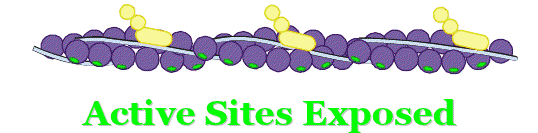
Active sites Exposed
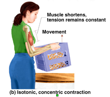
Isotonic Contractions

The type of contractions that move your limbs and move your body around.
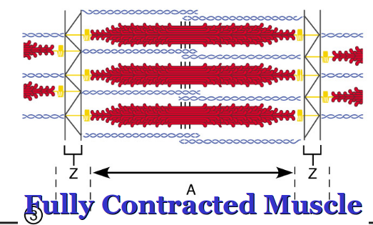
This contraction is explained by the Sliding Filament Theory of Contration, which says that: *Muscle contracts when the myosin heades pull on the actin filaments, causing them to slide inward, toward the center of the sarcomere.
In this contraction, the muscle shortens as it contracts (due to the sliding of the actin filaments.)
Isotonic means "same tension" because the tension in the muscle remains constant during the contraction.
Sliding Filament Theory
a. Sequence of Events:
1) Myosin heads attach to actin filaments at the active sites.
2) Mysoin heads bend at hinge regions.
3) The movememt of the myosin heads pulls the actin filaments toward each other.
4) When fully contracted, the actin filaments on either side of the sarcomere overlap each other slightly.
b. Results in the Sarcomere:
1) Sacromere length shortens
2) H Zone disappears
3) I Band length shortens
4) A Band length remains the same
Two Types of Isotonic Contractions
a. Concentric Contractions: Sliding of actin TOWARD the center of the sarcomere, shortening the sarcomere thus the muscle EX: Lifting a box
b. Eccentric Contractions: Sliding of actin AWAY from center of the sarcomere which lengthens the muscle to its original length (after a concentric contraction especially) EX: Putting box back down
Concentric Contractions

Sliding of actin TOWARD the center of the sarcomere, shortening the sarcomere thus the muscle EX: Lifting a box
Eccentric Contractions
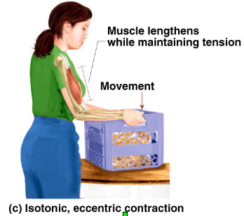
Sliding of actin AWAY from center of the sarcomere which lengthens the muscle to its original length (after a concentric contraction especially) EX: Putting box back down
How Isometric is Different From Isotonic
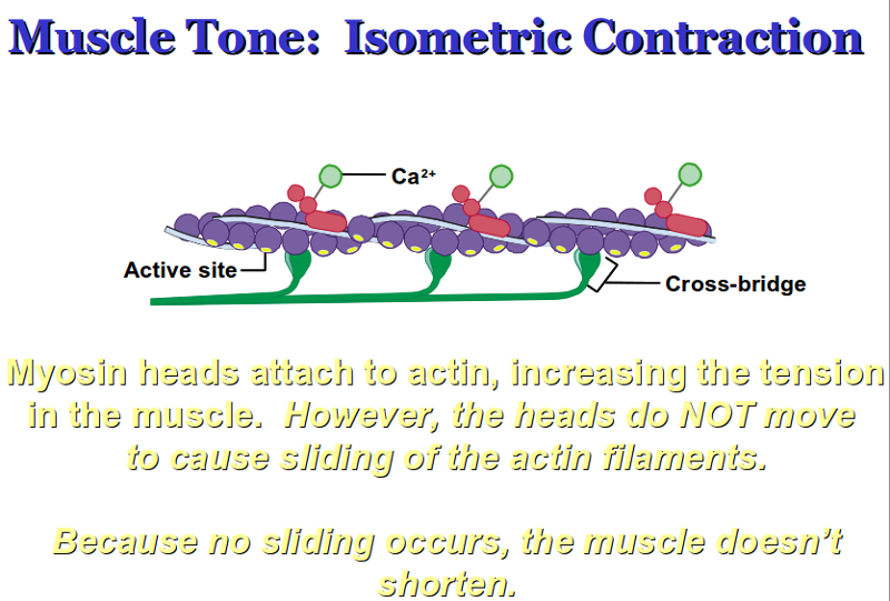
a. In Isometric contractions, there is an increase in TENSION which is caused when…Myosin heads attach to actin
b. In Isometric contractions, the muscle doesn’t change in length, which means that… Actin does not slide.
c. Isometric means “same length”… No shortening or lengthening of muscle.
Isometric Contractions
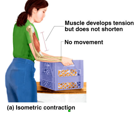
Isometric Contractions
a,These conractions do NOT move the limbs or body around.
b.These contractions are used in two Situations:
1) To stabilize joints/limbs after movement to hold them in position.
2) In postural muscles, to keep these muscles taunt so that:
i) Head is kept upright
ii) Back and abdomen are straight, flat
iii) Calf muscles are straight, flat
2. How Isometric is Different From Isotonic
a. In Isometric contractions, there is an increase in TENSION which is caused when…Myosin heads attach to actin
b. In Isometric contractions, the muscle doesn’t change in length, which means that… Actin does not slide.
c. Isometric means “same length”… No shortening or lengthening of muscle.
General Characteristics of Skeletal Muscle Fibers
a. Appearance ---Very large, long, thin, may extend to a foot or more
b. Nuclei---Hundreds per cell
c. Striations/Stripes---Due to actin & myosin in the sarcomere
d. Contraction---Stimulated by nerves
e. Control---Conscious/voluntary
f. There are two types of skeletal muscles cells/fibers in muscles
Slow Twitch Fibers "Dark Meat"
a. Thin fibers with little power; Have low sugar stores.
b. Contract slowly.
c. Designed to contract for long periods of time. Resistant to fatigue. Good for endurance sports like marathons and swimming.
d. Contain myoglobin, with makes them look dark red and which helps them turn food into energy well. (endurance)
Fast Twitch Fibers "White Meat"
a. Thick fibers with powerful contractions; Have high sugar stores.
b. Contract quickly for speed.
c. Designed to contract for short periods of time.
Fatigue much more easily than slow twitch. Good for short bouts of exercise, as in sprinting, jumping, weight lifting.
*Especially concentrated in finger & eye muscles to give quick, darting movements
d. Lack myoglobin, causing them to be pale or whitish in color and causing them to not be as good at making energy(speed)
e. Intermediate Fibers
* Fast twitch fiber that has the endurance of a slow twitch
* intensive training can cause regular fast twitch to turn into this type
CARDIAC MUSCLE---STRIATED INVOLUNTARY
General Characteristics of Muscle Fibers
a. Appearance---Intermediate length, thin, branched
b. Nuclei---One per cell
2. Specialization: Branched and connected via intercalated discs
-Intercalated discs allow the electrical current from nerves to pass directly from one fiber to another so that groups of fibers are stimulated simultaneously and therefore beat/contract in unison.
3. Action---Does not require stimulation from the nervous system. Instead, pacemaker cells stimulate
contraction.
*One type of smooth muscle is also controlled by pacemakers
4. Control---Unconscious/Involuntary
SMOOTH MUSCLE---NONSTRIATED INVOLUNTARY
General Characteristics of Smooth Muscle Fibers
a. Appearance---Small, shorter, spindle-shaped cells
b. Nuclei---One per cell
c. Lack Striations because the actin and myosin filaments do not form sarcomeres
*Instead of sarcomeres, actin & myosin form a diagonal pattern or “on the bias” pattern.
2. Types of Smooth Muscle
a. Multiunit
*Multiunit: Small bundles of cells that contract independently of each other
1) Found in ciliary muscle and iris of eye, large air passageways-->*Bronchi eye(ciliary muscle:controls focusing of lens)-->Iris-->contractions of the muscle controls amount of light coming into eye.
2) Contractions initiated by nervous system
b. Visceral/Unitary
*Peristalsis: SM contractions in digestive & urinary organs that moves food (or urine)
*Visceral/Unitary:
Sheets of cells that contract simultaneously, as one unit
Only Muscle Type Controlled by Hormones:Smooth Muscle
a. Many hormones affect blood flow and blood pressure:
1) Histamine—Causes vasodilation: blood vessel exppands, increasing blood flow
2) Angiotensin—Causes vasoconstriction: blood vessel gets smaller, decreasing blood flow
3) Adrenalin (Epinephrine)---Causes either vasodilation or vasoconstriction, depending upon location in body
b. Other hormone examples:
1) Gastrin: Juices in your stomach; cause contraction of the stomachfor digestion
2) Oxytocin: uterus contractions for labor
Multiunit: Type of Smooth Muscle
*Multiunit: Small bundles of cells that contract independently of each other
1) Found in ciliary muscle and iris of eye, large air passageways-->*Bronchi eye(ciliary muscle:controls focusing of lens)-->Iris-->
contractions of the muscle controls amount of light coming into eye.
Visceral/Unitary: Type of Smooth Muscle
*Visceral/Unitary:
Sheets of cells that contract simultaneously, as one unit
1) Found lining large blood vessels
*This smooth muscle is important in changing blood flow, controlling blood pressure.
2) Forms walls of many organs---digestive, urinary, reproductive tracts
*Peristalsis: SM contractions in digestive & urinary organs that moves food (or urine)
3) Contractions initiated by pacemaker cells (autorhythmic contractions)
Histamine
Causes vasodilation: blood vessel exppands, increasing blood flow
Angiotensin
Causes vasoconstriction: blood vessel gets smaller, decreasing blood flow
Adrenalin (Epinephrine)
Causes either vasodilation or vasoconstriction, depending upon location in body
Gastrin
Juices in your stomach; cause contraction of the stomachfor digestion
Oxytocin
uterus contractions for labor
smooth non-invol contin
1) Found lining large blood vessels
*This smooth muscle is important in changing blood flow, controlling blood pressur.
2) Forms walls of many organs---digestive, urinary, reproductive tracts
3) Contractions initiated by pacemaker cells
(autorhythmic contractions)