Aorta
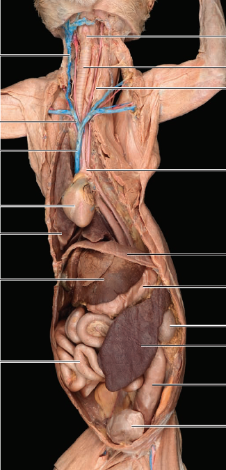
largest artery in body; issuing from left ventricle
superior vena cava
precava; largest, dark-colored vessel entering the base of the heart
inferior vena cava
post cava; enters right atrium
coronary arteries
supply myocardium; can be seen on surface of heart
heart
in mediastinum enclosed by pericardium
lungs
flanking the heart
thymus
superior to and partially covering the heart; large in young cats & replaced by fat in older cats
liver
posterior to diaphragm
stomach
dorsally located and to the left of liver
spleen
flattened, brown organ curving around the lateral aspect of the stomach
small intestine
continues posteriorly from stomach
large intestine
takes a U-shaped course around small intestine and terminates in rectum
aortic arch
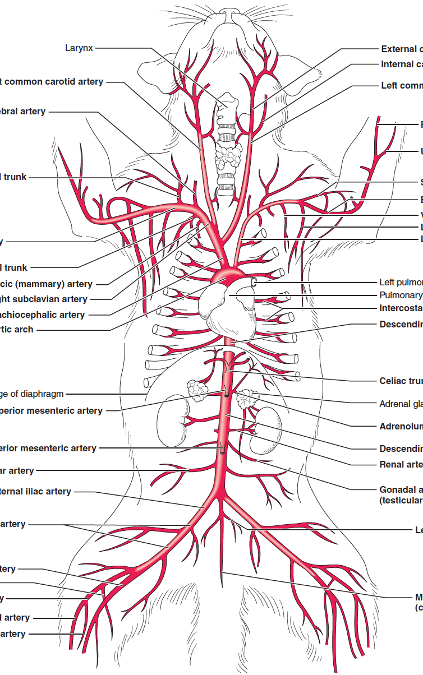
gives off two large vessels: left subclavian artery and brachiocephalic artery
brachiocephalic artery
has three major branches:
Right subclavian artery, and right/left common carotid arteries
Difference between origin of left common carotid arteries in humans & cats
In humans, the left common carotid artery & left subclavian artery are direct braches off aortic arch
right common carotid artery
gives off branches to neck muscles, thyroid gland, and trachea
branches to form external/internal carotid arteries
right subclavian artery
gives off four branches:
vertebral artery, costocervical trunk, thyrocervical trunk, and internal thoracic artery
vertebral artery
along with internal carotid artery supplies the arterial circulation of the brain
costocervical trunk
branches to the costal and cervical regions
thyrocervical trunk
branches to the shoulder
internal thoracic (mammary) artery
serving the ventral thoracic artery
axillary artery
when subclavian passes in front of first rib, it becomes this artery. Branches of this artery include: ventral thoracic artery, long thoracic artery, and subscapular artery which supply the trunk and shoulder muscles
ventral thoracic artery
to the pectoral muscles
long thoracic artery
to pectoral muscles and latissimus dorsi
subscapular artery
to trunk muscles
brachial artery
as axillary artery enters the arm, it becomes this artery which branches at the elbow to produce the radial and ulnar arteries
radial and ulnar arteries
serve the forearm and hand
celiac trunk
supplies the stomach, liver, gallbladder, pancreas, and the spleen
superior mesenteric artery
immediately posterior to celiac trunk
supplies the small intestine and most of large intestine
adrenolumnar arteries
paired arteries diverging from aorta slightly posterior to superior mesenteric artery
supply the muscles of the body wall and adrenal glands
renal arteries
paired arteries supplying kidneys
gonadal arteries
ovarian or testicular arteries that supply gonads
inferior mesenteric artery
unpaired; thin vessel arising from ventral surface of aorta posterior to the gonadal arteries
supplies the second half of large intestines
iliolumbar arteries
paired, rather large arteries that supply the body musculature in iliolumbar region
external iliac arteries
paired arteries which continue through the body wall & pass under the inguinal ligament to hindlimb
internal iliac arteries
two arteries which supply the pelvic viscera
Difference between iliac arteries in cat and humans
there is NO COMMON ILIAC ARTERY in cat
median sacral artery
descending abdominal aorta divides into the two internal iliac arteries and ___________
caudal artery
as median sacral artery enters tail, it becomes this artery
femoral artery
courses through thigh and gives off branches to thigh muscles
saphenous artery
branches off femoral artery to supply medial portion of the leg
popliteal artery
descends deep to knee to become this artery which gives off two branches:
sural artery and posterior tibial artery
sural, posterior tibial, and anterior tibial arteries
supply the leg and foot
Aygos vein
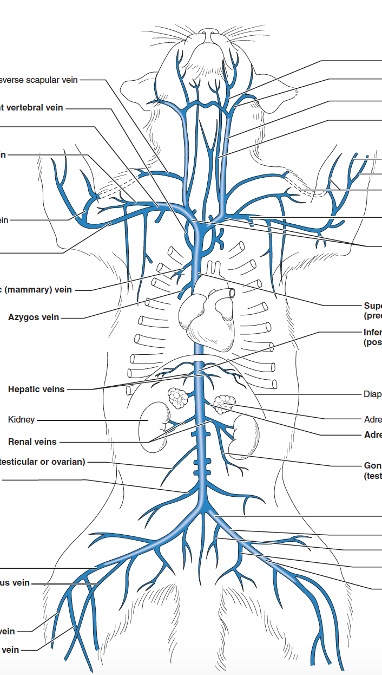
passing directly into tits dorsal surface
drains the thoracic intercostal muscles
internal thoracic (mammary) vein
drains chest and abdominal wall
right vertebral vein
drains spinal cord and brain
right and left brachiocephalic veins
form the precava by their union
Difference in formation of brachiocephalic veins in humans and cats
Humans: brachiocephalic vein is formed by union of internal jugular vein and subclavian veins
Cats: formed by union of external jugular vein and subclavian veins
external jugular vein
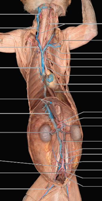
courses anteriorly along side of neck to point where its joined on medial surface by internal jugular vein
Difference in cat & human jugular veins
Human: internal jugular vein is considerably larger & drains into subclavian vein
Cat: External jugular vein is larger & internal jugular vein drains into it
subclavian vein
moves laterally toward the arm; becomes axillary vein
axillary vein
becomes this vein as it passes in front of first rib and runs through brachial plexus giving off subscapular vein
subscapular vein
drains the proximal part of the arm and shoulder
brachial vein
axillary vein becomes this vein as it enters the arm; receives radial/ulnar vein at the inner bend of elbow
radial and ulnar veins
drain the forelimb
cephalic vein
on dorsal side of the arm; communicates with brachial vein via median cubital vein in elbow then enters transverse scapular vein in shoulder
hepatic veins
entering postcava from liver
adrenolumbar veins
empty into postcava and drain adrenal glands and body wall
renal veins
drain the kidneys & empties into postcava
(common to find two right renal veins)
gonadal veins
testicular or ovarian veins
left vein of this venous pair enters the left renal vein anteriorly
Iliolumbar veins
drain muscles of the back & empties into postcava
common iliac veins
unite to form postcava
internal and external iliac veins
unite to form common iliac veins
internal iliac veins
receive branches from pelvic organs and gluteal region
external iliac veins
receives venous drainage from lower limb
deep femoral vein
drains the thigh and external genital region
femoral vein
receives blood from the thigh, leg, and foot
formed by union of great saphenous vein and popliteal vein
great saphenous vein
superficial vein that courses up inner aspect of calf & across inferior portion o gracilis muscle to enter femoral vein
popliteal vein
located deep in the thigh beneath the semimembranosus and semitendinosus muscles in popliteal spaces accompanying popliteal artery
posterior and anterior tibial veins
drain the leg
hepatic portal vein
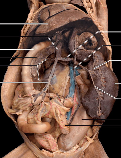
formed by the union of the gastrosplenic and superior mesenteric veins
Difference between formation of hepatic portal vein in cats and humans
Humans: formed by union of splenic and superior mesenteric veins
Cat: formed by union of gastrosplenic and superior mesenteric veins
Gastrosplenic vein
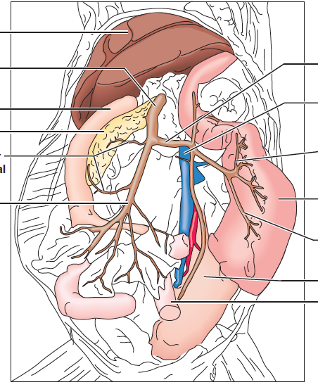
carries blood from spleen and stomach
located dorsal to stomach
superior mesenteric vein
large vein draining small and large intestine and pancreas
inferior mesenteric vein
parallels course of inferior mesenteric artery
empties into superior mesenteric vein
pancreaticoduodenal vein
anterior branch empties into hepatic portal vein
posterior branch empties into superior mesenteric vein