Lacrimal Apparatus (External Eye)
Contains lacrimal glands, lacrimal canals, lacrimal sac, lacrimal canaliculi, and nasolacrimal duct.
Lacrimal Sac (External Eye)
Dilated ends of the lacrimal ducts at the nasal ends of the eyes that fill with tears secreted by the lacrimal glands.
Lacrimal Glands (External Eye)
Paired almond-shaped glands, one for each eye, that secrete the aqueous layer of the tear film. They are situated in the upper, outer portion of each eye.
Lacrimal Canals (External Eye)
Also know as the lacrimal canaliculi.
Conjunctiva (External Eye)
Thin, protective mucous membrane lining the eyelids and covering the anterior surface of the eye itself.
Sclera - Fibrous Tunic (Non-vascular)
(Internal Eye)
White opaque portion of the fibrous layer of the eyeball.
Cornea - Fibrous Tunic (Non-vascular)
(Internal Eye)
Transparent anterior portion of the eyeball; part of the fibrous layer.
Choroid - Uvea Tunic (Vascular)
(Internal Eye)
The vascular middle layer of the eye.
Iris - Uvea Tunic (Vascular)
(Internal Eye)
Colored portion of the eye.
Ciliary Body (Internal Eye)
Muscle that controls lens shape, attached to lense
Pupil (Internal Eye)
Opening in the center of the iris through which light enters the eye.
Retina - Sensory Tunic (contains vitreous humor)(Internal Eye)
Neural layer of the eyeball; contains photoreceptors (rods, cones).
Rods (Internal Eye)
For dim light and black and white.
Cones (Internal Eye)
For color.
Bipolar Cells (Internal Eye)
Cells located in the retina the connect ganglion cells and photoreceptors.
Ganglion Cells (Internal Eye)
The output cells of the retina. They indirectly receive their inputs from the photoreceptors and send their outputs to the brain.
Optic Disc (Internal Eye)
Blind spot: the point where the optic nerve enters the retina; not sensitive to light
Macula Lutea (Internal Eye)
A small yellowish central area of the retina that is rich in cones and that mediates clear detailed vision
Fovea Centralis (Internal Eye)
Area on macula lutea that consists only of cones; for acute vision
Lens (Internal Eye)
A transparent, biconvex structure in the eye that, along with the cornea, helps to refract light to be focused on the retina.
Anterior Segment (Internal Eye)
Front third of eyeball, including the cornea, anterior chamber, iris and ciliary body.
Posterior Segment (Internal Eye)
Back two-thirds of the eye that includes the anterior hyaloid membrane and all of the optical structures behind it: the vitreous humor, retina, choroid, and optic nerve.
Aqueous Humor (Internal Eye)
Watery fluid in the anterior segment of the eye.
Vitreous Humor (Internal Eye)
Clear colorless transparent jelly that fills the posterior segment of the eye.
Visual Pathway
Pathway that light travels to the brain to become images.
OPTIC NERVE --> OPTIC CHIASMA --> LATERAL GENICULATE BODY --> OPTIC RADIATION --> PRIMARY VISUAL CORTEX
Optic nerve
2nd cranial nerve that is responsible for seeing
Optic Chiasma
The partial crossover of fibers of the optic nerves.
Optic Tract
A collection of retinal ganglion cell axons stretching from the optic chiasm to the brain stem.
Emmetropy
Normal condition of the eye in which visual images are in clear focus on the retina
Myopia
A condition in which visual images are focused in front of rather than on the retina; nearsightedness.
Hyperopia
A condition in which visual images are routinely focused behind rather than on the retina; commonly known as farsightedness
Presbyopia
A condition that results in the loss of near focusing ability; typical onset is around age 40.

What is A?
Ciliary Body

What is B?
Ciliary Zonule

What is C?
Cornea

What is D?
Iris

What is E?
Pupil

What is F?
Anterior Segment (Aqueous Humor)
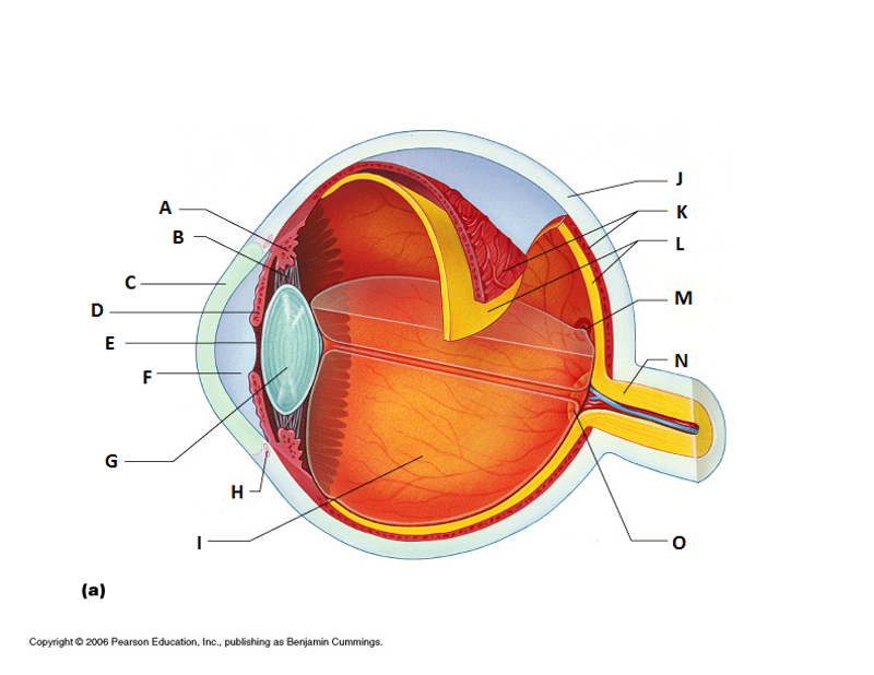
What is G?
Lens

What is H?
Scleral Venous Sinus

What is I?
Posterior Segment (Vitreous Humor)

What is J?
Sclera

What is K?
Choroid

What is L?
Retina

What is M?
Macila Lutea and Fovea Centralis

What is N?
Optic Nerve

What is O?
Optic Disc (blind spot)

What is A?
Lacrimal Gland

What is B?
Eye lid
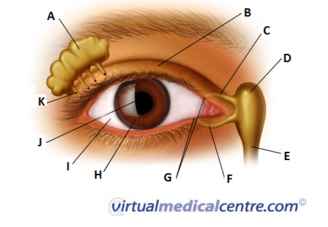
What is C?
Superior Lacrimal Canal

What is D?
Lacrimal Sac

What is E?
Lacrimal Duct

What is F?
Inferior Lacrimal Canal

What is G?
Lacrimal Puncta

What is H?
Iris

What is I?
Sclera

What is J?
Pupil

What is K?
Lacrimal Gland Duct