Instructions for Side by Side Printing
- Print the notecards
- Fold each page in half along the solid vertical line
- Cut out the notecards by cutting along each horizontal dotted line
- Optional: Glue, tape or staple the ends of each notecard together
Chapter 24: Special Senses: Vision
front 1 Lacrimal Apparatus (External Eye) | back 1 Contains lacrimal glands, lacrimal canals, lacrimal sac, lacrimal canaliculi, and nasolacrimal duct. |
front 2 Lacrimal Sac (External Eye) | back 2 Dilated ends of the lacrimal ducts at the nasal ends of the eyes that fill with tears secreted by the lacrimal glands. |
front 3 Lacrimal Glands (External Eye) | back 3 Paired almond-shaped glands, one for each eye, that secrete the aqueous layer of the tear film. They are situated in the upper, outer portion of each eye. |
front 4 Lacrimal Canals (External Eye) | back 4 Also know as the lacrimal canaliculi. |
front 5 Conjunctiva (External Eye) | back 5 Thin, protective mucous membrane lining the eyelids and covering the anterior surface of the eye itself. |
front 6 Sclera - Fibrous Tunic (Non-vascular)
| back 6 White opaque portion of the fibrous layer of the eyeball. |
front 7 Cornea - Fibrous Tunic (Non-vascular)
| back 7 Transparent anterior portion of the eyeball; part of the fibrous layer. |
front 8 Choroid - Uvea Tunic (Vascular)
| back 8 The vascular middle layer of the eye. |
front 9 Iris - Uvea Tunic (Vascular)
| back 9 Colored portion of the eye. |
front 10 Ciliary Body (Internal Eye) | back 10 Muscle that controls lens shape, attached to lense |
front 11 Pupil (Internal Eye) | back 11 Opening in the center of the iris through which light enters the eye. |
front 12 Retina - Sensory Tunic (contains vitreous humor)(Internal Eye) | back 12 Neural layer of the eyeball; contains photoreceptors (rods, cones). |
front 13 Rods (Internal Eye) | back 13 For dim light and black and white. |
front 14 Cones (Internal Eye) | back 14 For color. |
front 15 Bipolar Cells (Internal Eye) | back 15 Cells located in the retina the connect ganglion cells and photoreceptors. |
front 16 Ganglion Cells (Internal Eye) | back 16 The output cells of the retina. They indirectly receive their inputs from the photoreceptors and send their outputs to the brain. |
front 17 Optic Disc (Internal Eye) | back 17 Blind spot: the point where the optic nerve enters the retina; not sensitive to light |
front 18 Macula Lutea (Internal Eye) | back 18 A small yellowish central area of the retina that is rich in cones and that mediates clear detailed vision |
front 19 Fovea Centralis (Internal Eye) | back 19 Area on macula lutea that consists only of cones; for acute vision |
front 20 Lens (Internal Eye) | back 20 A transparent, biconvex structure in the eye that, along with the cornea, helps to refract light to be focused on the retina. |
front 21 Anterior Segment (Internal Eye) | back 21 Front third of eyeball, including the cornea, anterior chamber, iris and ciliary body. |
front 22 Posterior Segment (Internal Eye) | back 22 Back two-thirds of the eye that includes the anterior hyaloid membrane and all of the optical structures behind it: the vitreous humor, retina, choroid, and optic nerve. |
front 23 Aqueous Humor (Internal Eye) | back 23 Watery fluid in the anterior segment of the eye. |
front 24 Vitreous Humor (Internal Eye) | back 24 Clear colorless transparent jelly that fills the posterior segment of the eye. |
front 25 Visual Pathway | back 25 Pathway that light travels to the brain to become images.
|
front 26 Optic nerve | back 26 2nd cranial nerve that is responsible for seeing |
front 27 Optic Chiasma | back 27 The partial crossover of fibers of the optic nerves. |
front 28 Optic Tract | back 28 A collection of retinal ganglion cell axons stretching from the optic chiasm to the brain stem. |
front 29 Emmetropy | back 29 Normal condition of the eye in which visual images are in clear focus on the retina |
front 30 Myopia | back 30 A condition in which visual images are focused in front of rather than on the retina; nearsightedness. |
front 31 Hyperopia | back 31 A condition in which visual images are routinely focused behind rather than on the retina; commonly known as farsightedness |
front 32 Presbyopia | back 32 A condition that results in the loss of near focusing ability; typical onset is around age 40. |
front 33  What is A? | back 33 Ciliary Body |
front 34  What is B? | back 34 Ciliary Zonule |
front 35  What is C? | back 35 Cornea |
front 36  What is D? | back 36 Iris |
front 37  What is E? | back 37 Pupil |
front 38  What is F? | back 38 Anterior Segment (Aqueous Humor) |
front 39 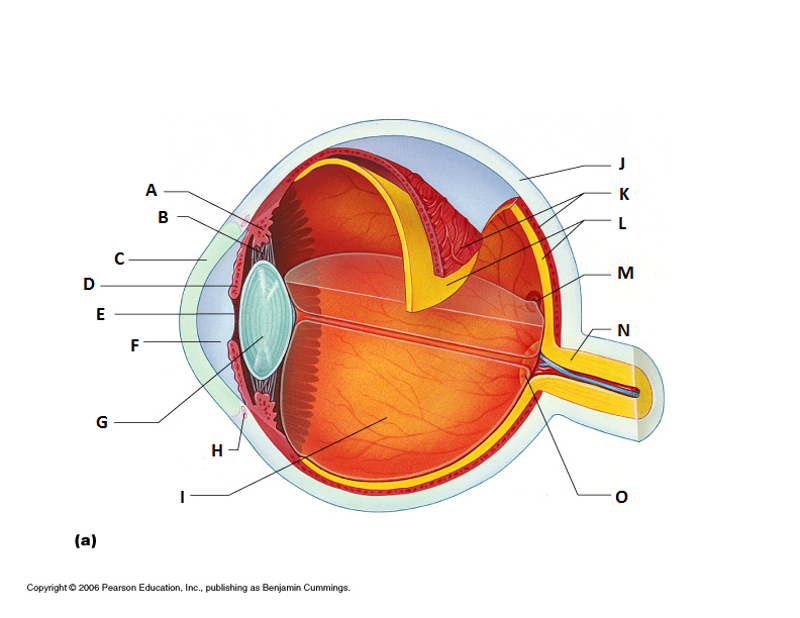 What is G? | back 39 Lens |
front 40  What is H? | back 40 Scleral Venous Sinus |
front 41  What is I? | back 41 Posterior Segment (Vitreous Humor) |
front 42  What is J? | back 42 Sclera |
front 43  What is K? | back 43 Choroid |
front 44  What is L? | back 44 Retina |
front 45  What is M? | back 45 Macila Lutea and Fovea Centralis |
front 46  What is N? | back 46 Optic Nerve |
front 47  What is O? | back 47 Optic Disc (blind spot) |
front 48  What is A? | back 48 Lacrimal Gland |
front 49  What is B? | back 49 Eye lid |
front 50 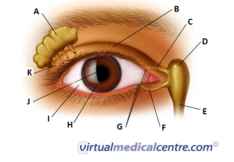 What is C? | back 50 Superior Lacrimal Canal |
front 51  What is D? | back 51 Lacrimal Sac |
front 52  What is E? | back 52 Lacrimal Duct |
front 53  What is F? | back 53 Inferior Lacrimal Canal |
front 54  What is G? | back 54 Lacrimal Puncta |
front 55  What is H? | back 55 Iris |
front 56  What is I? | back 56 Sclera |
front 57  What is J? | back 57 Pupil |
front 58  What is K? | back 58 Lacrimal Gland Duct |