Which property of muscle gives it the ability to stretch without damage?
extensibility
This is the outermost layer of connective tissue surrounding a skeletal muscle.
epimysium
The contractile organelles of a skeletal muscle fiber are thread-like structures called
Myofibrils
Release of calcium from these structures triggers skeletal muscle contraction.
terminal cisterns of sarcoplasmic reticulum
Which of the following regions of a sarcomere contain thin filaments?
Both I band and A band
Myofibrils contain
all of these answers are correct.
What regulatory proteins can be found in the thin filaments of skeletal muscle fibers?
tropomyosin and troponin
Which of the regions of a sarcomere contain titin?
from M line to Z disc
What energizes the myosin head?
ATP hydrolysis reaction
Skeletal muscle contraction will continue to occur as long as the following chemicals are available in the cytosol of the muscle fiber.
Calcium ions and ATP
This is the least powerful type of skeletal muscle fiber.
slow oxidative fiber
Which of the following microscopic structures is only found in the cardiac muscle tissue?
intercalated discs
Which of the following types of muscle tissue contract when excited by their own autorhythmic muscle fibers?
cardiac muscle
Smooth muscle tone is maintained by the prolonged presence of _____ in the muscle cell’s cytosol?
Calcium ions
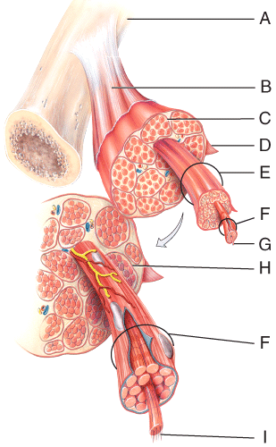
Which of the labeled structures on the diagram holds muscles with similar functions together, allows free movement of muscles, carries nerves, blood vessels and lymphatic vessels, and fills spaces between muscles?
B
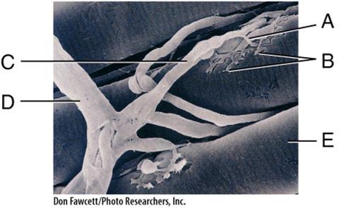
In the diagram, the neurotransmitter, acetylcholine, is released from this area.
B
In the diagram, what is the basic functional unit of a myofibril?
F
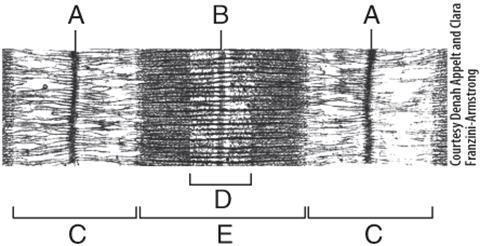
In the diagram, where are thick filaments found?
E
Which of the following types of muscle tissue is capable of undergoing the stress-relaxation response when they are stretched?
single-unit smooth muscle fibers
Cross bridges are formed during muscle contraction when _____ on the thick filaments binds to _____ on the thin filaments.
myosin; actin
In a neuromuscular junction, the effect of acetylcholine (ACh) binding to receptors on the motor end plate lasts only briefly due to
rapid destruction of ACh in the synaptic cleft by acetylcholinesterase.
Which of following is a common characteristic of fast glycolytic (FG) skeletal muscle fibers?
high amount of glycogen in the sarcoplasm.
Which of the following is NOT a characteristic commonly used to name skeletal muscles?
Thickness of fibers

Where is the latissimus dorsi?
A

Where is the rectus femoris?
C
Where is the deltoid?
F

Where is the masseter?
E

Where is the gastrocnemius?
D
Where is the soleus?
E

Where is the external oblique?
G

Where is the masseter?
B
Where is the orbicularis oculi?
D

Where is the lateral rectus?
E

Where is the serratus anterior?
G

Where is the external oblique?
E
Where is the transverse abdominus?
H
Where is the infraspinatus?
G

Where is the rhomboid major?
I

Where is the brachioradialis?
C
Where is the flexor carpi radialis?
E
Where is the abductor digiti minimi?
E

Where is the quadratus lumborum?
A

Where is the sartorius?
C
Where is the vastus intermedius?
F

Where is the gracilis?
I

Which three muscles make up the hamstring?
L, M, N
Where is the semitendinosus?
L
A muscle that has three origins is called a
triceps
A muscle that raises or elevates a body part is called a
levator
A muscle that decreases the size of an opening is a
sphincter
Which of the following is a muscle whose insertion is found on the clavicle and acromion process of the scapula within the pectoral girdle?
trapezius
Which of the following types of joints lacks a joint cavity and is
held together by a fibrous connective tissue?
1. Fibrous joints
2. Cartilaginous joints
3. Synovial joints
1 only
Which of the following types of joints do NOT have a synovial cavity?
1. Fibrous joints
2. Cartilaginous joints
3. Synovial joints
1 and 2
Which of the following types of joints do NOT have a synovial cavity?
1. Fibrous joints
2. Cartilaginous joints
3. Synovial joints
3 only
Moving the humerus laterally at the shoulder joint is an example of which type of movement?
Abduction
Which type of movement involves a continuous sequence of flexion, abduction, extension, and adduction resulting in a distal body part moving in a circle?
Circumduction
What type of special movement occurs in your clavicles at your acromioclavicular and sternoclavicular joints when you cross your arms in front of your body?
Protraction
Which special movement occurs when you bend your foot at the ankle in the direction of the foot’s superior surface as would occur when you stand on your heels?
Dorsiflexion
Which special movement involves moving your thumb across the palm to touch the tips of the fingers on the same hand?
Opposition
Which of the joints shown in the figure is classified as a multiaxial joint?
F
Which type of joint permits this type of movement?
1. Synovial joint
2. Cartilaginous joint
3. Fibrous joint
1 only
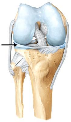
The line is pointing to the _____ ligament.
anterior cruciate
The line is pointing to the _____.
lateral meniscus

The line is pointing to the _____ ligament.
coracohumeral