lhow many facets make up the subtakar joint
three
another term for osteochondroma is
exostosis
what is one advantage of the lateromedial projection of the foot?
the foot assumes a more true lateral position
to properly visualize the joint spaces with the AP projection of the foot, the CR must be:
perpendicular to the metatarsals
situation: a patient enters radiology with a possible ligament tear to the lateral aspect of the ankle. initial ankle radiographs are negative for fracture or dislocation. because the clinic is ina rural setting, the patient cant have an MRI performed to evaluate the ligaments of the ankle. Which of the following techniques may provide an assessment of the soft-tissue structures of the ankle?
AP stress projections
where would the interphalangeal joint be in the foot?
between the phalanges of the first digit
what is the major disadvantage of using 45 degrees of flexion for the lateral projection of the knee?
draws the patella into the intercondylar sulcus
which term describes the top or anterior surface of the foot?
dorsum
a correctly positioned AP 45-degree medial oblique ankle projection frequently may also demonstrate a fracture of the base of the 5th metatarsal if present
true
situatuion: a patient enters the ED with an injury near the base of the first and second metatarsals. the basic foot projections are inconclusive on demonstrating a fracture to the medial cuneiform. Which of the following projections would best demonstrate this bone?
AP oblique with lateral rotation

(picture) which of these labeled structures or bones identifies the navicular?
b
situation: a patient comes to the radiology department for a knee study with special interest in the region of the proximal tibiofibular joint and the lateral condyle of the tibia. Which of the following positioning routines should the technologist obtain?
AP,lateral,and medial oblique knee
to reduce scatter radiation during tabletop procedures, the bucky tray should not be positioned directly under the lower limb being radiographed
true
where is the CR placed for a mediolateral projection of the calcaneus?
1 inch distal to the medial malleolus
a radiograph of a lateral projection of the patella reveals that the femoropatellar joint space isnt open. the patella is superimposed over the distal femur. The most likely cause of this is:
excessive flexion of the knee
the foot must be force dorsiflexed so the long axis of the foot is perpendicular to the image receptor for AP and mortise projections of the ankle.
false
what CR angle should be used for a lateral projection of the knee on a short, wide-pelvis patient?
7-10 degrees cephalad
how much rotation from an AP of the ankle will typically produce a mortise view?
15-20 degrees
a correctly positioned lateral ankle will demonstrate the lateral malleolus superimposed over the posterior half of the tibia.
true
another term for the intercondylar sulcus is the:
patellar surface

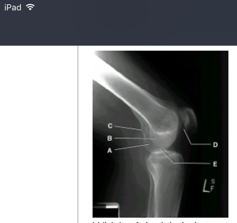
(picture) which of the labeled structures is the lateral condyle?
b
situation: a patient comes to radiology for an evaluation of the longitudinal arch of the foot. Which of the following projections would provide the best information about the arch?
lateral weight-bearing projections
a radiograph of an AP projection of the second toe reveals that the interphalangeal joints are not open. What is the most likely cause for this radiographic outcome?
incorrect CR centering or angle
follow-up radiographs for a fractured tibia and fibula may include only the joint closest to the site of injury
true
a radiographic appearance of a highly malignant and extensive destructive lesion that usually occurs in long bones and produces a sunburst pattern describes:
an osteogenic sarcoma
which metatarsal bone of the foot has a prominent tuberosity frequently fractured?
fifth
which of the following imaging modalities/procedures will provide the best assessment for osteomyelitis of the foot?
nuclear medicine
to decrease the angle between the antrior surface of the foot and antrior surface of the lower leg is described as
dorisiflexion
the best method of evaluating injuries to the menisci and ligaments of the knee joint involves:
and MRI procedure
a radiograph of an AP ankle projection reveals that the lateral joint space is not open(lateral malleolus is partially superimposed by the talus).The superior and medial joint spaces are open. WHat should the technologist do to correct the problem and improve the image?
nothing; this is an acceptable image
placing two images on the same computed radiographic imaging plate is not permitted with newer CR systems
false
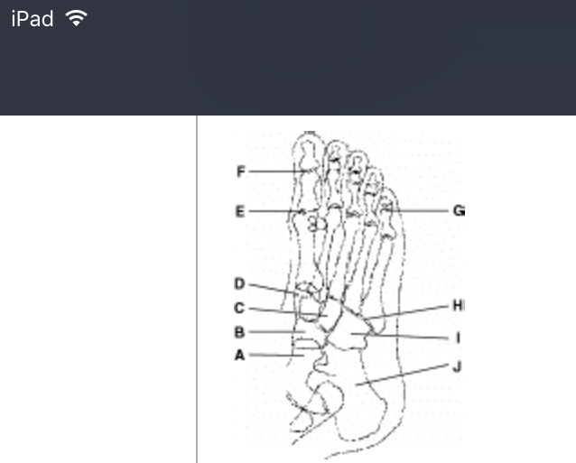
which of these labeled structures or bones identifies the talus?(picture)
a
which projection of the knee will best demonstrate the styloid process of the fibula?
AP oblique with medial rotation
a radiograph of a plantodorsal (axial) projection of the calcaneus reveals foreshortening. The technologist used 60 kv, 6 MAS,40-inch (100cm) SID, and a 30-degree cephalad angle from the long axis of the foot. Which of the following modifications will produce a more diagnostic image of the calcaneus?
increase CR angulation
how much CR angulation (if any) should be used for an AP projection of the toes?
10-15 degrees toward calcaneus
this radiograph represents which of the following positions?(picture)
lateral knee under-rotated toward the IR
the adductor is present on the lateral femoral condlye and can be used to determine possible rotation of a lateral projection.
false
how much flexion of the knee is recommened for the lateral projection of the patella?
5 to 10 degrees or less
the adductor tubercle is located on the posterior aspect of the medial femoral condyle.
true
which structure or bone contains the sustentaculum tali?
calcaneus
a radiograph of an oblique foot with medial rotation demonstrates considerable superimposition of the third through fifth metatarsals. How must the original position be changed to eliminate this problem?
decrease obliquity of the foot
extending the ankle joint or pointing the and toes downward is called:
plantar flexion
what CR angulation is required for the AP oblique projection of the foot?
CR is perpendicular to the image receptor
the correct CR placement for an AP projection of the knee is midpatella?
false

which of the labeled structures is the adductor tubercle? (picture)
c
when using computed radiography, lead masking should not be placed on the regions of the imaging plate not within the collimation field.
false
the calcaneus articulations with the talus and the:
cuboid
how much CR angulation to the long axis of the foot is required for the plantodorsal (Axial) projection of the calcaneus?
40 degrees

which of these labeled structures or bones identifies the metatarsophalangeal joint? (picture)
e
a lateral knee radiograph that is over-rotated toward the image receptor can be recognized by which of the following?
the fibular head will appear less superimposed by the tibia than a true lateral

which one of the labeled structures is the medial condyle? (picture)
a
to ensure that both joints are included on an AP projection of the tibia and fibula on an adult, the technologist should:
turn image receptor diagonally
a radiograph of a mortise view of the ankle reveals that the lateral malleolus is slightly superimposed over the talus and the lateral joint space is not open. What is most likely the cause for this radiographic outcome?
insuffcient medial rotation of the foot and ankle
the patella is drawn into the intercondylar sulcus when the knee is overextended.
false
which position of the foot will best demonstrate the lateral (third) cuneiform?
AP oblique with medial rotation
situation: a patient enters the ED with a possible transverse fracture of the patella. Which of the following routines would safely provide the best images of the patella?
AP and horizontal beam lateral,no flexion
situation: A patient has a lower leg fracture reduced and cast in the ED. The following factors were used initially: 65kv,10 mAS,40 inch SID,detail screens. a large plaster cast is applied. Which of the following exposure factors should be used on the postreduction study?
65kv, 20 MAS, 40inch SID, detail-speed screens
saclike structures found in the knee joint that allow smooth articulation between ligaments and tendons are called:
bursae
a radiograph of an AP knee reveals rotation with almost total superimposition of the fibular head and the proximal tibia. What must the technologist do to correct this positioning error on the repeat exposure?
nothing;this is an acceptable image
a 3-to 5-degree caudad should be used for an AP knee projection for patients with thick thighs
false
what CR angulation is required for an AP projection of the knee on a patient with an ASIS-to-tabletop measurement of 18cm?
5 to 5 degrees caudad

which of these labeled structures or bones identifies the lateral cuneiform?(picture)
c
which projection of the ankle best demonstrates the distal tibiofibular joint?
AP oblique with 45-degree rotation
a radiograph appearance of a well-circumscribed lucency within bones describes:
a bone cyst
how many tarsal bones are found in the foot?
7
the posterior visibility of the adductor tubercle on a lateral knee projection indicates:
under-rotation of knee toward the IR
a radiograph of an AP medial oblique projection of the foot, if positioned correctly, should demonstrate:
third through fifth metatarsals free of superimposition
which tendon attaches directly to the tibial tuberosity?
patellar
what is the purpose for the AP stress views of the ankle?
to demonstrate possible joint seperations or ligament tear
which joint surfaces of the ankle joint are open with a true AP projection of the ankle?
medial and superior

which projection/position of the foot is represented by this drawing of the foot?(picture)
AP oblique, 45 degrees medial rotation
what are the two arches of the foot?
longitudinal and transverse
which of the following routines should be performed for a study of the second toe?
AP,AP oblique with medial rotation, lateromedial projection
the medial malleolus is part of the:
tibia