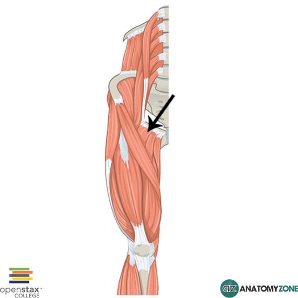
A doctor tells a soccer player that he has "a pulled hamstring" muscle. This results from a tearing of the origination of a hamstring muscle from the ___________.
ischial tuberosity
If the femoral nerve is cut, which muscle compartment would be most affected?
anterior thigh
what is the deep tissue that invests the thigh?
fascia lata
What is the iliotibial tract?
which muscles attach here?
thickened portion of the fascia lata. attachment site for two muscles. --> gluteus maximus and tensor fascia latae
O I A INN of PECtinius
O superior ramus of pubis
I pectineal line of femur, just inferior to lesser trochanter
A adduct hip, flex hip. (assists with medial rotation of hip joint.)
INN femoral (L2, L3) and may receive branch from obturator nerve
Origin, insertion, action and innervation of gluteus maximus?

O posterior ilium, dorsal surface of sacrum and coccyx and sacrotuberous ligament
I gluteal tuberosity of the femur and iliotibial tract
A extend hip/thigh and lateral rotation of hip/thigh; steadies thigh and helps rise from sitting position
Inn inferior gluteal nerve (L5, S1, S2)
O I A INN of gluteus medius?

O outer surface and crest of ilium between anterior and posterior gluteal lines
I lateral surface of greater trochanter of femur
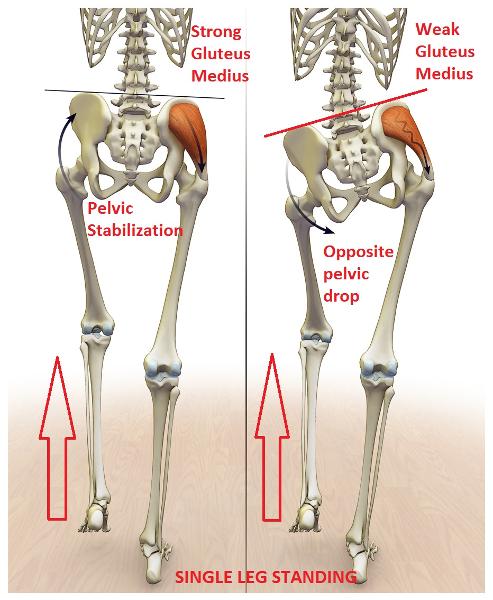
A abduct hip/thigh. anterior portions medially rotate hip joint. keep pelvis level when opposite limb is elevated.
INN superior gluteal nerve (L4, L5, S1)
O I A INN of gluteus minimus?
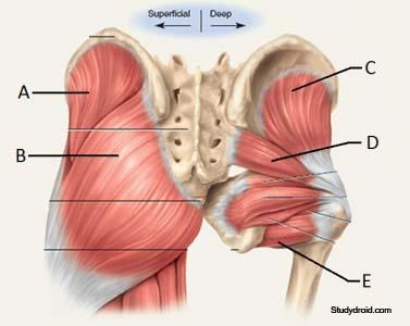
O outer surface and crest of ilium between anterior and inferior gluteal lines
I anterior surface of greater trochanter of femur
A abduct hip/thigh. anterior portions medially rotate hip joint. keep pelvis level when opposite limb is elevated. INN superior gluteal nerve (L4, L5, S1)
where does the iliotibial tract insert?
name of structure and the bone that it is on
gerdy's tubercle on lateral surface of tibia
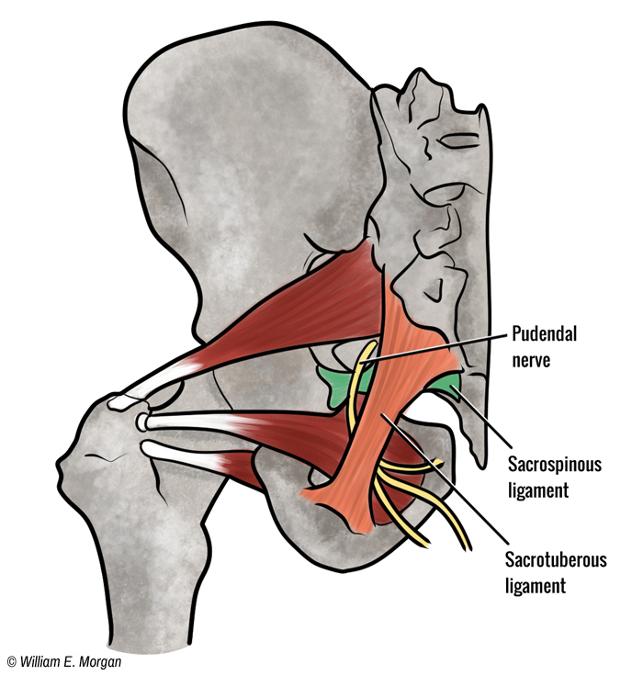
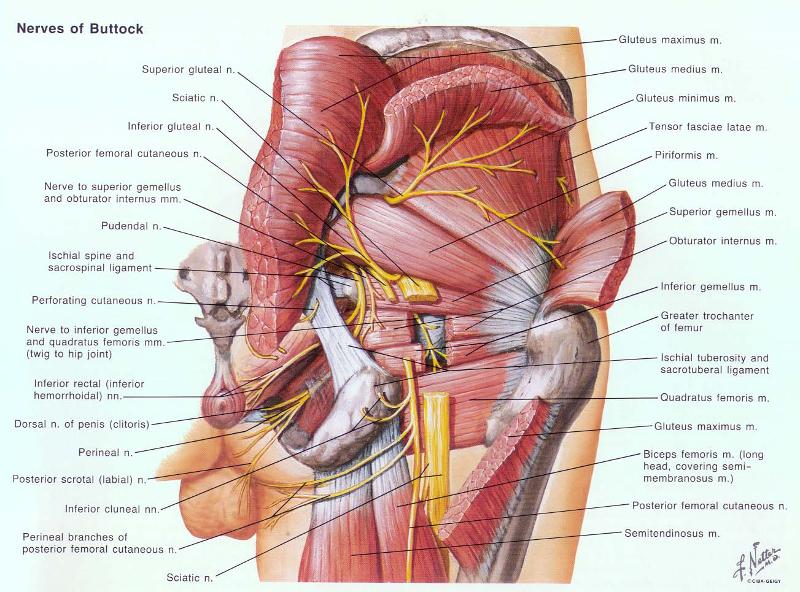
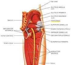
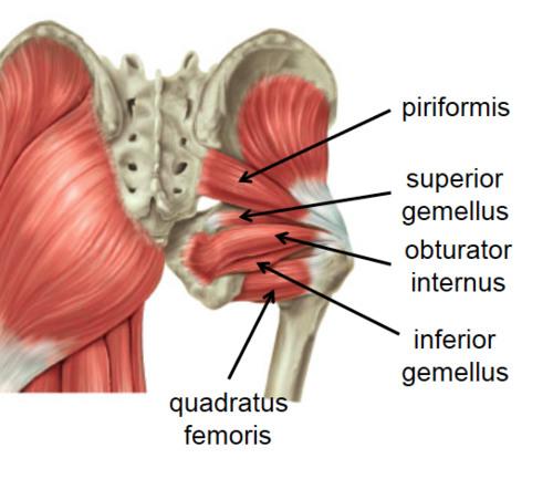
What is the O I A INN of piriformis
O anterior sacrum 2nd-4th sacral segments, superior margin of the greater sciatic notch, and sacrotuberous ligament
I superior border of the greater trochanter of the femur
A laterally rotate extended hip joint, abduct flexed hip joint, stabilize femoral head in acetabulum
INN branches of anterior rami of S1,S2
Which nerve may be damaged if someone display excessive pelvic tilt while walking?
superior gluteal nerve
If the right superior gluteal nerve is damaged, the pelvic drop will be toward which side of the body?

left side of the body
O I A INN of tensor fasciae latae?
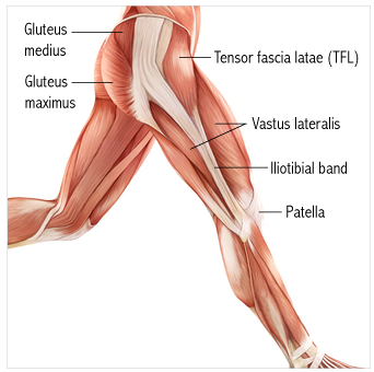
O anterior superior iliac spine; anterior part of iliac crest
I iliotibial tract which attaches to lateral condyle of tibia (Gerdy tubercle)
A flex hip, abduct hip. Stabilizes the extended knee joint
Inn superior gluteal nerve (L4, L5, S1)
What action are the deep gluteal muscles responsible for?
lateral rotation also help stabilize the hip joint
What is the O I A INN of piriformis
O anterior sacrum 2nd-4th sacral segments, superior margin of the greater sciatic notch, and sacrotuberous ligament
I superior border of the greater trochanter of the femur
A laterally rotate extended hip joint, abduct flexed hip joint, stabilize femoral head in acetabulum
INN branches of anterior rami of S1,S2
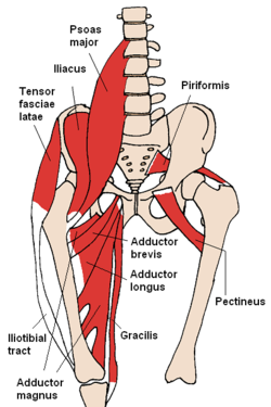
What are the two major anterior hip muscles?
psoas major and iliacus
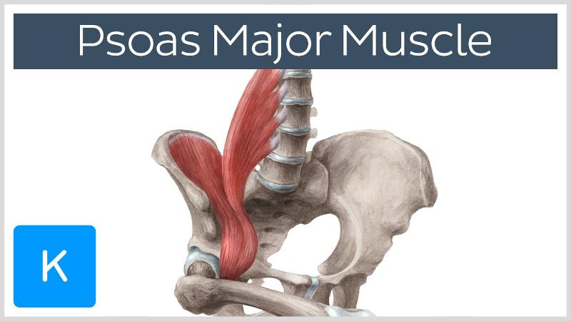
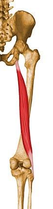
O I A INN of psoas major?

O sides of T12-L5 vertebrae and discs between them. transverse processes of all lumbar vertebrae
I lesser trochanter of the femur
A acting conjointly with iliacus in flexing and stabilizing hip joint, psoas major is also a postural muscle that helps control deviation of the trunk and is active during standing
INN Anterior rami of lumbar nerves (L1, L2, L3)
which ligament spans pubic tubercle and anterior superior iliac spine?

inguinal ligament
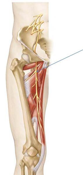
which is the largest nerve of the body and exits inferior to pirifomis?
sciatic nerve
O I A INN of iliacus?
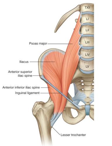
O iliac fossa, iliac crest, ala of sacrum, and anterior sacro-iliac ligaments
I tendon of psoas major, lesser trochanter, and femur distal to lesser trochanter
A acting conjointly with psoas major in flexing and stabilizing hip joint.
INN femoral n (L2, L3)
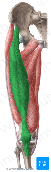
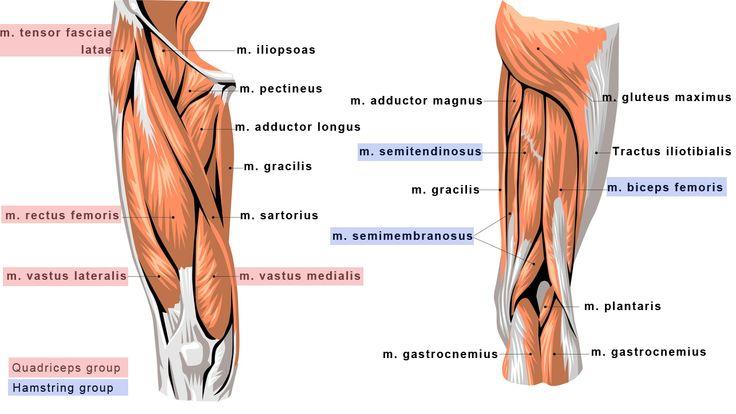
what are the anterior thigh muscles?
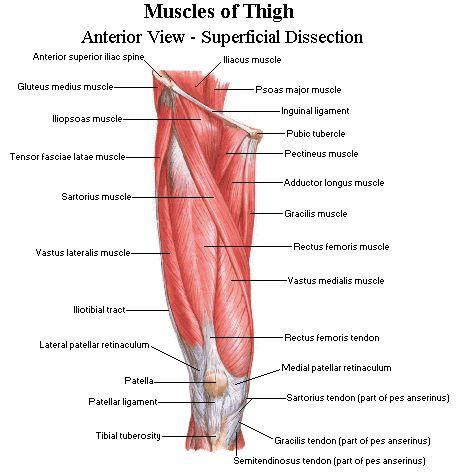
sartorius, rectus femoris, vastus medialis, vastus lateralis, vastus intermedius
vastus medialis O I A INN
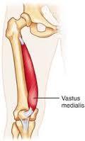
intertrochanteric line and medial lip of linea aspera
Via common tendinous (quadriceps tendon) and independent attachments to the base of the patella; indirectly via patellar ligament to tibial tuberosity. attaches to tibia and patella via aponeurosis (medial and lateral patellar retinacula)
extend leg
femoral nerve (L2, L3, L4)
O I A INN of sartorius?
anterior superior iliac spine and superior part of notch inferior to it
medial surface of proximal tibia
flexes, abducts and laterally rotates hip joint. flexes knee joint.
femoral n (L2, L3)
all anterior thigh muscles are innervated by which nerve?
femoral nerve
rectus femoris O I A IN

anterior inferior iliac spine and ilium superior to acetabulum
Via common tendinous (quadriceps tendon) and independent attachments to the base of the patella; indirectly via patellar ligament to tibial tuberosity.
extends knee joint; rectus femoris also stabilizes hip joint and helps iliopsoas flex hip joint
femoral nerve (L2, L3, L4)
O I A INN of quadratus femoris
O lateral border of ischial tuberosity
I quadrate tubercle on the intertrochanteric crest of femur and area inferior to it
A laterally rotates hip joint; also pulls femoral head into acetabulum to stabilize hip joint/pelvis
Inn nerve to quadratus femoris L5 S1
vastus medialis O I A INN
intertrochanteric line and medial lip of linea aspera
Via common tendinous (quadriceps tendon) and independent attachments to the base of the patella; indirectly via patellar ligament to tibial tuberosity. attaches to tibia and patella via aponeurosis (medial and lateral patellar retinacula)
extend leg
femoral nerve (L2, L3, L4)
vastus intermedius O I A INN
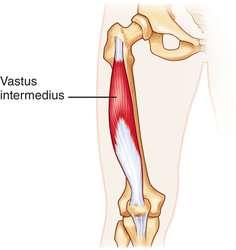
anterior and lateral surfaces of shaft of fermur
Via common tendinous (quadriceps tendon) and independent attachments to the base of the patella; indirectly via patellar ligament to tibial tuberosity.
extend leg
femoral nerve (L2,L3,L4)
what is the main action of the medial thigh muscles?
adductors
the majority of medial thigh muscles are innervated by which nerve? What are the exceptions?
obturator;
pectinius is innervated by both femoral and obturator
the hamstring part of adductor magnus is innervated by tibial division of sciatic n (L4)
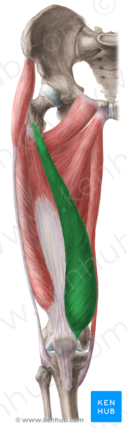
what are the medial thigh muscles?
gracilis, pectinius, adductor longus, adductor brevis, adductor magnus
O I A INN of gracilis
body and inferior ramus of pubis
medial surface of proximal tibia
adduct hip, flex knee and helps rotate knee medially
obturator n (L2, L3)
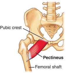
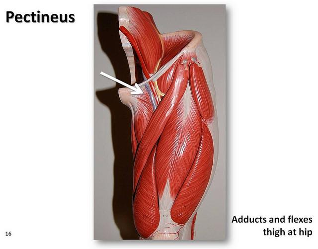
O I A INN of pectinius

O superior ramus of pubis
I pectineal line of femur, just inferior to lesser trochanter
A adduct hip, flex hip. (assists with medial rotation of hip joint.)
INN femoral (L2, L3) and may receive branch from obturator nerve
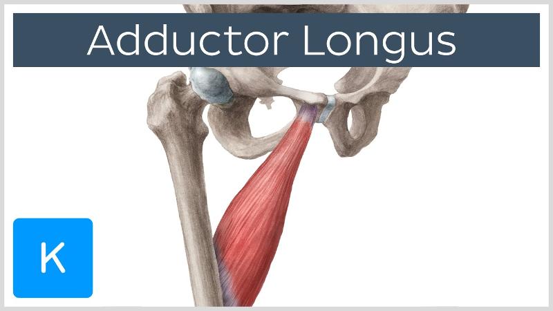
O I A INN of adductor longus
O body of pubis inferior to pubic crest
I middle third of linea aspera of femur
A adduct hip
INN obturator n (L2, L3, L4)
O I A INN of adductor brevis?
O body and inferior ramus of pubic bone
I pectineal line and proximal part of linea aspera of femur
A adduct hip and to some extent flexes hip
INN obturator nerve (L2, L3, L4)
O I A INN of adductor magnus?
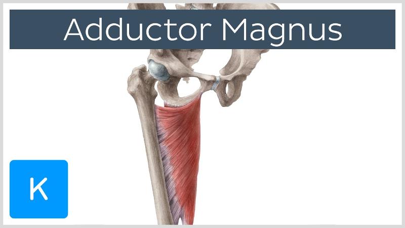
O adductor part: inferior ramus of pubis, and ramus of ischium
hamstring part: ischial tuberosity
I adductor part: gluteal tuberosity, linea aspera, and medial supracondylar line
hamstring part: adductor tubercle of femur
A adduct hip. Adductor part also flexes hip joint and hamstring part extends it.
INN adductor part: obturator n (L2, L3, L4)
hamstring part: tibial division of sciatic n (L4)
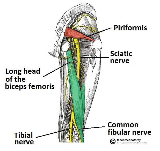
What are the posterior thigh muscles?
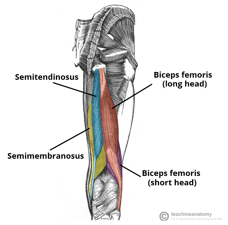
biceps femoris (long head and short head)
semitendinosis
semimembanosus
What nerve innervates the majority of posterior thigh muscles? what is the exception?
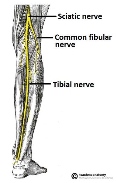
tibial division of sciatic nerve (L4-S3). the short head of biceps femoris is innervated by the common fibular nerve (L4-S2)
what is the deepest and largest medial thigh muscle?
adductor magnus

O I A INN of biceps femoris, long head
O ischial tuberosity
I lateral side of head of fibula; tendon is split at this site by fibular collateral ligament of the knee
A flexes knee joint and rotates it laterally when knee is flexed; extends hip joint (like when starting to walk)
INN tibial division of the sciatic nerve (L5, S1, S2)
O I A INN of PECtinius
O superior ramus of pubis
I pectineal line of femur, just inferior to lesser trochanter
A adduct hip, flex hip. (assists with medial rotation of hip joint.)
INN femoral (L2, L3) and may receive branch from obturator nerve
O I A INN of semitendinosis
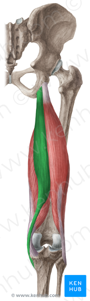
O ischial tuberosity
I medial surface of proximal tibia
A extend hip, flex knee, and rotate leg medially when knee is flexed. when hip and knee are flexed, can extended trunk.
INN tibial division of sciatic n (L5, S1, S2)
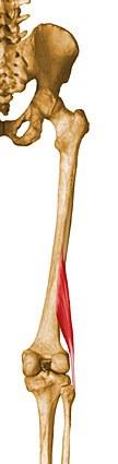
O I A INN of semimembranosis
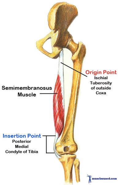
O iscial tuberosity
I posterior surface of medial condyle of tibia
A extend hip, flex knee, and rotate leg medially when knee is flexed. when hip and knee are flexed, can extended trunk.
INN tibial division of sciatic n ( L5, S1, S2)
Spinal cord levels of inferior gluteal nerve?
L5, S1, S2
O I A INN of obturator internus
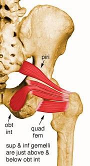
O pelvic surface of ilium and ischium; obturator membrane
I medial surface of greater trochanter (trochanteric fossa) of femur
A laterally rotate extended hip joint, abduct flexed hip joint, steady femoral head in acetabulum
Inn nerve to obturator internus (L5, S1)
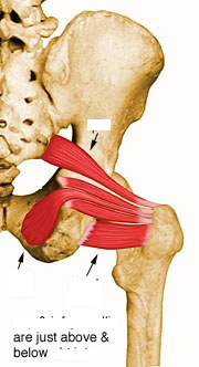
O I A INN of inferior and superior gemelli
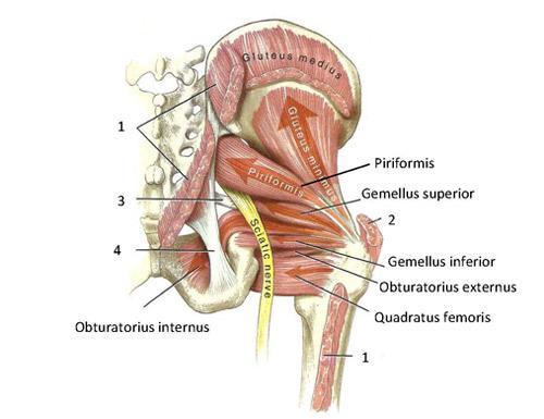
O superior: ischial spine
inferior: ischial tuberosity
I medial surface of greater trochanter (trochanteric fossa) of femur
A laterally rotate extended hip joint, abduct flexed hip joint, steady femoral head in acetabulum
Inn superior: nerve to obturator internus L5, S1
inferior: nerve to quadratus femoris L5, S1
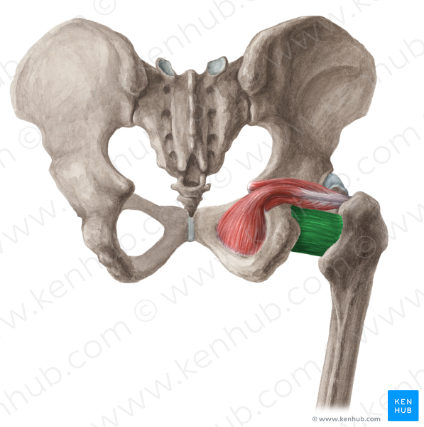
O I A INN of quadratus femoris
O lateral border of ischial tuberosity
I quadrate tubercle on the intertrochanteric crest of femur and area inferior to it
A laterally rotates hip joint; also pulls femoral head into acetabulum to stabilize hip joint/pelvis
Inn nerve to quadratus femoris L5 S1
what are the quadriceps femoris muscles?
rectus femoris, vastus lateralis, vastus medialis, and vastus intermedius
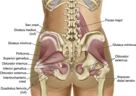
O I A INN of obturator externus
O margins of obturator foramen and obturator membrane
I trochanteric fossa of femur
A laterally rotates hip joint, pulls head of femur into acetabulum holding pelvis steady; adduction
Inn obturator nerve (L3, L4)
what structures pass through adductor hiatus
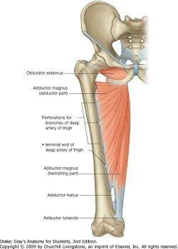
femoral artery and vein from the anterior compartment of the thigh to the popliteal fossa posterior to the knee
what are the borders of the adductor hiatus?
distal aponeurotic attachment of the adductor part of the adductor magnus and tendon of hamstring part.
where does the iliotibial tract insert?
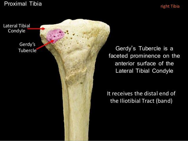
gerdy's tubercle on lateral surface of tibia