What three things make up the pelvic girdle?
Ilium, Ischium, Pubis
made up of to coxae (hip or innominate bones)
What three things form the pelvis?
sacrum, coccyx, and pelvic girdle
what does the femoral head of the femur articulate with?
acetabulum
Fovea Capitis Properties
Depression in the femoral head
what part of the femur is a frequent location for fractures?
femoral neck
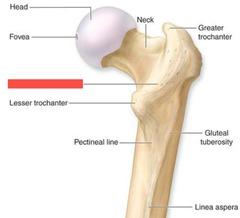
Greater trochanter
Lesser trochanter
Intertrochanteric crest (posterior)
Linea aspera
adductor tubercle
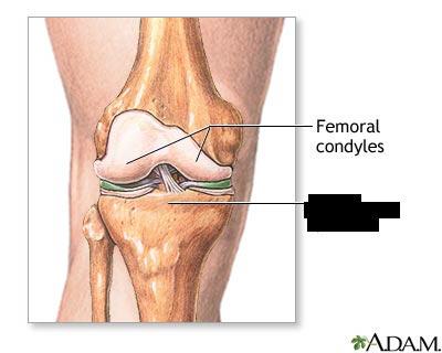
Medial & Lateral condyles
Medial & Lateral epicondyles
patella
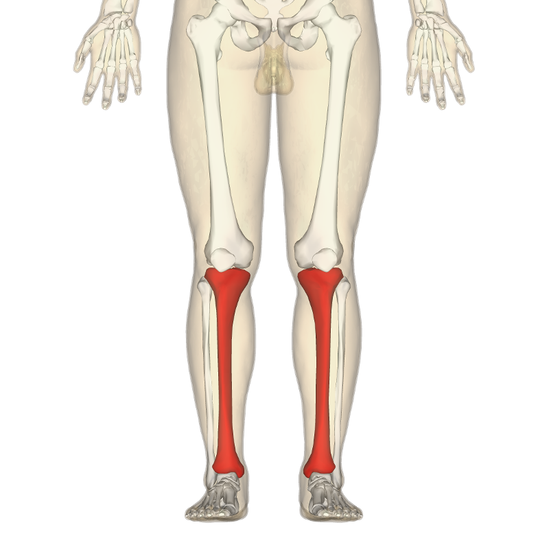
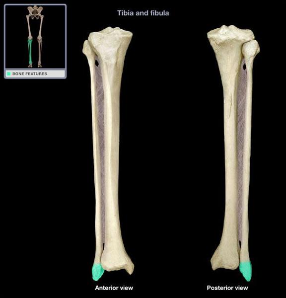
Tibia
Tibial plateau
Medial & lateral condyles of tibia
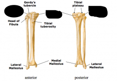
tibial tuberosity
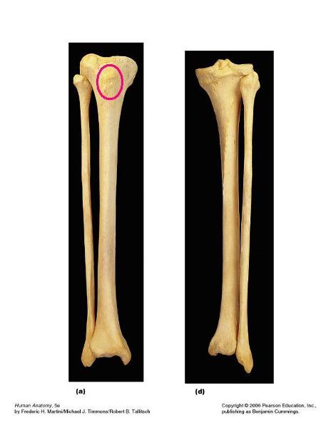
pes anserine
gerdy's tubercle
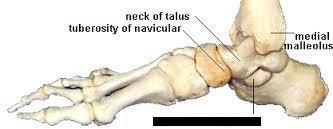
medial malleolus

Fibula
head of fibula
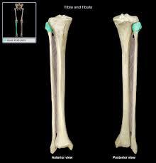
lateral malleolus
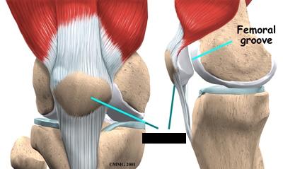
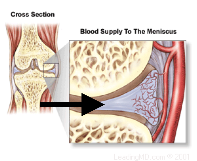
Meniscus is composed of what
-Semilunar cartilages
-medial- "C shaped"
-lateral- "O shaped"
-only 3-5mm thick on average
Functions of the meniscus
absorbs shock/jolts
improve stability of tibiofemoral joint
-deepens the condylar surface
Which part of the meniscus is more firmly attached?
the medial meniscus is more firmly attached than the lateral
meniscotibial (coronary) ligaments
?
Why do lesions occur?
it is a result of femoral rotation with foot planted (fixed tibia)
With a cross-sectional appearance, menisci are wedged shaped and have three zones. What are they?
red zone (highly vascular)
red-white zone (mild vascularity)
white zone (avascular)
What are the three types of knee alignments?
genu varum "bow legged"
genu valgum "knock knees"
genu recurvatum "hyperextension"
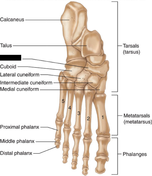
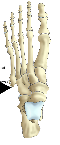
What are the 7 tarsal bones?
Talus
Calcaneus
Navicular
Cuboid
Lateral, intermediate & medial cuneiforms
Talus
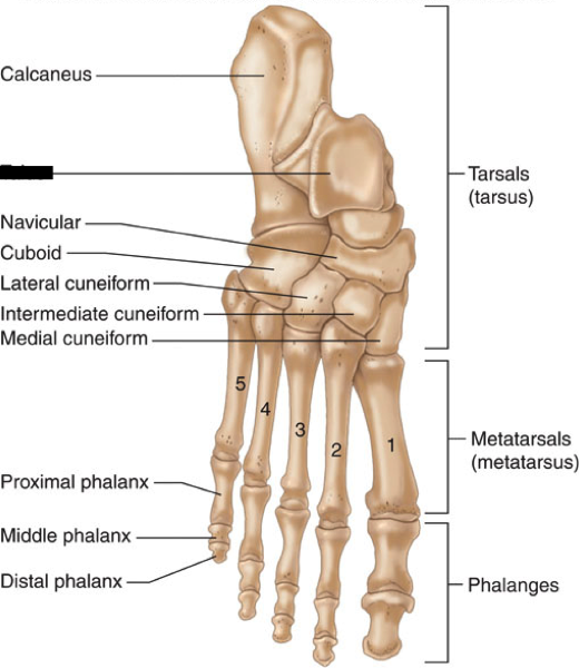
Calcaneus
Navicular
Cuboid
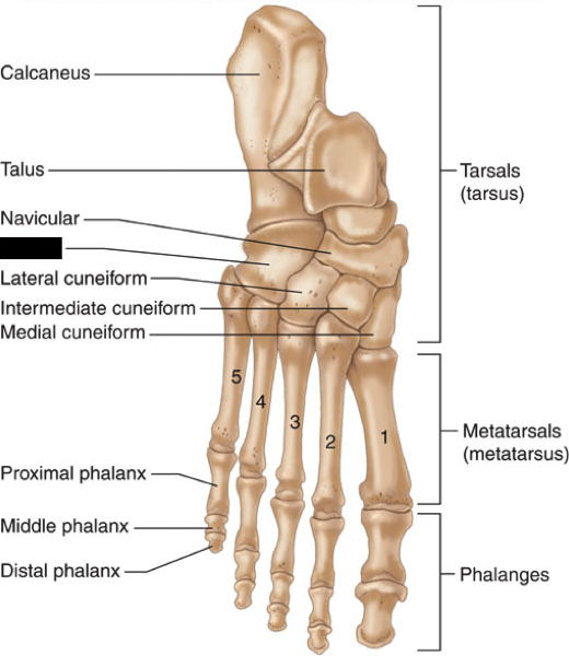
Lateral cuneiform
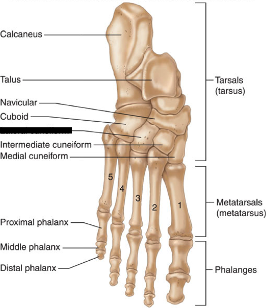
intermediate cuneiform
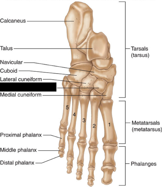
medial cuneiform
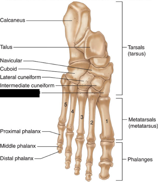
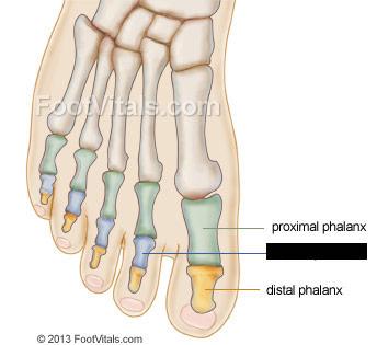
1st-5th metatarsals
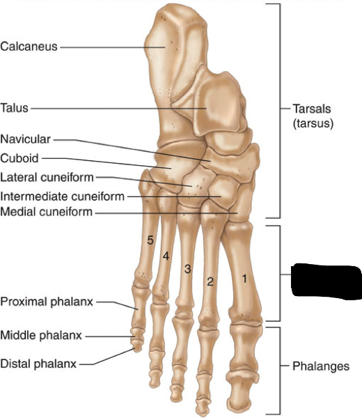
Distal phalanx of great toe
Proximal phalanx of great toe
middle phalanx of second toe
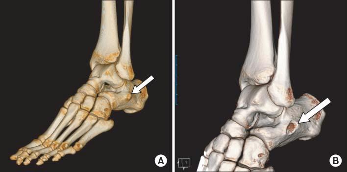
Two landmarks found on the calcaneus?
sustentaculum tali- medial
peroneal tubercle- lateral
navicular tubercle

5th metatarsal styloid process
Sesamoid bones in foot
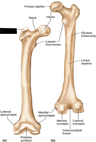
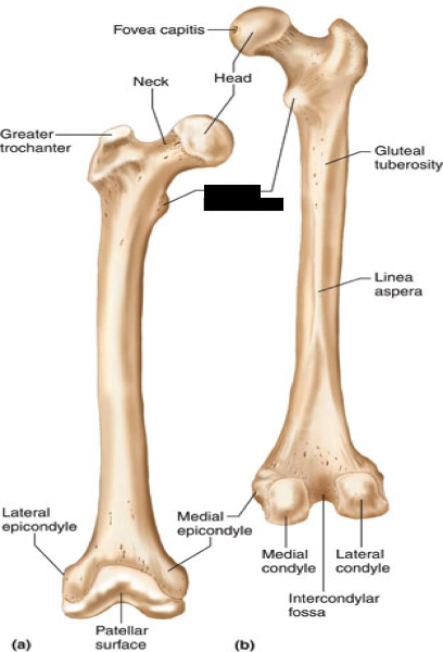
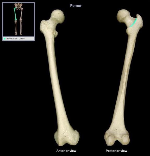
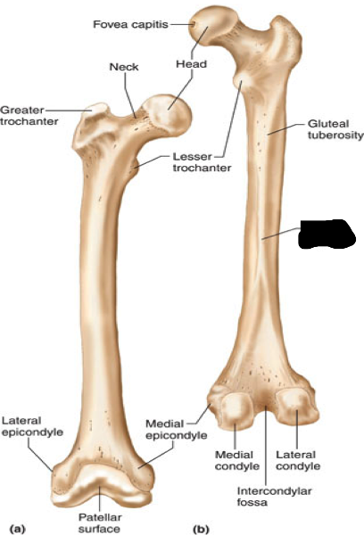
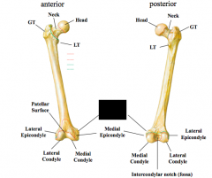
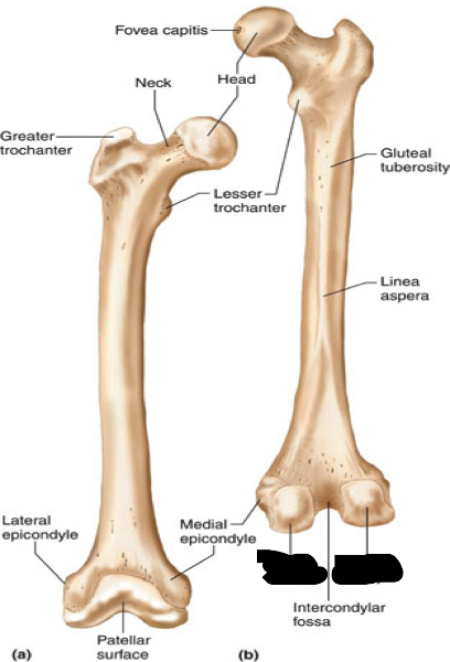
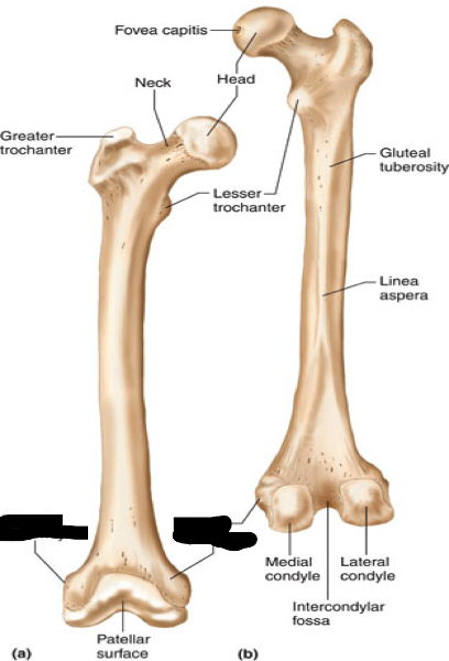
Femur Properties
Longest & strongest bone in the body
Landmarks of the proximal femur
-Femoral head
-Fovea capitis
-Femoral neck
-Greater trochanter
-Lesser trochanter
-Intertrochanteric crest
Attachment site for fovea capitis
Ligamentum Teres
Fovea capitis
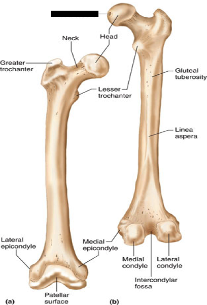
Landmarks of the distal femur
-linea aspera
-adductor tubercle
-medial & lateral epicondyles
-medial & lateral condyles
What do the medial and lateral condyles of the femur articulate with
Tibia
Advantages of a sesamoid bone ("free-floating")
Improves leverage of quadriceps and their ability to extend knee
what is the patella embedded in
Within the quadriceps and patellar tendon
Kansas patella fracture, how did it occur?
the rectus femurs tendon and patellar tendon tore causing the patella to shatter
Proximal end of tibia expands into
-Tibial Plateau
-Medial and lateral condyles
-Tibial tuberosity
-Pes anserine
-Gerdy's tubercle
Distal end of tibia forms into
medial malleolus
Insertion site of quadriceps
Tibial tuberosity
Limited weight-bearing during movement
Lateral Malleolus Property
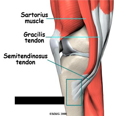
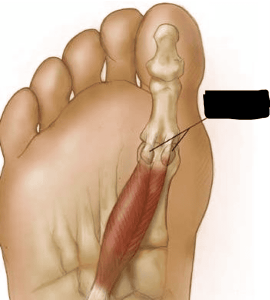
What 3 muscles insert at the pes anserine
-sartorius
-gracilis
-semitendinous
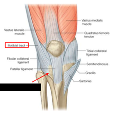
What band inserts at the Gerdy's tubercle?
Iliotibial band (IT)
Intertrochanteric line (anterior)
Red Zone
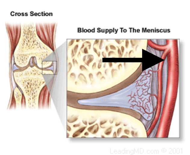
red-white zone
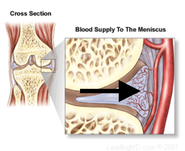
White zone
Patellar Tendon properties
continuation of the quadriceps tendon
tibia and fibula articulates with what bone
talus
Largest tarsal bone
Calcaneus (heel)
-weight bearing bone
Navicular location
medial
Cuboid location
lateral
Property of the metatarsals
form the arches of foot with ligamentous and muscular support
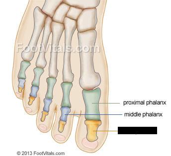
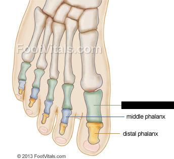
Property of the phalanges
-each toe has has 3 phalanges (proximal, middle, distal) except big toe/first digit
-first digit only has a proximal and distal phalanx
sustentaculum tali- medial
peroneal tubercle- lateral