Instructions for Side by Side Printing
- Print the notecards
- Fold each page in half along the solid vertical line
- Cut out the notecards by cutting along each horizontal dotted line
- Optional: Glue, tape or staple the ends of each notecard together
Lower Extremity
front 1 What three things make up the pelvic girdle? | back 1 Ilium, Ischium, Pubis made up of to coxae (hip or innominate bones) |
front 2 What three things form the pelvis? | back 2 sacrum, coccyx, and pelvic girdle |
front 3 what does the femoral head of the femur articulate with? | back 3 acetabulum |
front 4 Fovea Capitis Properties | back 4 Depression in the femoral head |
front 5 what part of the femur is a frequent location for fractures? | back 5 femoral neck |
front 6 Greater trochanter | back 6 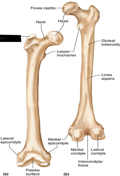 |
front 7 Lesser trochanter | back 7 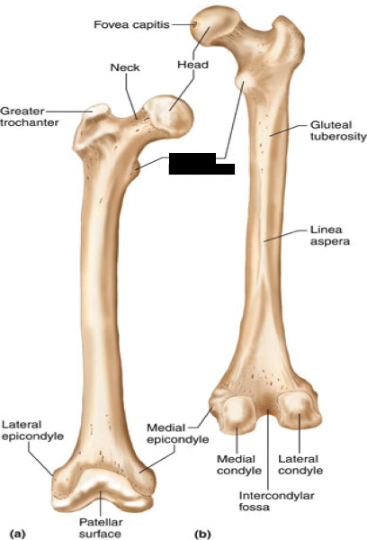 |
front 8 Intertrochanteric crest (posterior) | back 8 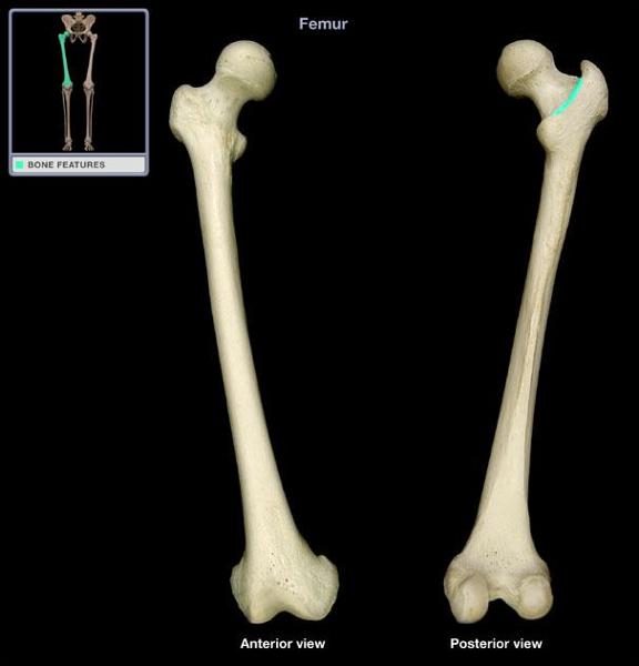 |
front 9 Linea aspera | back 9 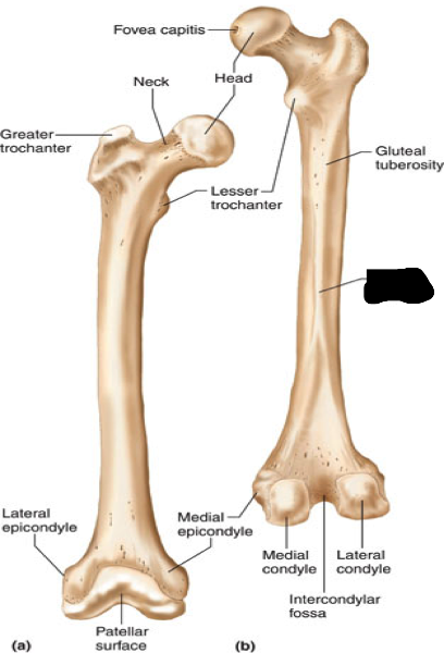 |
front 10 adductor tubercle | back 10 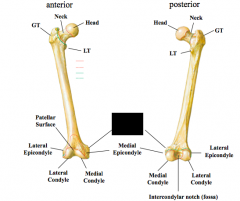 |
front 11 Medial & Lateral condyles | back 11 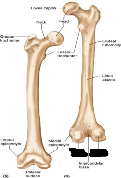 |
front 12 Medial & Lateral epicondyles | back 12 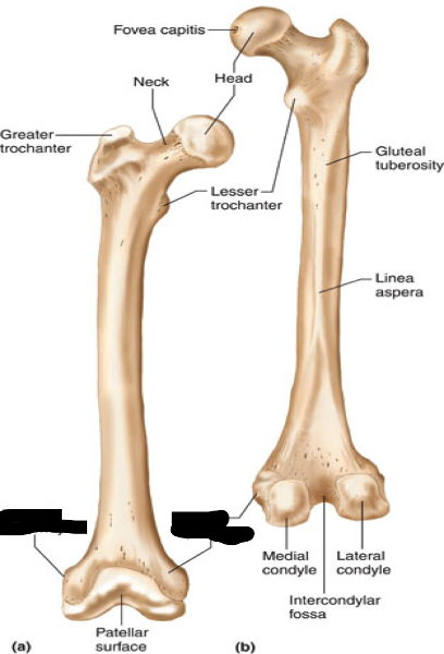 |
front 13 patella | back 13 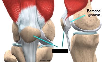 |
front 14 Tibia | back 14 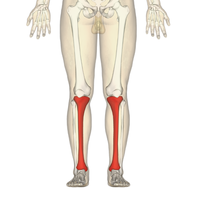 |
front 15 Tibial plateau | back 15 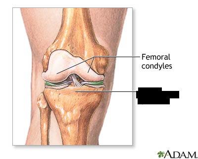 |
front 16 Medial & lateral condyles of tibia | back 16 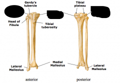 |
front 17 tibial tuberosity | back 17 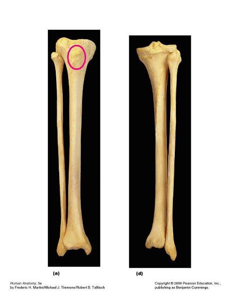 |
front 18 pes anserine | back 18 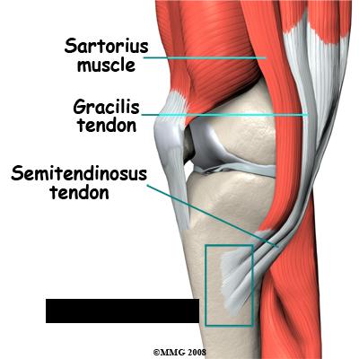 |
front 19 gerdy's tubercle | back 19 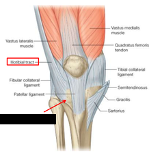 |
front 20 medial malleolus | back 20  |
front 21 Fibula | back 21 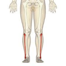 |
front 22 head of fibula | back 22 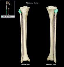 |
front 23 lateral malleolus | back 23 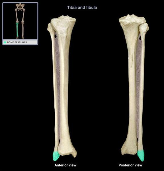 |
front 24 Meniscus is composed of what | back 24 -Semilunar cartilages -medial- "C shaped" -lateral- "O shaped" -only 3-5mm thick on average |
front 25 Functions of the meniscus | back 25 absorbs shock/jolts improve stability of tibiofemoral joint -deepens the condylar surface |
front 26 Which part of the meniscus is more firmly attached? | back 26 the medial meniscus is more firmly attached than the lateral |
front 27 meniscotibial (coronary) ligaments | back 27 ? |
front 28 Why do lesions occur? | back 28 it is a result of femoral rotation with foot planted (fixed tibia) |
front 29 With a cross-sectional appearance, menisci are wedged shaped and have three zones. What are they? | back 29 red zone (highly vascular) red-white zone (mild vascularity) white zone (avascular) |
front 30 What are the three types of knee alignments? | back 30 genu varum "bow legged" genu valgum "knock knees" genu recurvatum "hyperextension" |
front 31 What are the 7 tarsal bones? | back 31 Talus Calcaneus Navicular Cuboid Lateral, intermediate & medial cuneiforms |
front 32 Talus | back 32 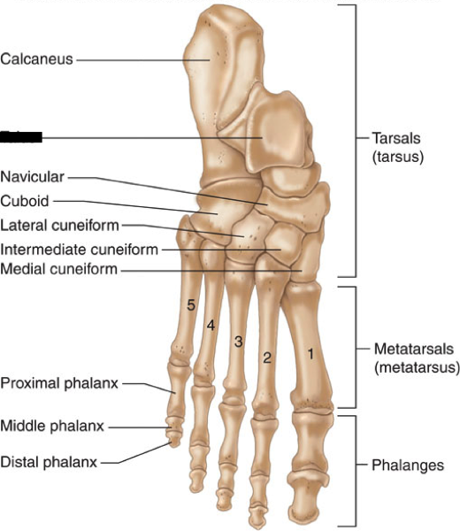 |
front 33 Calcaneus | back 33 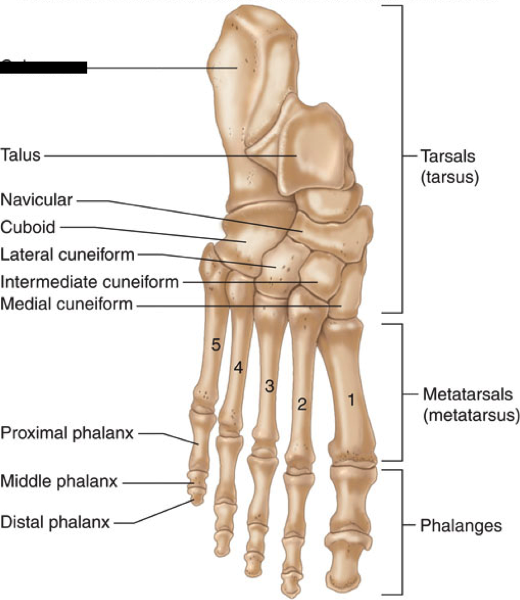 |
front 34 Navicular | back 34 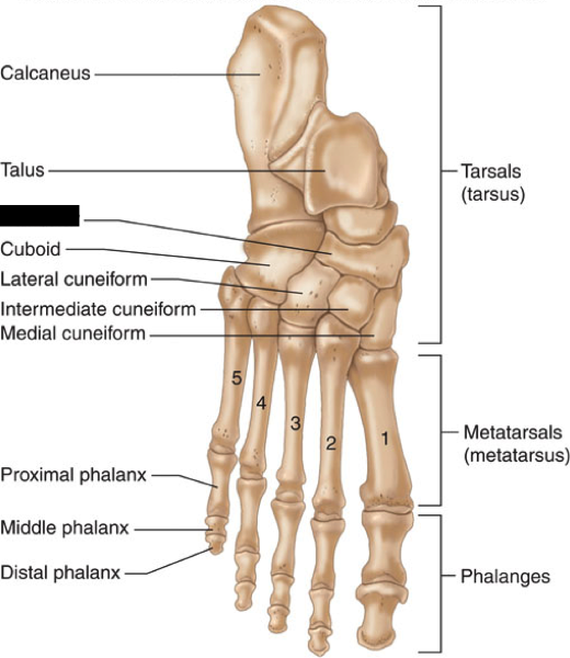 |
front 35 Cuboid | back 35 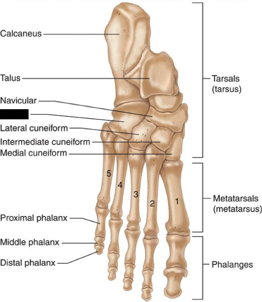 |
front 36 Lateral cuneiform | back 36 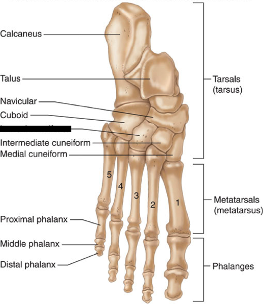 |
front 37 intermediate cuneiform | back 37 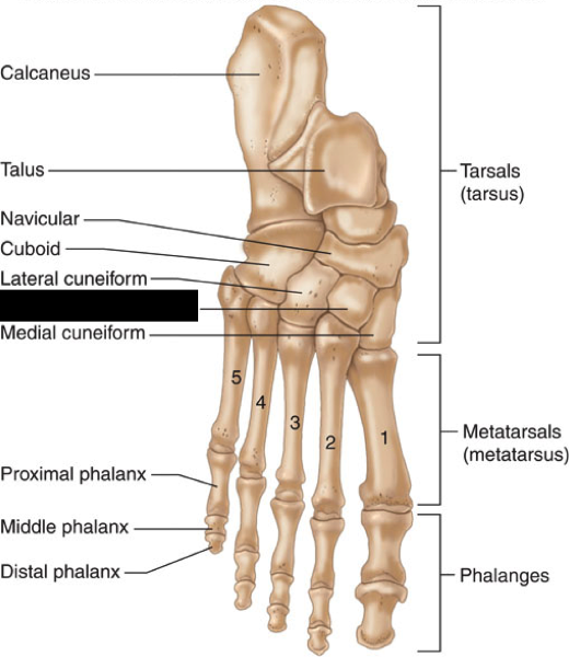 |
front 38 medial cuneiform | back 38 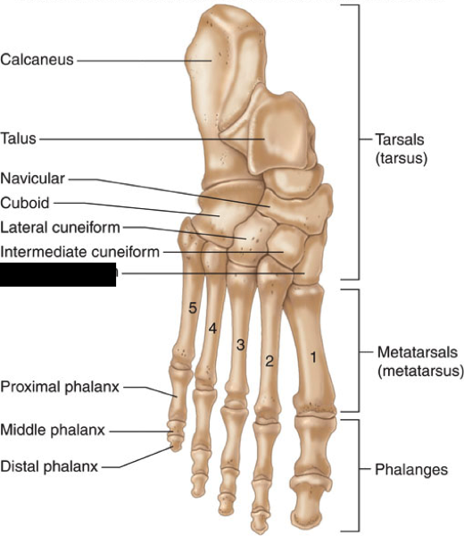 |
front 39 1st-5th metatarsals | back 39 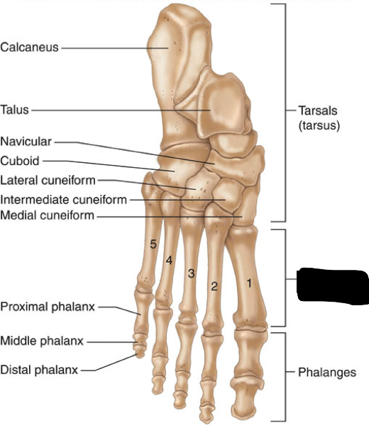 |
front 40 Distal phalanx of great toe | back 40 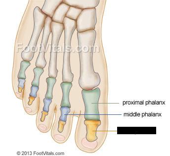 |
front 41 Proximal phalanx of great toe | back 41 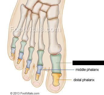 |
front 42 middle phalanx of second toe | back 42 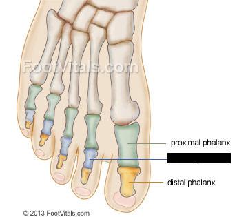 |
front 43 Two landmarks found on the calcaneus? | back 43 sustentaculum tali- medial peroneal tubercle- lateral |
front 44 navicular tubercle | back 44  |
front 45 5th metatarsal styloid process | back 45 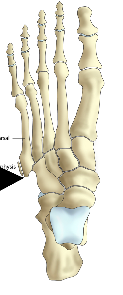 |
front 46 Sesamoid bones in foot | back 46 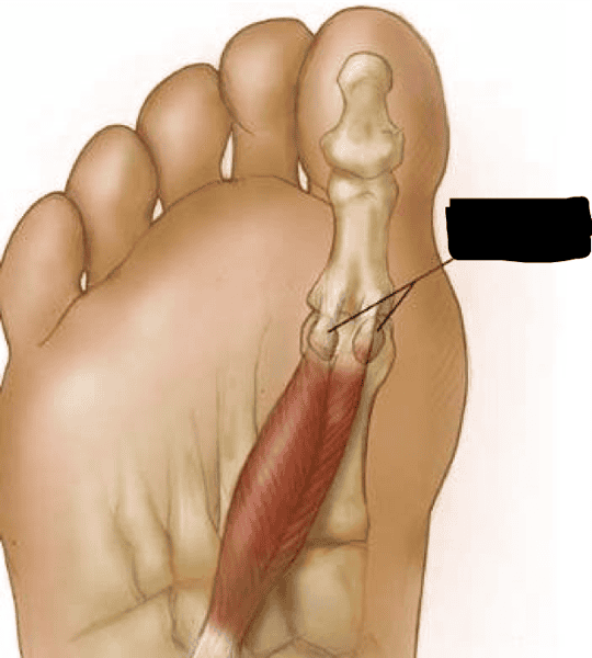 |
front 47 Femur Properties | back 47 Longest & strongest bone in the body |
front 48 Landmarks of the proximal femur | back 48 -Femoral head -Fovea capitis -Femoral neck -Greater trochanter -Lesser trochanter -Intertrochanteric crest |
front 49 Attachment site for fovea capitis | back 49 Ligamentum Teres |
front 50 Fovea capitis | back 50 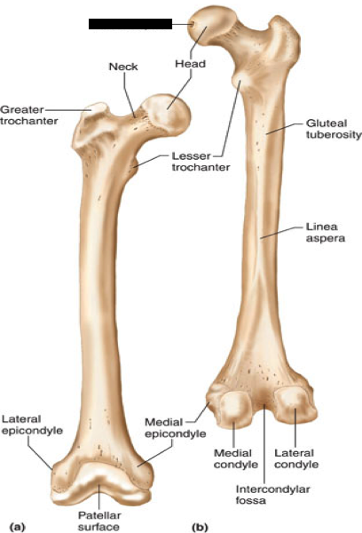 |
front 51 Landmarks of the distal femur | back 51 -linea aspera -adductor tubercle -medial & lateral epicondyles -medial & lateral condyles |
front 52 What do the medial and lateral condyles of the femur articulate with | back 52 Tibia |
front 53 Advantages of a sesamoid bone ("free-floating") | back 53 Improves leverage of quadriceps and their ability to extend knee |
front 54 what is the patella embedded in | back 54 Within the quadriceps and patellar tendon |
front 55 Kansas patella fracture, how did it occur? | back 55 the rectus femurs tendon and patellar tendon tore causing the patella to shatter |
front 56 Proximal end of tibia expands into | back 56 -Tibial Plateau -Medial and lateral condyles -Tibial tuberosity -Pes anserine -Gerdy's tubercle |
front 57 Distal end of tibia forms into | back 57 medial malleolus |
front 58 Insertion site of quadriceps | back 58 Tibial tuberosity |
front 59 Limited weight-bearing during movement | back 59 Lateral Malleolus Property |
front 60 What 3 muscles insert at the pes anserine | back 60 -sartorius -gracilis -semitendinous |
front 61 What band inserts at the Gerdy's tubercle? | back 61 Iliotibial band (IT) |
front 62 Intertrochanteric line (anterior) | back 62 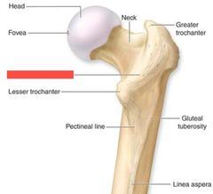 |
front 63 Red Zone | back 63 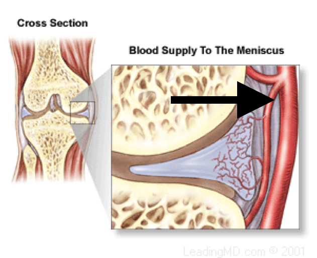 |
front 64 red-white zone | back 64 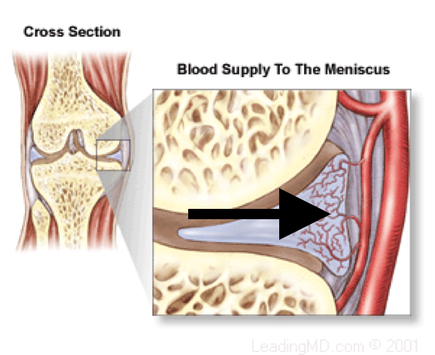 |
front 65 White zone | back 65 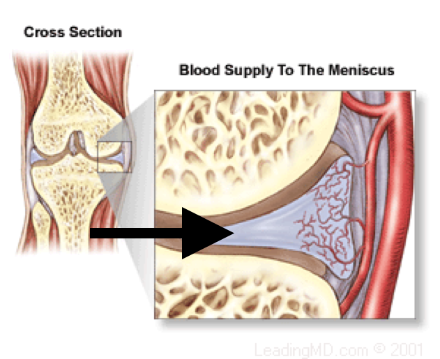 |
front 66 Patellar Tendon properties | back 66 continuation of the quadriceps tendon |
front 67 tibia and fibula articulates with what bone | back 67 talus |
front 68 Largest tarsal bone | back 68 Calcaneus (heel) -weight bearing bone |
front 69 Navicular location | back 69 medial |
front 70 Cuboid location | back 70 lateral |
front 71 Property of the metatarsals | back 71 form the arches of foot with ligamentous and muscular support |
front 72 Property of the phalanges | back 72 -each toe has has 3 phalanges (proximal, middle, distal) except big toe/first digit -first digit only has a proximal and distal phalanx |
front 73 sustentaculum tali- medial | back 73 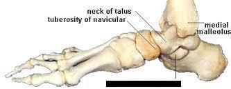 |
front 74 peroneal tubercle- lateral | back 74  |