Blood main functions
Transport oxygen and nutrients, remove wastes, immune defense, maintain homeostasis
Blood components
Plasma, erythrocytes, leukocytes, thrombocytes
Arteries
Carry blood away from the heart
Veins
Return blood to the heart
Capillaries
Site of gas, nutrient, and waste exchange
Blood cells formed in RED bone marrow
Hemocytoblast (early stem cell), Proerythroblast (RBC), Myeloblast (Granulocytic WBC), Lymphoblast (lymphocytes/agranulocytes), Monoblast (monocytes/agranulocytes), Megakaryoblast (thrombocytes/platelets)
Erythrocyte characteristics
Biconcave flexible discs, no nucleus, glucose is the primary nutrient source, contains hemoglobin. Erythropoietin produced in the kidney stimulates erythrocyte production. Folic acid and Vitamin B12 are essential for RBC maturation.
Breakdown of hemoglobin
1. Hemolysis of erythrocyte->Hemoglobin->Heme and Globin. 2. Heme->Iron that is recycled to bone marrow or stored->Hematopoiesis. 3. Heme->Bilirubin/unconjugated (transported bound to serum albumin)->Blood->Liver (conjugated with glucuronic acid)->Bilirubin/conjugated-> Bile 4. Globin-> Amino acids recycled
Hemostasis steps
Vasoconstriction or vascular spasm after injury, platelet plug, coagulation fibrin mesh formation
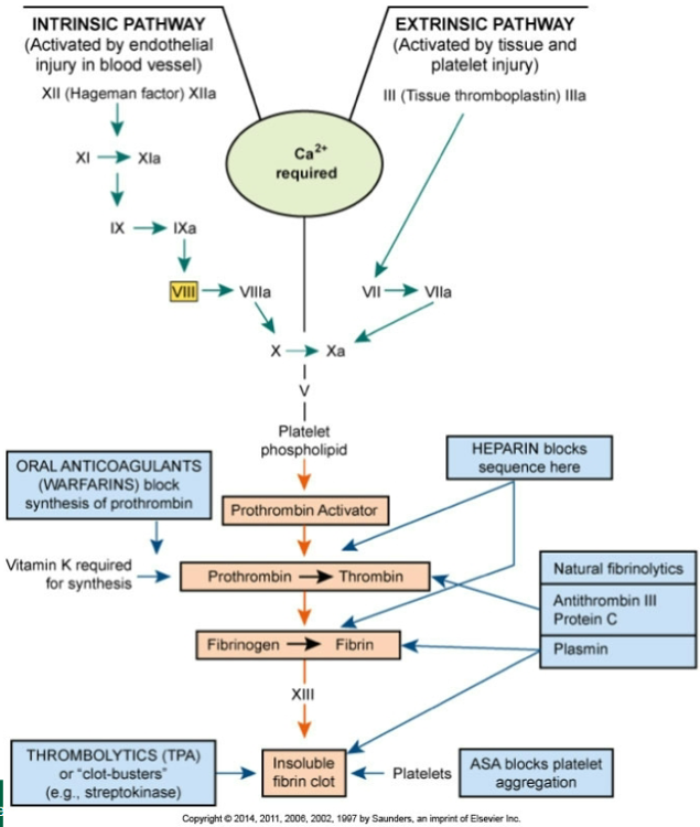
The Common Pathway of Clot Formation and Anticoagulant Drugs
CBC test
Measures RBC, WBC, platelet counts hemoglobin and hematocrit.
Leukocytosis
Increased WBCs associated with inflammation or infection
Leukopenia
Decreased WBCs associated with some viral infections, radiation, chemotherapy
Increased eosinophils
Common in allergic responses
Types of diagnostic tests
Morphology (shows size, shape, uniformity, maturity of cells), Hematocrit (Percent by volume of cellular elements in blood), Hemoglobin (Amount of hemoglobin per unit volume of blood. Mean corpuscular volume (MCV) indicates the oxygen-carrying capacity of blood).
Reticulocyte count
Indicates bone marrow production of RBCs
Chemical analysis
Determines serum levels of components such as iron, vitamin B12, folic acid, cholesterol, urea, glucose
Bleeding time
Measures platelet function
PT and PTT
Tests measuring blood clotting factor function
Whole blood, packed RBCs, packed platelets
For severe anemia or thrombocytopenia
Plasma or colloid volume-expanding solutions
To maintain blood volume. Crystalloids (intravenous saline; increase intravascular and interstitial volume) and Colloid (contain glucose or hydroxyethyl starch to increase intravascular volume)
Artificial blood products
O2 and CO2 transport. Compatible with all blood types. None of them can perform all the complex functions of normal whole blood
Epoetin Alpha
Artificial form of erythropoietin. Boost RBC production. Before certain surgical procedures, anemia related to cancer, chronic renal failure
Bone marrow or stem cell transplantation
Close tissue matches necessary. Treatment of some cancers, severe immunodeficiency, severe blood cell diseases
Drug treatment
Aids in the clotting process
Anemia general definition
Reduced oxygen transport due to low hemoglobin or RBCs
In anemia oxygen deficit leads to what?
Less energy production, Compensation mechanisms (Tachycardia and vasoconstriction=Increased HR and BP), General signs of anemia (fatigue, pallor, dyspnea/shortness of breath), Decreased regeneration of epithelial cells, Angina and Congestive Heart Failure
Iron deficiency anemia cause
Low iron from blood loss, poor intake, malabsorption, liver disease, which impairs hemoglobin synthesis.
Iron deficiency RBC type
Microcytic (small) hypochromic (reduced hemoglobin content) erythrocytes
Iron deficiency manifestations
Cold intolerance, irritability, fatigue, pallor, stomatitis/glossitis (inflammation of the mouth mucous membranes and tongue), tachycardia, syncope (fainting)
Iron deficiency treatment
Iron replacement and treatment of underlying cause
Pernicious anemia cause
Lack of intrinsic factor prevents vitamin B12 absorption possibly due to genetic factors and often accompanied by chronic gastritis
Pernicious anemia RBC type
Large immature megaloblastic erythrocytes that carry less hemoglobin and have a shorter life span
Pernicious anemia manifestations
Red enlarged sore smooth tongue, Paresthesia (tingling due to nerve damage), digestive discomfort (nausea and diarrhea)
Pernicious anemia diagnosis
Microscopic examination (erythrocytes), Bone marrow examination (hyperactive), Low serum B12, hypochlorhydria/achlorhydria (low levels of stomach acid), gastric atrophy (stomach cells atrophy)
Pernicious anemia treatment
Vitamin B12 replacement injections
Aplastic anemia definition
Bone marrow failure causing pancytopenia (low levels of RBCs, WBCs, and platelets.) Normal appearing RBCs.
Aplastic anemia causes
Often idiopathic but could be due to Myelotoxins (Radiation, chemicals, drugs), Viruses, Genetic disorders (Myelodysplastic syndrome and Fanconi's anemia)
Aplastic anemia treatment
Removal of any bone marrow suppressants and supportive care bone marrow transplant
Hemolytic anemia definition
Excessive destruction of red blood cells
Hemolytic anemia causes
Immune reactions, changes in blood chemistry, infections, toxins, incompatible transfusion, genetic defects
Sickle cell anemia cause
Genetic condition (Autosomal, incomplete dominance, homozygous recessive) Inherited abnormal hemoglobin HbS (Amino acid, Valine, is replacing glutamic acid on the Beta chain)
Sickle cell crisis trigger
Low oxygen causes RBC sickling and vessel obstruction because they are too large to pass through microcirculation.
Sickle cell complications
Infarctions (obstruction of blood flow), Hyperbilirubinemia, jaundice, gallstones (high rate of hemolysis)
Sickle cell manifestations
Vascular occlusion/infarctions (lungs: acute chest syndrome and smaller blood vessels: Hand-foot syndrome), necrosis, ischemia, severe pain, Hyperbilirubinemia (Jaundice), splenomegaly (enlarged spleen), Congestive heart failure
Sickle cell diagnosis
Hemoglobin electrophoresis, Blood test (abnormal RBC morphology), Prenatal DNA analysis
Sickle cell treatment
Hydroxyurea, folic acid, immunizations, bone marrow transplant
Thalassemia cause
Genetic defect (Autosomal dominant inheritance) reducing alpha or beta globin chains. Abnormal globin damages RBCs causing hemolysis (lower RBC numbers) and anemia
Thalassemia manifestations
Pallor, tachycardia, vasoconstriction, increased hemolysis, impaired growth, hyperactivity in the bone marrow, and heart failure.
Thalassemia diagnosis
Microcytic (small) hypochromic (reduced hemoglobin content) RBCs, increased erythropoietin, iron overload
Thalassemia treatment
Blood transfusions, iron chelation (removes excess iron), bone marrow transplant
Primary polycythemia
Neoplastic bone marrow disorder causing excessive RBC production with low erythropoietin
Secondary polycythemia
Increased RBC production from chronic hypoxia with high erythropoietin
Polycythemia manifestations
Thick sluggish blood, increased BP, thrombosis, hypertrophied heart, headaches, dyspnea (shortness of breath), splenomegaly, hepatomegaly, visual disturbances
Polycythemia diagnostic tests
Tests to determine increased cell counts, increased hemoglobin and hematocrit, hypercellular bone marrow, hyperuricemia (elevated uric acid levels)
Polycythemia treatment
Identify specific cause (Primary: low erythropoietin levels or secondary: elevated erythropoietin levels). Drugs or radiation, suppressions of bone marrow activity, periodic phlebotomy (bloodletting)
Indications of blood clotting disorders
Bleeding from gums, epistaxis (nosebleed), hemarthrosis (joints), hematuria (feces), Hemoptysis (cough), and Hematemesis (vomit). Petechiae, purpura, anemia, low BP and rapid pulse.
Hemophilia A cause
Inherited x-linked recessive trait (manifested in men and carried in woman) where there is a factor VIII deficiency.
Hemophilia A manifestations
Prolonged bleeding, hemarthrosis (bleeding into joints), hematuria (blood in feces)
Hemophilia A diagnostic tests
Normal PT and bleeding time, prolonged PTT, low serum levels of factor VIII
Hemophilia A treatment
Factor VIII replacement, desmopressin
Petechiae
Small pinpoint hemorrhages under skin
Purpura
Medium sized bleeding spots in skin
Ecchymosis
Large bruising from bleeding into tissue
Von Willebrand's Disease cause
Most common hereditary clotting disorder. Type 1: most common (85% of cases), mildest form, low levels of VWF and factor VIII. Type 2: normal amounts of VWF, but non-functional; 4 subtypes (A: wrong size, B: attaches at the wrong time, M: doesn't attach to platelets, N: cannot attach to factor VIII)
Von Willebrand's Disease manifestations
Skin rashes, frequent nosebleeds, easy bruising, bleeding of gums, abnormal menstrual bleeding, longer than normal bleeding
Von Willebrand's Disease Treatment
Desmopressin Acetate (mild forms/type 1) and Factor replacement therapy (more serious forms/type 2&3)
Disseminated intravascular coagulation definition
Excessive bleeding and clotting and consume clotting factors causing both thrombosis and severe bleeding
DIC causes
Septicemia, childbirth complications, severe burns, trauma
DIC manifestations
Ischemia, infarctions, excessive bleeding and hemorrhage, organ failure
DIC treatment
Treat underlying cause replace clotting factors supportive care
Thrombophilia
Group of inherited or acquired disorders. Risk of abnormal clots in veins or arteries. Blood testing for clotting factor levels and abnormal antibody levels. Causative condition should be treated.
Leukemia definition
Malignant proliferation of abnormal white blood cells in bone marrow
Leukemia pathophysiology
Immature nonfunctional leukocytes crowd out normal blood cell production
Acute leukemia characteristics
Rapid onset many immature blast cells severe symptoms common in children
Chronic leukemia characteristics
Slow onset more mature cells milder symptoms common in older adults
Leukemia manifestations
Frequent infections, anemia, bleeding, bone pain, enlarged spleen liver lymph nodes
Leukemia complications
Opportunistic infections, sepsis, congestive heart failure, hemorrhage, liver and renal failure, CNS depression and coma
Leukemia diagnosis
Blood smear abnormal leukocytes bone marrow biopsy confirmation
Leukemia treatment
Chemotherapy, biological therapy, stem cell transplant
Arteriosclerosis
General term for arterial thickening loss of elasticity, lumen narrowing, and increased BP
Atherosclerosis
Plaque formation of lipids calcium and clots in large arteries. Related to diet, exercise, and stress
LDL role in atherosclerosis
Transports cholesterol to tissues promotes plaque formation
HDL role in atherosclerosis
Removes cholesterol from tissues and carries it to liver where it is catabolized and excreted
Nonmodifiable atherosclerosis risks
Age, sex, genetic factors
Modifiable atherosclerosis risks
Smoking, obesity, sedentary lifestyle, diabetes, hypertension
Atherosclerosis treatment (no cure)
Weight loss, exercise, diet, reduce Na+ intake, control hypertension, stop smoking, antilipidemic drugs (lowers LDL levels), surgical intervention
Peripheral arterial disease definition
Atherosclerosis in arteries outside the heart causing reduced blood flow
Peripheral arterial disease symptom
Intermittent claudication (leg pain) during exercise due to ischemia
Peripheral arterial disease signs
Weak pulses, numbness, pale, or cyanotic skin, dry, hairless skin
Peripheral arterial disease treatment
Smoking cessation, exercise, control diabetes, reduce cholesterol, anticoagulants
Aneurysm definition
Localized dilation and weakening of arterial wall
Aneurysm types
Saccular (bulging wall on the side), fusiform (Circumferential dilation along a section of artery), and dissecting (develops when there is a tear in the intima of the wall and blood continues to dissect or separate tissues)
Aneurysm cause
Atherosclerosis, trauma, syphilis, congenital (present from birth) defects
Aneurysm manifestations
Murmurs may be heard on auscultation, pulse felt on palpation of abdomen, frequently asymptomatic until it ruptures
Aneurysm diagnosis
Ultrasound, CT, MRI, radiography
Aneurysm treatment
Blood pressure control, surgical repair, avoid strain
Varicose veins
Dilated tortuous superficial veins from valve weakness and venous pressure
Varicose vein treatment
Elevation compression stockings avoid prolonged standing
Thrombophlebitis
Thrombus formation in inflamed vein (IV site)
Phlebothrombosis
Thrombus formation without prior inflammation loosely attached
Venous thrombosis manifestations
Often unnoticed, Aching, burning, tenderness, fever, malaise, leukocytosis (high WBC count)
Major complication venous thrombosis
Pulmonary embolism
Venous thrombosis treatment
Anticoagulants, exercise, leg elevation, surgery if needed