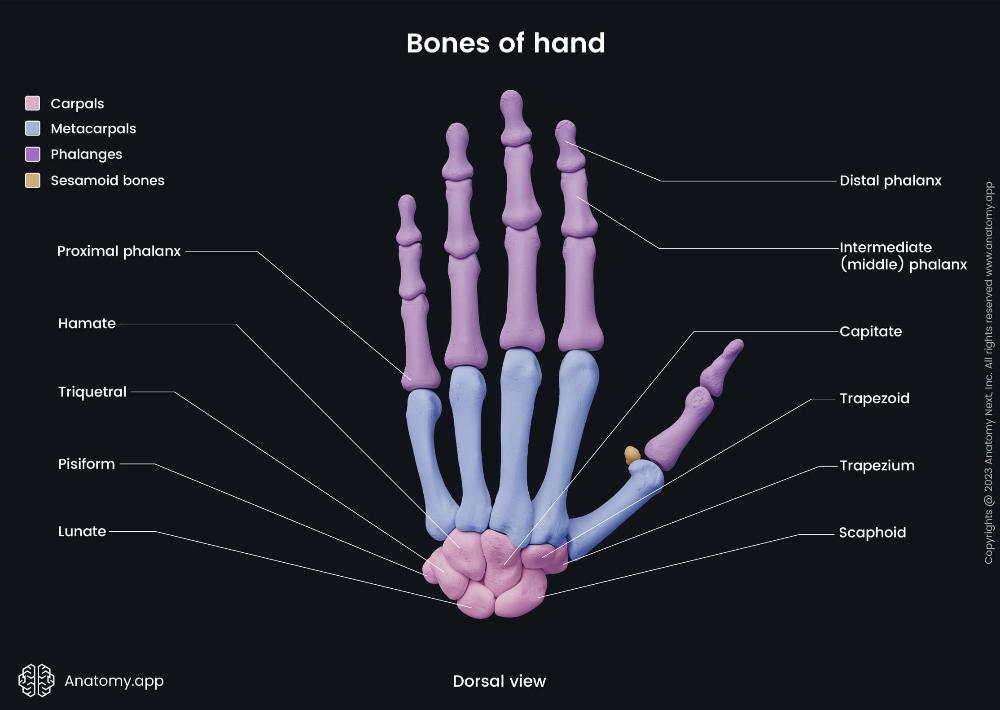
Hand (distal phalanx)
Articulates with the middle phalanx at the distal interphalangeal (DIP) joint and features a rough, horseshoe-shaped tuberosity on the palmar side.
Hand (intermediate phalanx)
Its main features include a base with two concave articular facets to connect with the proximal phalanx.
Hand (proximal phalanx)
The main features of the proximal phalanx are its structure, which includes a base, a diaphysis (body), and a head, and its specific articulations at each end
Hand (Hamate)
Wedge-shaped carpal bone with a hook; articulates with capitate, triquetral, 4th and 5th metacarpals; forms ulnar carpal tunnel boundary.
Hand (triquetral)
Pyramidal carpal bone; articulates with pisiform, lunate, and hamate; on ulnar side; forms part of wrist joint capsule.
Hand (Capitate)
Largest carpal bone; centrally located; articulates with third metacarpal, scaphoid, lunate, trapezoid, and hamate bones.
Hand (Trapezoid)
Small wedge-shaped carpal bone; articulates with second metacarpal, trapezium, capitate, and scaphoid; located lateral to capitate.
Hand (Trapezium)
Saddle-shaped carpal bone; articulates with first metacarpal, scaphoid, and trapezoid; allows thumb opposition and mobility.
Hand (Scaphoid)
Boat-shaped carpal bone; articulates with radius, lunate, trapezium, and capitate; has tubercle; commonly fractured bone.
Hand (Pisiform)
Small pea-shaped sesamoid bone; lies in flexor carpi ulnaris tendon; articulates only with triquetral; palpable on wrist.
Hand (Lunate)
Crescent-shaped carpal bone; articulates with radius, scaphoid, triquetral, and capitate; central wrist bone aiding flexion-extension.
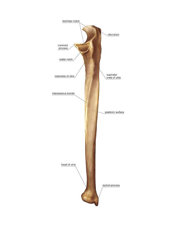
Ulna (Trochlear notch)
Large curved notch of ulna; articulates with humeral trochlea; enables elbow hinge movement (flexion and extension).
Ulna (Olecranon)
Proximal ulna prominence; forms elbow tip; articulates with humerus trochlea; attachment for triceps tendon; enables elbow extension leverage.
Ulna (Coronoid process)
Anterior ulna projection; fits humeral trochlea; stabilizes elbow; attachment for brachialis and ulnar collateral ligament.
Ulna (Styloid Process)
A small, pointed projection at the distal end of the ulna, providing attachment for ligaments of the wrist.
Ulna (Head)
The distal, rounded end of the ulna; articulates with the ulnar notch of the radius
Ulna (Posterior surface)
The back surface of the ulna shaft, providing attachment for several forearm muscles.
Ulna (Interosseous border)
The sharp ridge along the lateral side of the ulna where the interosseous membrane attaches, connecting the ulna to the radius
Ulna (Supinator crest)
A ridge located below the radial notch; attachment site for part of the supinator muscle.
Ulna (Tuberosity)
A roughened area just below the coronoid process; site of attachment for the brachialis muscle.
Ulna (Radial notch)
A small depression on the lateral side of the coronoid process where the head of the radius articulates with the ulna.
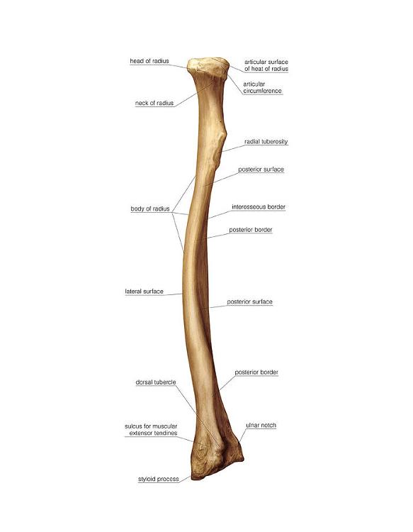
Radius (Head)
proximal end
The disc-shaped top part that articulates with the capitulum of the humerus and the radial notch of the ulna.
Radius (Articular surface of head of radius )
proximal end
The concave superior surface of the head that articulates with the capitulum of the humerus.
Radius (Articular circumference )
proximal end
The smooth, rounded area around the edge of the head that articulates with the radial notch of the ulna, allowing rotation.
Radius (Neck)
proximal end
The narrow area just below the head; it supports the head and serves as an attachment site for ligaments.
Radius (Radial tuberosity)
proximal end
A bony prominence below the neck on the medial side; attachment site for the biceps brachii tendon.
Radius (shaft) of radius
body
The long, central portion of the bone.
Radius (Lateral surface)
body
The outer surface; relatively smooth and gives attachment to muscles such as the supinator.
Radius (Posterior surface)
body
The back surface of the shaft; gives attachment to the abductor pollicis longus and extensor muscles.
Radius (Interosseous border)
body
The sharp medial ridge where the interosseous membrane attaches, connecting the radius and ulna.
Radius (Styloid process)
Distal End
The pointed projection on the lateral side of the distal radius; provides attachment for ligaments of the wrist.
Radius (ulnar notch)
Distal end
The concave depression on the medial side of the distal radius that articulates with the head of the ulna
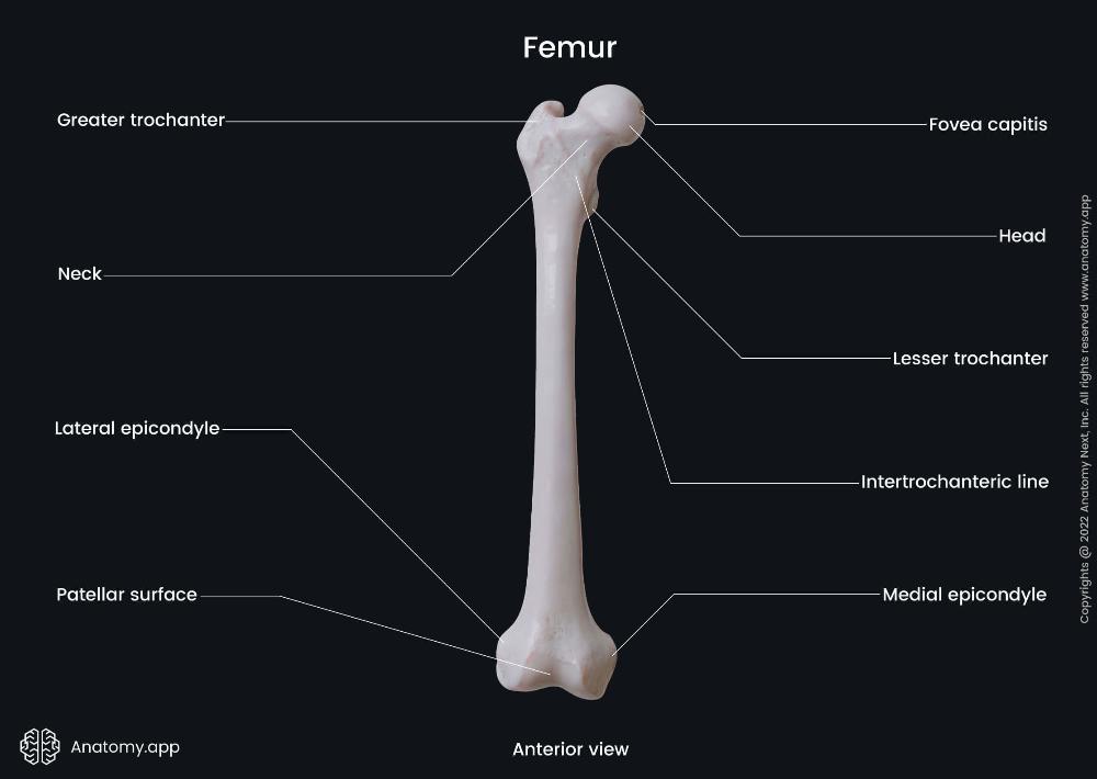
Femur (Head)
Proximal End
The rounded, smooth portion that articulates with the acetabulum of the pelvis to form the hip joint.
Femur (Neck)
Proximal End
The constricted region just below the head; connects the head to the shaft and allows range of motion at the hip.
Femur (Greater trochanter)
Proximal End
The large, lateral projection that serves as an attachment site for gluteal muscles (gluteus medius, minimus, and piriformis).
Femur (Lesser trochanter)
Proximal End
The smaller, posteromedial projection where the iliopsoas muscle attaches.
Femur (Lateral epicondyle)
Distal End
A projection on the outer side of the distal femur; provides attachment for the lateral collateral ligament of the knee.
Femur (Medial epicondyle)
Distal End
A projection on the inner side of the distal femur; provides attachment for the medial collateral ligament of the knee.

Femur (Patellar surface)
Distal End
The smooth anterior surface between the condyles where the patella (kneecap) articulates with the femur.
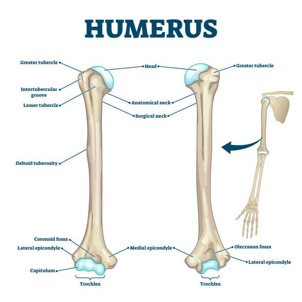
Humerus (Head)
Proximal End
<p data-start="295" data-end="430">The rounded upper part of the humerus that articulates with the glenoid cavity of the scapula, forming the shoulder joint.</p> <br>
Humerus (Greater tubercle)
Proximal End
Large projection on the lateral side of the humerus; attachment site for supraspinatus, infraspinatus, and teres minor muscles
Humerus (Lesser tubercle)
Proximal End
Smaller projection on the anterior surface; attachment site for the subscapularis muscle.
Humerus (Intertubercular groove)
Proximal End
The groove between the greater and lesser tubercles where the tendon of the long head of the biceps brachii runs.
Humerus (Deltoid tuberosity)
Body
Roughened area on the lateral surface of the shaft; insertion point for the deltoid muscle.
Humerus (Medial epicondyle)
Distal End
Large projection on the medial side; attachment site for forearm flexor muscles.
Humerus (Lateral epicondyle )
Distal End
Smaller projection on the lateral side; attachment site for forearm extensor muscles
Humerus (Capitulum)
Distal End
Rounded knob on the lateral side of the distal humerus that articulates with the head of the radius.
Humerus (Trochlea)
Distal End
Spool-shaped surface on the medial side that articulates with the trochlear notch of the ulna
Humerus (Coronoid fossa )
Distal End
Depression above the trochlea on the anterior side; receives the coronoid process of the ulna during elbow flexion.
Humerus (Olecranon fossa )
Distal End
Large depression on the posterior side; receives the olecranon of the ulna when the elbow is extended.
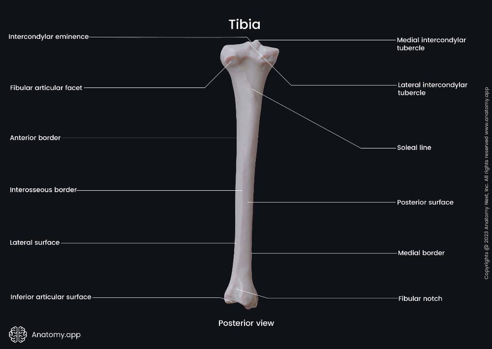
Tibia
Proximal End
intercondylar eminence – The raised ridge between the medial and lateral condyles; provides attachment for the cruciate ligaments and menisci of the knee.
Medial intercondylar tubercle – A prominence on the medial side of the intercondylar eminence; attachment site for ligaments and menisci.
Lateral intercondylar tubercle – A prominence on the lateral side of the intercondylar eminence; also serves as a ligament and meniscus attachment point.
Tibia
Body
Anterior border (shin) – The sharp ridge along the anterior surface; easily palpable under the skin (the “shin bone”).
Posterior surface – The back surface of the tibial shaft; provides attachment for several leg muscles, including the popliteus.
Medial border – The inner ridge of the tibial shaft, forming the medial contour of the leg.
Lateral surface – The outer surface of the shaft; attachment site for muscles like the tibialis anterior.
Tibia
Distal End
Inferior articular surface – The smooth surface at the bottom of the tibia; articulates with the talus bone of the ankle join
Fibular notch – A shallow depression on the lateral side of the distal tibia; articulates with the distal end of the fibula to form the tibiofibular joint.

scapula features
Spine of scapula – The prominent ridge running across the posterior surface of the scapula; divides it into the supraspinous and infraspinous fossae.
Acromion – The broad, flat projection extending laterally from the spine; articulates with the clavicle to form the acromioclavicular (AC) joint.
Coracoid process – A hook-like projection on the anterior surface of the scapula (visible from posterior in part); provides attachment for muscles like the pectoralis minor, short head of biceps brachii, and coracobrachialis.
Scapular notch (suprascapular notch) – A small indentation on the superior border; allows passage of the suprascapular nerve.
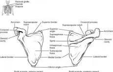
scapula (Fossae Depressions))
Supraspinous fossa – The shallow depression above the spine; origin site for the supraspinatus muscle
Infraspinous fossa – The larger depression below the spine; origin site for the infraspinatus muscle.
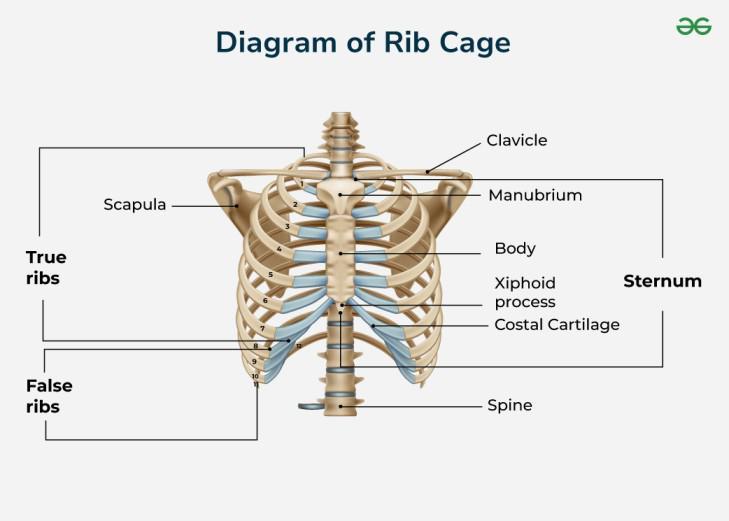
Ribs (sternum)
Located in the center of the chest; it connects to the ribs via costal cartilages.
Ribs (Manubrium)
Upper part of the sternum; articulates with the clavicles (collarbones) and first pair of ribs.
Ribs (Body)
The long, middle portion; articulates with ribs 2–7 via costal cartilages.
Ribs (Xiphoid Process)
Small, cartilaginous lower tip that ossifies (turns to bone) with age.
Ribs (True Ribs (1–7)
Attach directly to the sternum through their own costal cartilages.
Ribs (False Ribs (8-10)
Attach indirectly to the sternum via the cartilage of the rib above them.
Ribs (Floating Ribs (11-12)
A subset of false ribs that do not attach to the sternum at all; they end in the posterior abdominal wall.
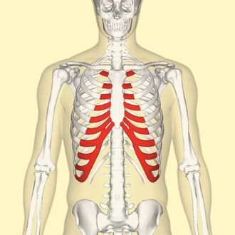
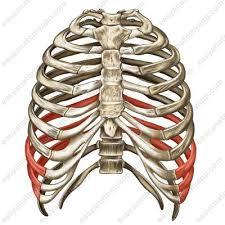
Costal Cartilage
- Bars of hyaline cartilage that connect the ribs to the sternum.
- Provide elasticity and flexibility to the rib cage for breathing movements.
Rib Cage Function
- Protection: Shields vital organs (heart, lungs).
- Support: Anchors muscles of respiration, back, chest, and shoulder.
- Respiration: Expands and contracts to assist breathing.
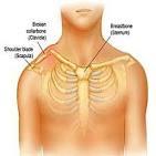
Clavicle (Collarbone)
The clavicle is a long, S-shaped bone that connects the arm to the trunk, with key features including its S-shape, two ends, and lack of a medullary cavity
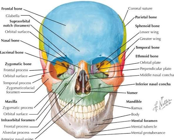
Maxilla
Upper jaw; alveolar process, infraorbital foramen, palatine process.
Zygomatic
Cheekbone; temporal, frontal, maxillary processes, zygomatic arch.
Nasal
Bridge of nose; forms upper nasal structure.
Lacrimal
Medial orbit wall; lacrimal fossa for tear duct.
Palatine
Posterior hard palate; horizontal and perpendicular plates.
Vomer
Lower nasal septum; thin vertical plate.
Mandible
Lower jaw; body, ramus, condylar process, mental foramen.
<p data-start="73" data-end="178">Cervical Vertebrae (C1–C7)</p> <br>
–Small body, transverse foramen, bifid spinous process (except C1, C7).
Atlas (C1) –
No body; anterior/posterior arches; supports skull.
<ul> <li data-start="253" data-end="316">Axis (C2) – Dens (odontoid process) for head rotation.</li> </ul> <br>
Dens (odontoid process) for head rotation.
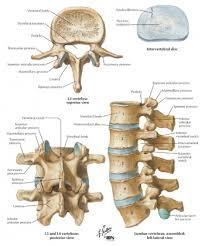
Thoracic Vertebrae (T1–T12)
Heart-shaped body; costal facets for ribs; long spinous process.