The external ear consists of the __________, also called the __________, and the __________ __________ __________.
auricle, pinna, external auditory meatus
The middle ear contains the __________ __________ and the __________ __________.
tympanic cavity, epitympanic recess
The inner ear is made up of the __________ __________ and the __________ __________.
bony labyrinth, membranous labyrinth
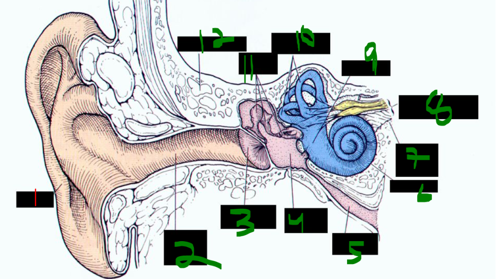
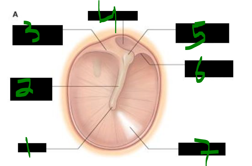
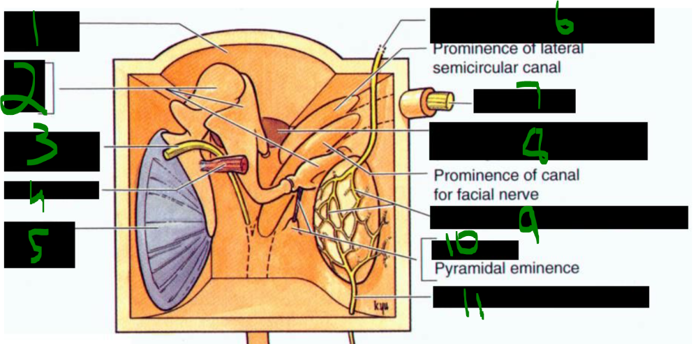
What is 1?
auricle
What is 2?
external acoustic meatus
What is 3?
tympanic membrane
What is 4?
tympanic cavity
What is 5?
auditory tube
What is 6?
cochlea
What is 7?
internal acoustic meatus
What is 8?
vestibulocochlear nerve
What is 9?
vestibule
What is 10?
semicircular canals
What is 11?
auditory ossicles
What is 12?
temporal bone
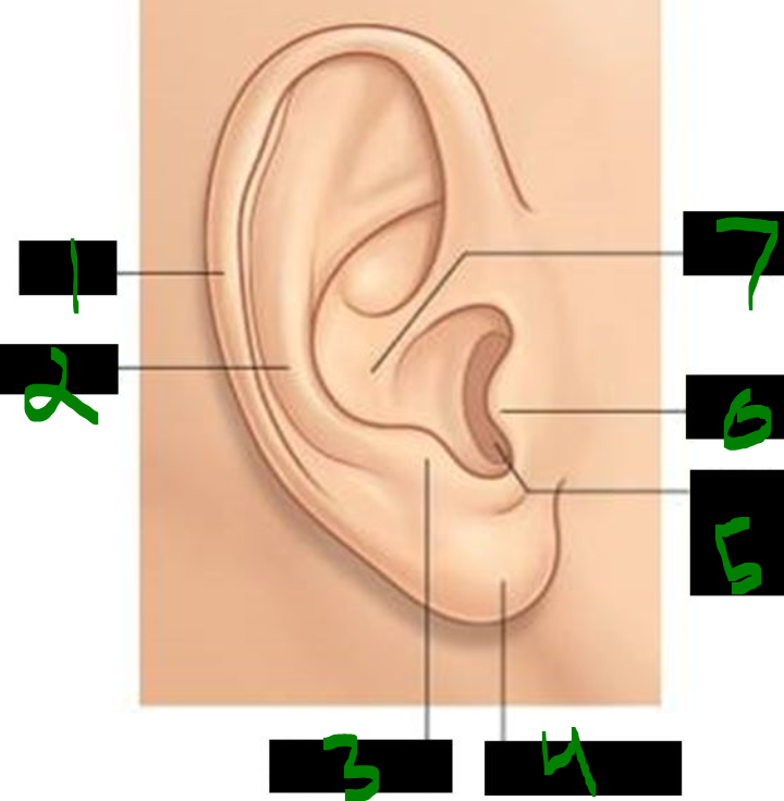
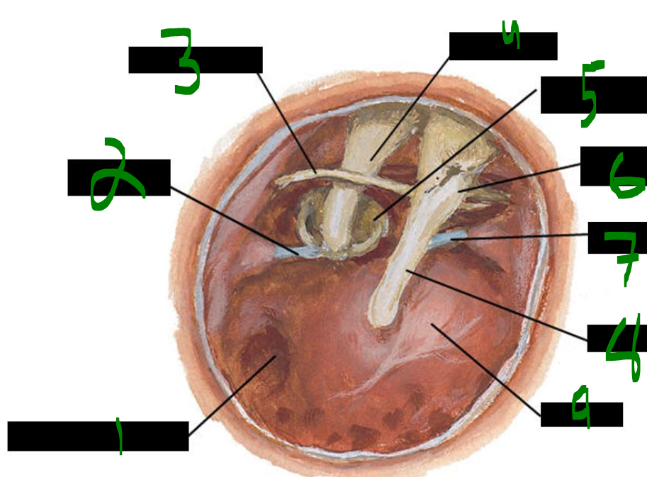
What is 1?
helix
What is 2?
antihelix
What is 3?
antitragus
What is 4?
lobule
What is 5?
external acoustic meatus
What is 6?
concha
What is 7?
tragus
The auricle, also called the __________, functions in __________ __________ and __________.
pinna, sound localization, amplification
Dr. Liuzzi suggests the majority of auricular innervation comes from the __________ __________ nerve, __________ __________ nerve, and some innervation from the __________ nerve.
lesser occipital, great auricular, vagus
The pinna, also called the __________, is innervated by multiple nerves including the __________ __________ nerve (C2), __________ __________ nerve (C2, C3), and the __________ branch of CN __________.
auricle, lesser occipital, great auricular, auriculotemporal, V3
Additional innervation of the auricle comes from cranial nerves __________ and __________, corresponding to the __________ nerve and the __________ nerve.
X, VII, vagus, facial
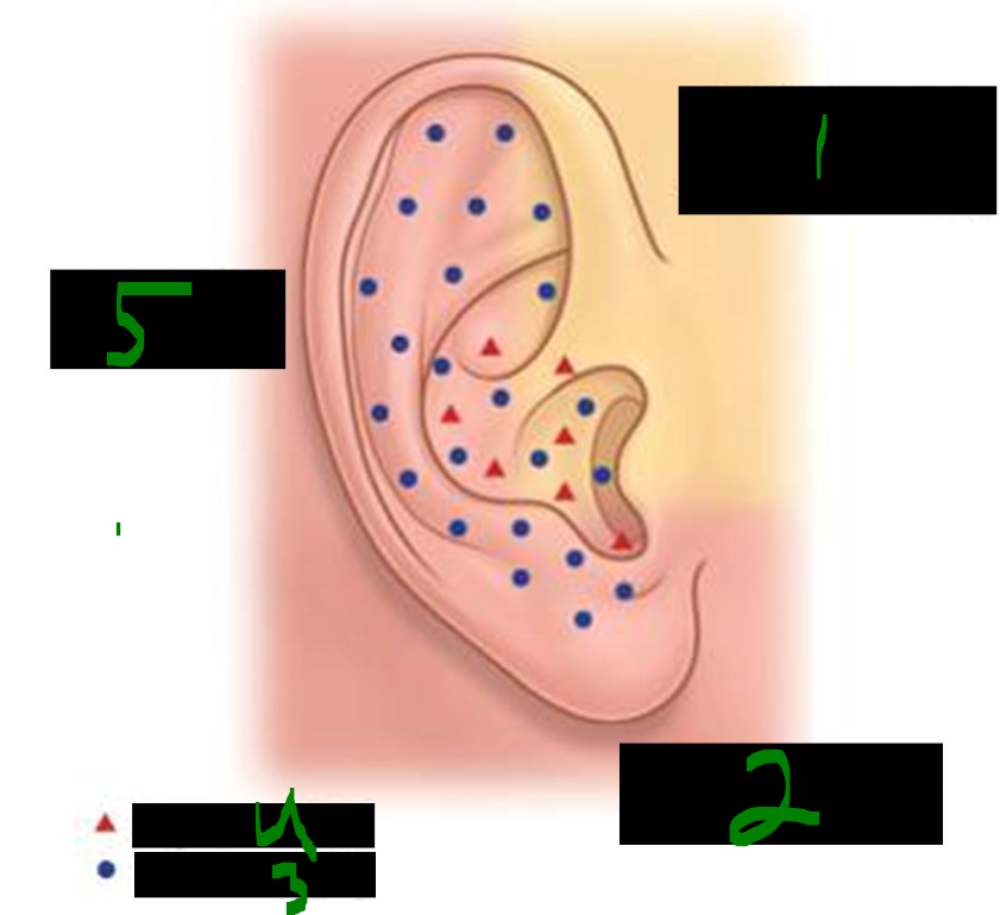
What is 1?
auriculotemporal branch of mandibular nerve v3
What is 2?
great auricular nerve c2 c3
What is 3?
facial nerve (VII)
What is 4?
Vagus nerve (X)
What is 5?
lesser occipital nerve (c2)
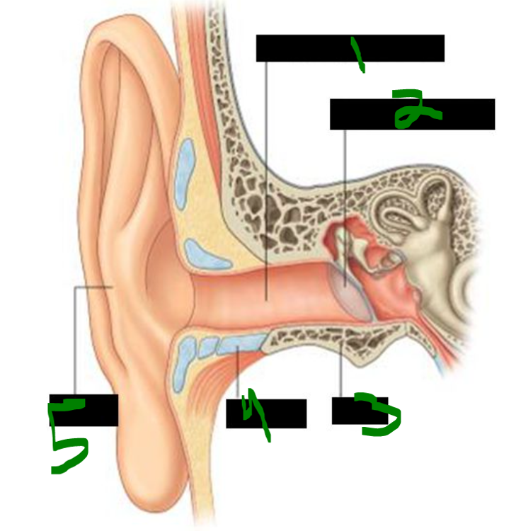
What is 1?
external acoustic meatus
What is 2?
tympanic membrane
What is 3?
bone
What is 4?
cartilage
What is 5?
auricle
The lateral ______ of the external auditory meatus is ______, while the medial ______ is made of ______ from the ______ bone.
one-third, cartilaginous, two-thirds, bone, temporal
The lateral part of the external auditory meatus is lined by skin containing ______, ______ glands, and ______ glands, which are modified ______ ______ glands that secrete ______.
hairs, sebaceous, ceruminous, apocrine sweat, cerumen
The lateral external auditory meatus contains no ______ ______ glands.
eccrine sweat
Cerumen, also known as ______ ______, is secreted by ______ glands, which are modified ______ ______ glands.
ear wax, ceruminous, apocrine sweat
Cerumen is found in the ______ part of the ______ ______ ______.
lateral, external auditory meatus
The lateral part of the external auditory meatus contains ______ glands and ______ glands, which are modified ______ ______ glands that secrete ______.
sebaceous, ceruminous, apocrine sweat, cerumen
The external auditory meatus does not contain any ______ ______ glands.
eccrine sweat
The external auditory meatus is mainly innervated by the ______ nerve, with additional minor contributions from the ______ nerve (CN ______) and ______ branches of the ______ nerve (CN ______).
auriculotemporal, facial, VII, vagal, vagus, X
Stimulation of the external auditory meatus can cause a gag reflex in some individuals due to ______ innervation from the ______ nerve (CN ______).
sensory, vagus, X
Some people gag when cleaning their ears because of ______ innervation from the ______ nerve (CN ______), which supplies part of the external auditory meatus.
vagal, vagus, X
The outer surface of the tympanic membrane is mainly innervated by the ______ nerve (branch of CN ______) and has a small contribution from the ______ branch of the ______ nerve (CN ______).
auriculotemporal, V3, auricular, vagus, X
The inner surface of the tympanic membrane is innervated by the ______ nerve (CN ______).
glossopharyngeal, IX
What is 1?
umbo
What is 2?
handle of malleus
What is 3?
posterior malleolar fold
What is 4?
pars flaccida
What is 5?
lateral process of malleus
What is 6?
anterior malleolar fold
What is 7?
cone of light
Otitis media is a middle ear infection characterized by a ______, ______ tympanic membrane.
bulging, red
Otitis media may cause ______ of the tympanic membrane.
perforation
Otitis media is more common in younger children because their ______ tubes have a ______ angle, impairing drainage from the middle ear to the ______.
pharyngotympanic, narrower, pharynx
If otitis media is severe, a common treatment is an ______ of the ______ part of the tympanic membrane to avoid damaging the ______ ______, followed by placement of a ______ ______.
incision, posteroinferior, chorda tympani, tympanostomy tube
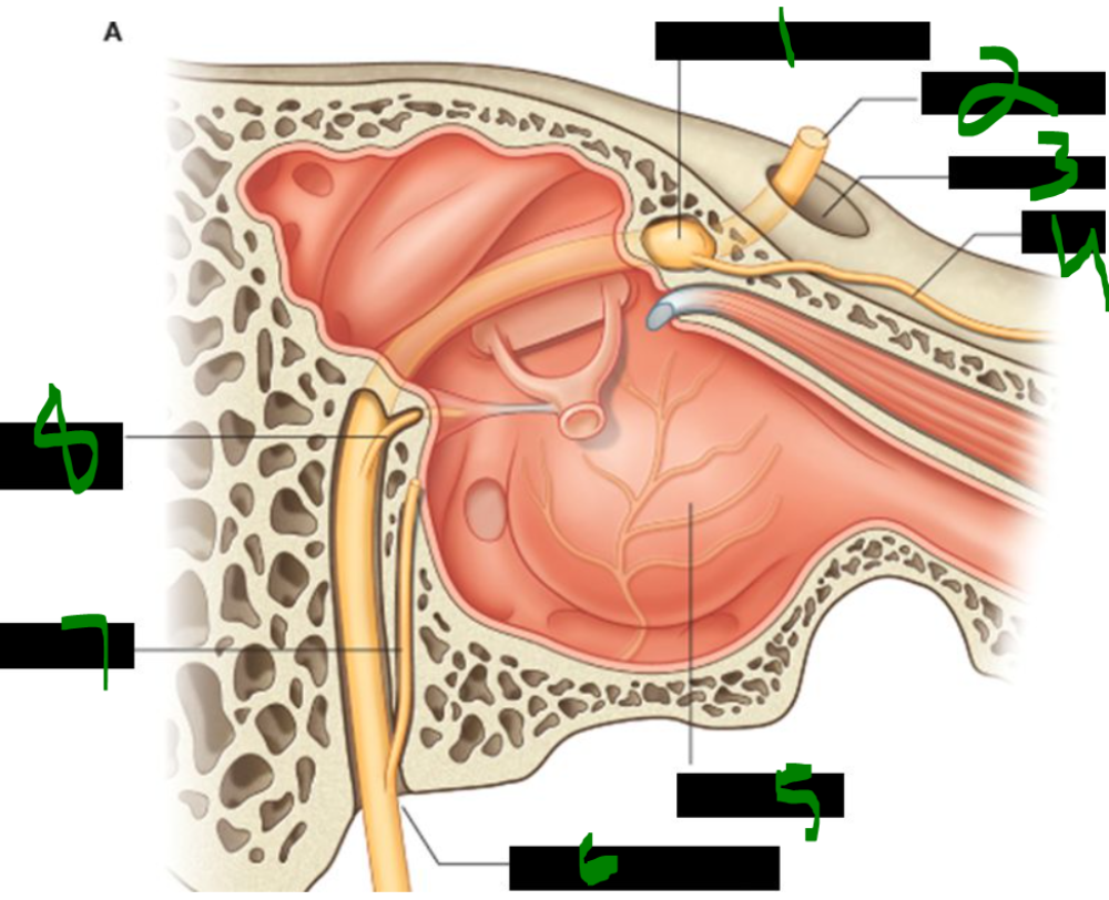
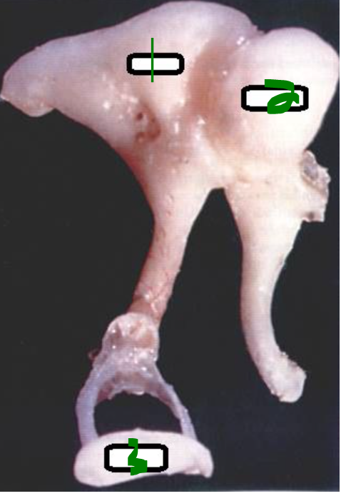
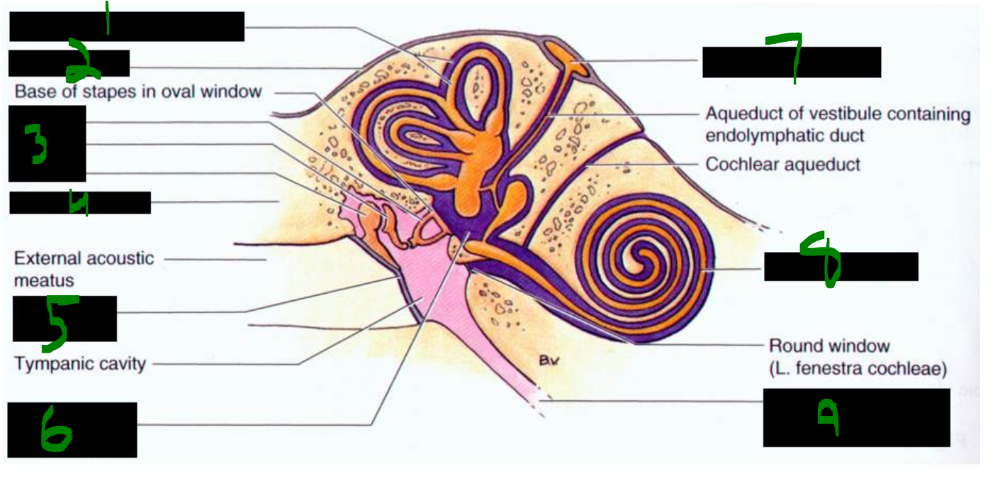
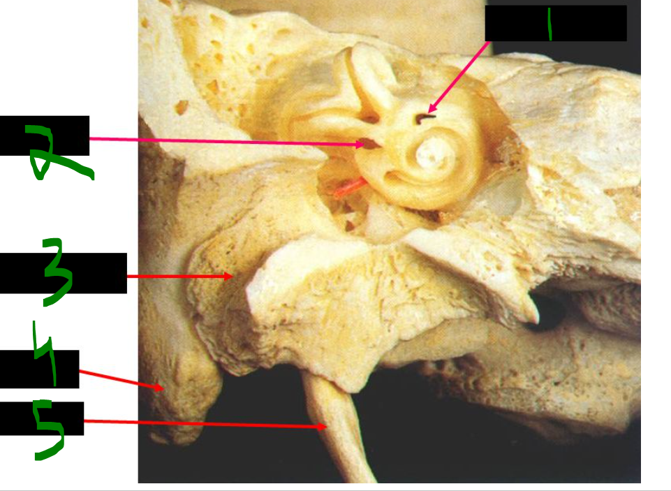
What is 1?
fossa of round (cochlear) window
What is 2?
tendon of stapedius muscle
What is 3?
chorda tympani nerve
What is 4?
long limb of incus
What is 5?
stapes in oval (vestibular) window
What is 6?
lateral process of malleus
What is 7?
tendon of tensor tympani muscle
What is 8?
handle of malleus
What is 9?
promontory
The chorda tympani passes just behind/medial to the tympanic membrane between the long limb of the ______ and the ______.
incus, malleus
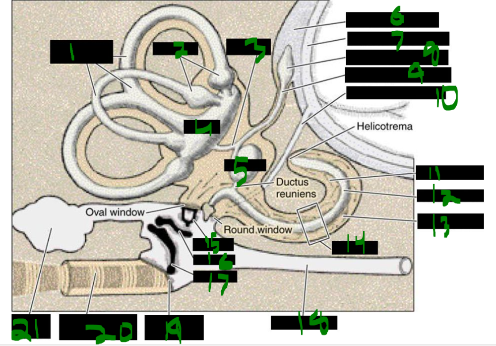
What is 1?
semicircular ducts
What is 2?
ampullae
What is 3?
vestibule
What is 4?
utricle
What is 5?
saccule
What is 6?
subdural space
What is 7?
subarachnoid space
What is 8?
endolymphatic space
What is 9?
endolymphatic duct
What is 10?
perilymphatic duct
What is 11?
scala vestibuli
What is 12?
scala media
What is 13?
scala tympani
What is 14?
cochlea
What is 15?
stapes
What is 16?
incus
What is 17?
malleus
What is 18?
auditory tube
What is 19?
tympanic membrane
What is 20?
external acoustic meatus
What is 21?
mastoid cavities
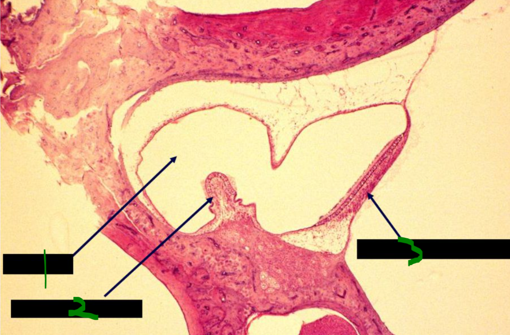
The middle ear includes the ______ cavity and the ______ recess, where the ______ are located.
tympanic, epitympanic, ossicles
Anteriorly, the middle ear connects to the ______ via the ______ ______ tube.
pharynx, pharyngotympanic (eustachian)
Posteriorly, the middle ear connects to the ______ air cells via the ______ ______.
mastoid, mastoid antrum
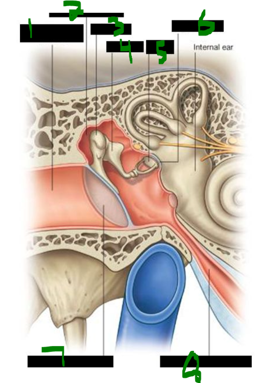
What is 1?
external acoustic meatus
What is 2?
epitympanic recess
What is 3?
malleus
What is 4?
incus
What is 5?
stapes
What is 6?
oval window
What is 7?
tympanic membrane
What is 8?
pharyngotympanic tube
The middle ear, including the inner surface of the tympanic cavity, is innervated by the ______ nerve (CN ______), also called ______ nerve.
tympanic, IX, Jacobson's
Jacobson’s nerve, also known as the ______ nerve (CN ______), innervates the ______ ear cavity, including the inner surface of the ______ cavity.
tympanic, IX, middle, tympanic
What is 1?
epitympanic recess
What is 2?
malleus incus stapes
What is 3?
chorda tympani nerve
What is 4?
tensor tympani
What is 5?
tympanic membrane
What is 6?
lesser petrosal nerve (preganglionic parasympathetics)
What is 7?
facial nerve
What is 8?
aditus to mastoid antrum
What is 9?
tympanic plexus on promontory
What is 10?
stapedius
What is 11?
tympanic nerve (from CN IX)
what vessel has a very close relationship to the middle ear?
_____ _____ _____
internal carotid a.
What is 1?
geniculate ganglion
What is 2?
facial neve (VII)
What is 3?
internal acoustic meatus
What is 4?
greater petrosal
What is 5?
middle ear
What is 6?
stylomastoid foramen
What is 7?
chorda tympani
What is 8?
nerve to stapedius muscle
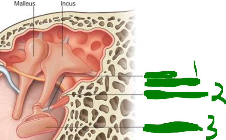
What is 1?
malleus
What is 2?
incus
What is 3?
chorda tympani
What is 4?
tympanic membrane
The ______ ______ is attached to the handle of the ______, functions to tense the ______ membrane to attenuate sound, and is innervated by the ______ nerve, mandibular division (CN ______).
tensor tympani, malleus, tympanic, trigeminal, V3
The ______ muscle is attached to the neck of the ______, functions to prevent excessive movement of the ______, and is innervated by the ______ nerve (CN ______).
stapedius, stapes, stapes, facial, VII
Paralysis of the facial nerve can cause ______.
hyperacusis
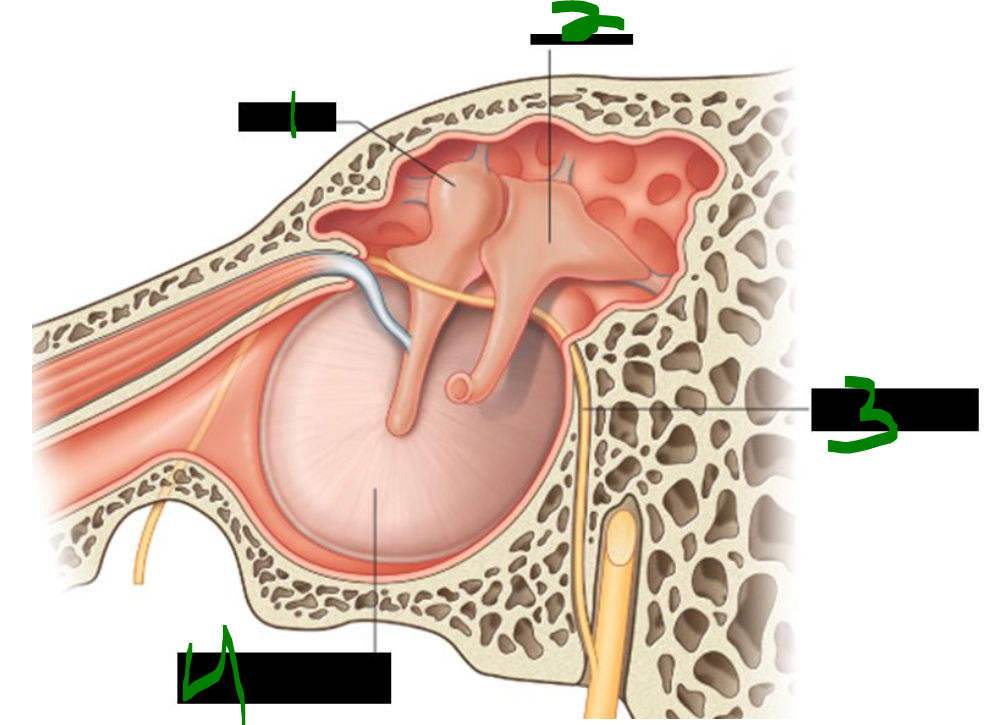
What is 1?
tendon of stapedius muscle
What is 2?
pyramidal eminence
What is 3?
footplate of stapes
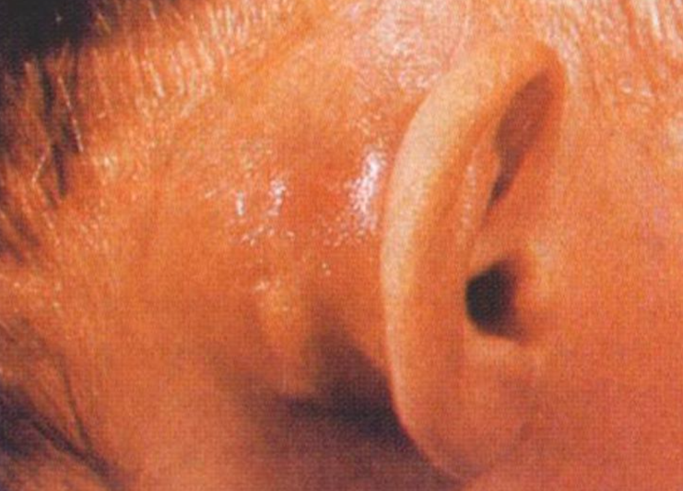
What is this?
Mastoiditis
Mastoiditis is inflammation of the ______ ______ and ______ ______, often resulting from ______ ______.
mastoid antrum, air cells, otitis media
Mastoiditis can spread superiorly into the ______ ______ cavity, potentially causing ______ meningitis.
middle cranial, bacterial
The otic vesicle divides into two parts during development:
- The ______ component forms the ______ and ______ duct.
- The ______ component forms the ______, ______ ducts, and the ______ duct.
ventral, saccule, cochlear
dorsal, utricle, semicircular, endolymphatic
what is the external auditory meatus derived from (development/embryo)?
______ ______ ______
1st pharyngeal cleft
what are the ossicles derived from (development/embryo)?
______ ______
mesenchymal condensation
what is the tympanic membrane derived from (development/embryo)?
outer part - ______
inner part - ______
ectoderm
endoderm
what is the embryonic origin of the middle ear cavity?
______
pharynx
The ______ and ______ develop from the 1st pharyngeal arch cartilage, also known as ______ cartilage.
malleus, incus, Meckel's
The ______ develops from the 2nd pharyngeal arch cartilage, also known as ______ cartilage.
stapes, Reichert
What is 1, 2, 3?
incus
malleus
stapes
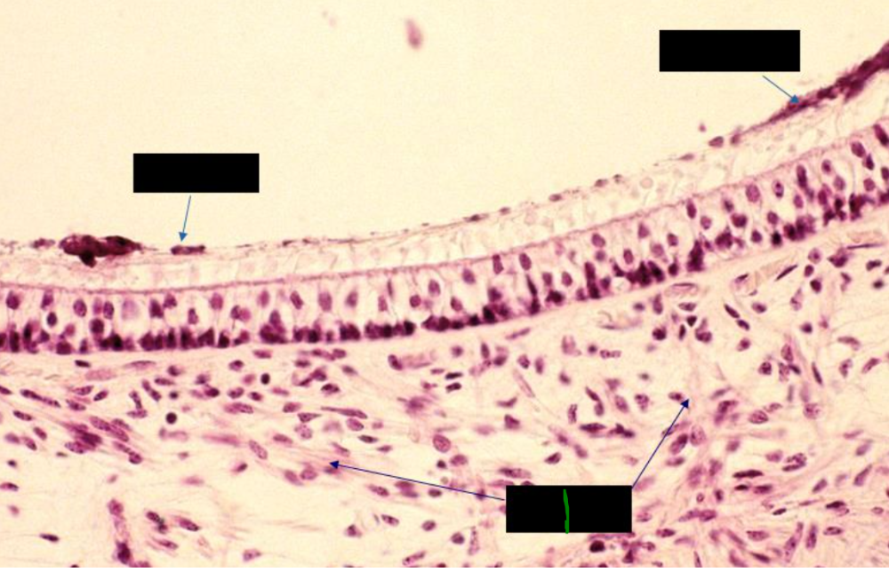
The bony labyrinth, located within the ______ portion of the ______ bone, includes the ______ canals, the ______, and the ______, and it contains ______.
petrous, temporal, semicircular, vestibule, cochlea, perilymph
The membranous labyrinth includes the ______ duct, ______, ______, ______ ducts, and the ______ duct, and it contains ______.
cochlear, saccule, utricle, semicircular, endolymphatic, endolymph
The vestibular apparatus detects ______ position (______ acceleration) and ______ movement (______ acceleration), orients us in ______ space, and serves as a ______ stabilizer for the ______ system.
head, linear, head, angular, 3-D, gyroscopic, visual
What is 1?
semicircular duct and canal
What is 2?
dura mater
What is 3?
stapes
incus
malleus
What is 4?
tympanic bone
What is 5?
tympanic membrane
What is 6?
vestibule of bony labyrinth
What is 7?
endolymphatic sac
What is 8?
duct of cochlea
What is 9?
pharyngotympanic tube
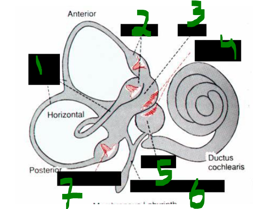
What is 1?
semicircular canals
What is 2?
ampullae
What is 3?
utricle
What is 4?
maculae and statoconia
What is 5?
saccule
What is 6?
ductus endolymphaticus
What is 7?
crista ampullae
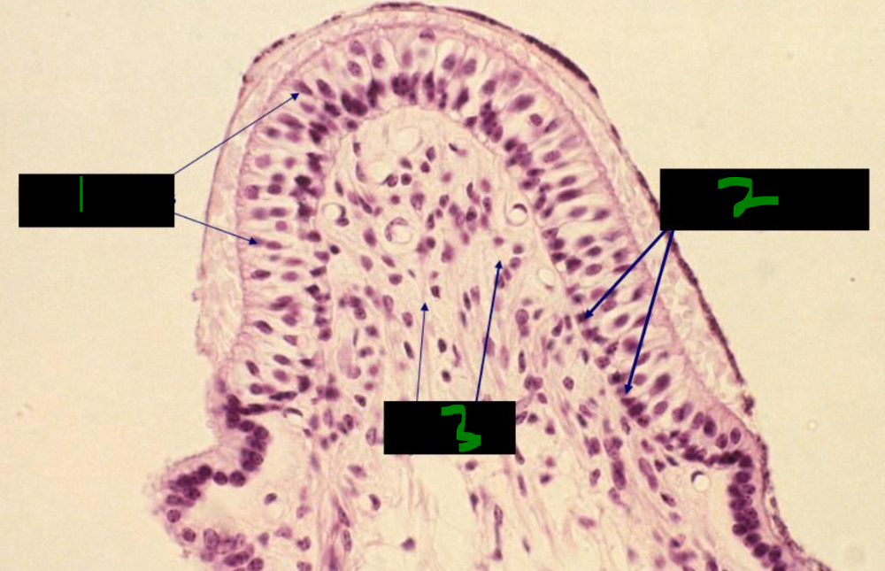
Vestibular hair cells have a single ______ called the ______ and numerous ______ (about 60), all embedded within a ______ matrix.
cilium, kinocilium, stereocilia, gelatinous

what is shown here?
vestibular apparatus
What is 1?
ampulla
What is 2?
crista ampullaris
What is 3?
macula of the utricle
Head position (gravity and ______ acceleration) is detected by specialized regions of neuroepithelial cells called ______.
linear, maculae
The maculae are located in the ______ and ______, and are oriented ______ to one another.
utricle, saccule, perpendicular
The maculae, located in the ______ and ______, function to detect ______ position by sensing ______ and ______ acceleration. They are oriented ______ to one another.
utricle, saccule, head, gravity, linear, perpendicular
Embedded within the surface of the gelatinous matrix of the maculae are ______, which are ______ ______ crystals. Gravity causes their weight to ______ the gelatinous matrix, leading the ______ cells to fire.
otoliths, calcium carbonate, distort, receptor
Otoliths are ______ ______ crystals found embedded in the surface of the ______ matrix of the ______. Their weight, influenced by ______, distorts the matrix and stimulates the ______ cells.
calcium carbonate, gelatinous, maculae, gravity, receptor
what is this?
macula
What is 1?
nerves
The semicircular canals detect head ______ by sensing ______ acceleration.
movements, angular
The ampullae of the semicircular canals contain a ridge of neuroepithelial cells called the ______ ______, whose cilia and stereocilia are embedded in a gelatinous matrix that forms the ______.
crista ampullaris, cupula
The ______ ______ detect head movements (angular acceleration). They have specialized dilations called ______, which contain neuroepithelial hair cells called the ______ ______. The hair cells’ cilia and stereocilia are embedded in a gelatinous matrix called the ______.
semicircular canals, ampullae, crista ampullaris, cupula
What is 1?
receptor cells
What is 2?
nerves
What is 3?
supporting cells
Nystagmus is a rhythmic eye movement where the eyes smoothly follow a moving object and then quickly ______ back to fixate on a new object.
snap
During nystagmus, the eyes follow a moving object at the same ______ as the object’s movement relative to the ______.velocity, head
velocity, head
Vestibular nuclei in the brainstem connect to the oculomotor nuclei (CN ______, ______, and ______) via the ______ ______ fasciculus (MLF), coordinating eye movements in nystagmus.
VI, IV, III, medial longitudinal
Damage to the ______ system or its connections with the ______, and use of ______ or other intoxicants can cause pathologic nystagmus.
In vestibular nystagmus, the eyes move slowly toward the side of ______ damage and then rapidly snap back.
vestibular, cerebellum, alcohol, damage
Meniere’s disease causes recurrent attacks of ______, ______ loss, and ______.
vertigo, hearing, tinnitus
Meniere’s disease may be accompanied by ______ and most commonly affects people in their ______ and ______ decades.
nystagmus, fourth, fifth
Attacks in Meniere’s disease last from a few ______ to several ______.
moments, hours
A consistent feature of Meniere’s disease is ______ ______, which is an increase in the volume of ______.
endolymphatic hydrops, endolymph
function of middle ear ossicles:
convert ______ ______to ______ ______
sound waves
fluid waves
The auditory apparatus captures and conducts ______.
sound
Fluid waves travel through ______, which is not ______ and transmits waves with relative ______.
perilymph, compressible, fidelity
The ______ is the central core of the spiral cochlea and contains the ______ ______ and the acoustic portion of the ______ nerve (CN ______).
modiolus, spiral ganglion, vestibulocochlear, VIII
how big is the human cochlea?
______ turns over distance of ______ mm
2.5 turns over distance of 35 mm
what is suspended wtihin the bony cochlea?
______ ______ duct aka ______ ______
membranous cochlear duct aka scala media
What is 1?
oval window
What is 2?
round window
What is 1?
facial canal
What is 2?
oval window
What is 3?
external auditory meatus
What is 4?
mastoid process
What is 5?
styloid process
What is 1?
1) scala media/cochlear duct
What is 2?
2) scala vestibuli (above scala media)
What is 3?
3) scala tympani (below scala media)
What is 4?
4) spiral ganglion
What is 5?
5) vestibular nerve
The ______ vestibuli and ______ contain perilymph and are part of the ______ labyrinth.
scala, tympani, bony
The ______ media contains endolymph and is part of the ______ labyrinth.
scala, membranous
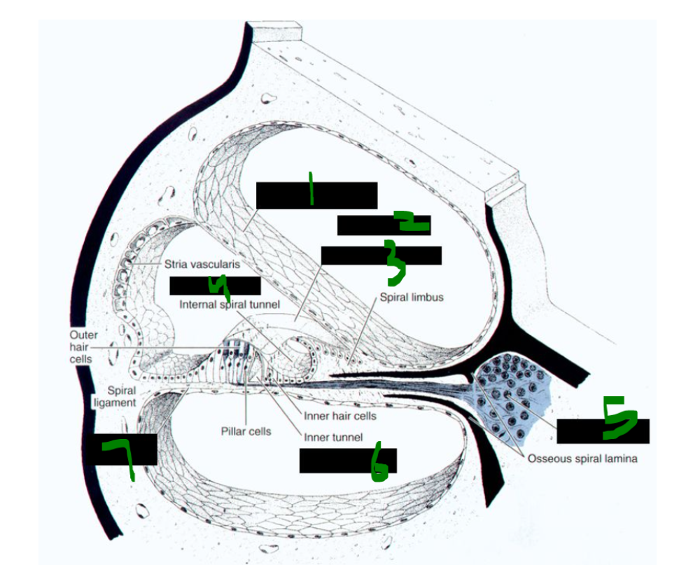
What is 1?
vestibular membrane
What is 2?
scala vestibuli
What is 3?
tectorial membrane
What is 4?
scala media
What is 5?
spiral ganglion
What is 6?
scala tympani
What is 7?
basilar membrane
what is this?
cochlea
what is 1?
scala vestibuli
what is 2?
vestibular membrane
what is 3?
modiolus
what is 4?
spiral limbus
What is 5?
spiral ganglion
What is 6?
basilar membrane
What is 7?
scala tympani
What is 8?
spiral ligament
What is 9?
stria vascularis
What is 10?
scala media
The ______ ______ is found within the ______ duct (scala media) and is the only ______ epithelium in the human body. It contains ______ and produces ______.
stria vascularis, cochlear, vascular, blood vessels, endolymph
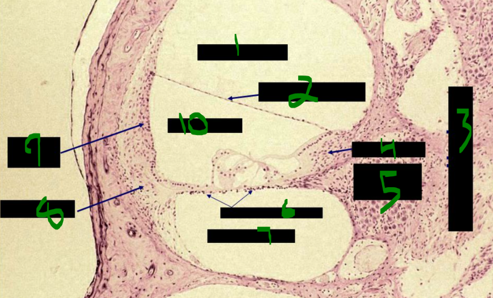
what is this?
organ of corti
The auditory apparatus contains ______ hair cells arranged in 3 rows with a ______-shaped array of stereocilia. It also contains ______ hair cells arranged in 1 row with 50–60 ______-shaped stereocilia. Auditory hair cells do not have ______.
outer, W, inner, V, kinocilia
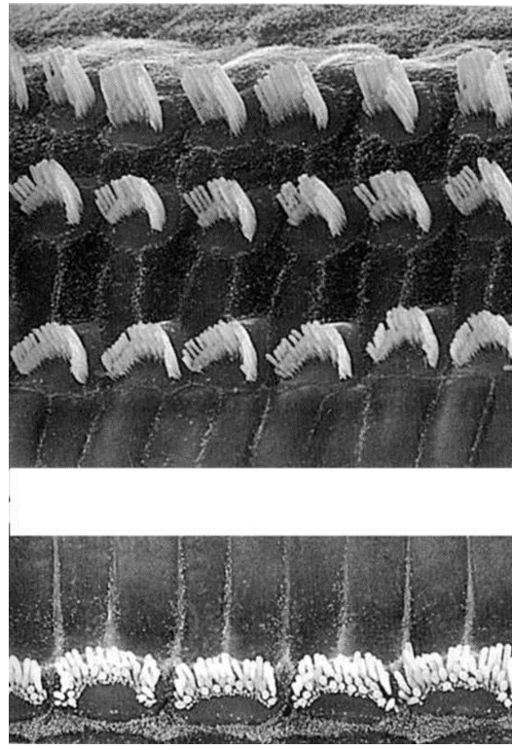
Which hair cell is which?
outer- top
inner- bottom
Constant exposure to loud sounds at specific frequencies can damage ______ cells, which do not ______.
The ______ nerve may be injured at its entrance into the brainstem by a ______ (acoustic neuroma), which may also involve the ______ nerve causing ______ palsy.
hair, regenerate
vestibulocochlear, Schwannoma, facial, facial