The posterior triangle is bounded anteriorly by the posterior border of the _____, posteriorly by the anterior border of the _____, and inferiorly by the middle third of the _____.
SCM, trapezius, clavicle
The superior boundary of the posterior triangle is the _____ bone, just posterior to the _____ process.
occipital, mastoid
The roof of the posterior triangle is formed by the _____ layer of the _____ fascia.
investing, cervical
The floor of the posterior triangle is formed by the _____ layer of deep cervical fascia covering the deep _____ muscles.
prevertebral, neck
what are the muscles of the posterior triangle?
-_____ _____ , _____ _____ , _____ _____ , _____ _____ , _____ _____ , _____ _____ _____
-splenius capitus, levator scapulae, posterior scalene, middle scalene, anterior scalene, omohyoid inferior belly
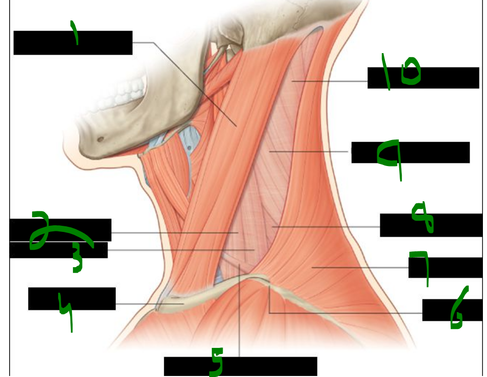
What is 1?
sternocleidomastoid muscle
What is 2?
anterior scalene muscle
What is 3?
middle scalene muscle
What is 4?
clavicle
What is 5?
inferior belly of omohyoid muscle
What is 6?
acromion
What is 7?
trapezius muscle
What is 8?
posterior scalene muscle
What is 9?
levator scapulae muscle
What is 10?
splenius capitus muscle
what fascial layer covers/encloses the muscles of the posterior triangle?
____ ____
prevertebral fascia
what structure subdivides the posterior triangle & into what?
____ ____ of ____ ____
inferior belly of omohyoid muscle
The inferior belly of the omohyoid divides the posterior triangle into the _____ triangle and the _____ triangle (also called the omoclavicular or subclavian triangle).
occipital, supraclavicular
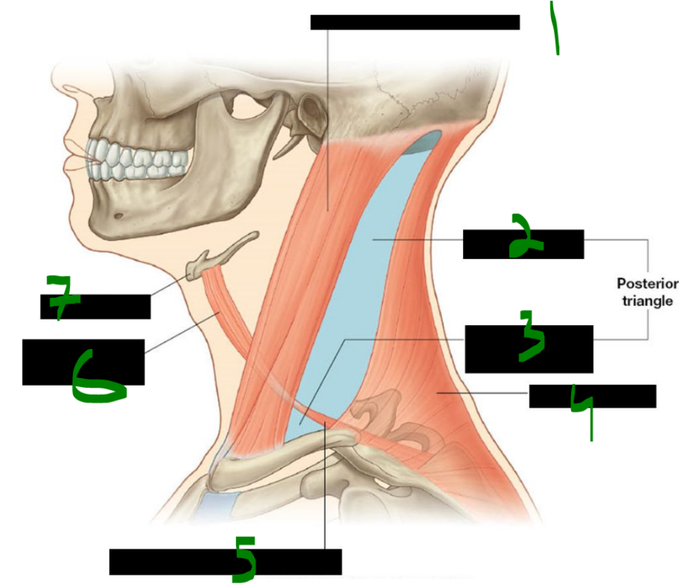
What is 1?
sternocleidomastoid muscle
What is 2?
occipital triangle
What is 3?
omoclavicular or subclavian triangle
What is 4?
trapezius muscle
What is 5?
inferior belly of omohyoid muscle
What is 6?
superior belly of omohyoid muscle
What is 7?
hyoid bone
The omohyoid muscle originates from the superior border of the _____ and inserts onto the inferior border of the _____ bone.
scapula, hyoid
The omohyoid is innervated by the _____ _____, which arises from the anterior rami of spinal nerves _____ to _____.
ansa cervicalis, C1, C3
The omohyoid muscle functions to _____ and _____ the hyoid bone.
depress, stabilize
The omohyoid has two _____ connected by an intermediate _____ and separated by a fascial _____.
bellies, tendon, sling
The scalene muscles originate from the _____ of the cervical _____ processes.
tubercles, transverse
The anterior and middle scalenes insert on the _____ rib, while the posterior scalene inserts on the _____ rib.
first, second
The scalene muscles are innervated by the anterior rami of spinal nerves _____ through _____.
C3, C7
The scalene muscles act to _____ the ribs during inspiration and to _____ the neck.
elevate, flex
The subclavian vein lies _____ to the anterior scalene, while the subclavian artery lies _____ to the anterior scalene.
anterior, posterior
The subclavian _____ is more accessible because it is not 'protected' by the anterior scalene muscle, unlike the subclavian _____.
vein, artery
The _____ nerve travels along the _____ surface of the anterior scalene muscle.
phrenic, anterior
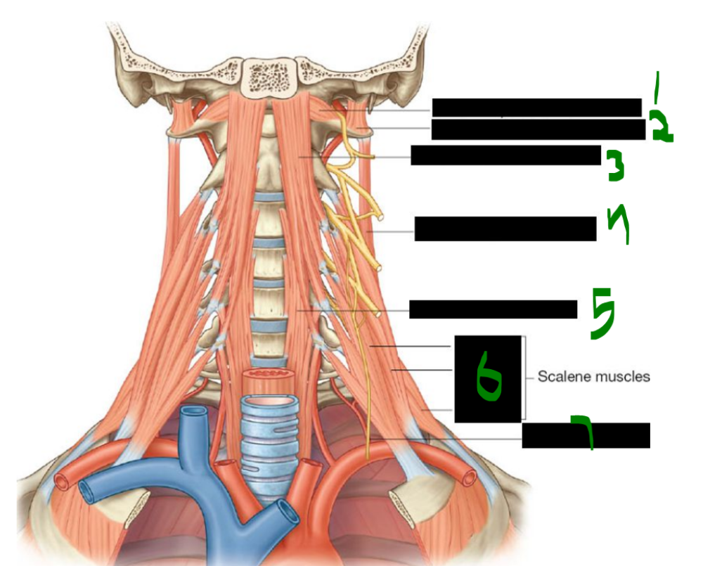
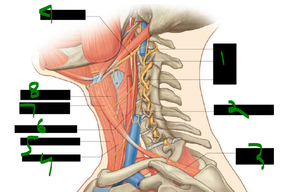
What is 1?
rectus capitis anterior muscle
What is 2?
rectus capitis lateralis muscle
What is 3?
longus capitis muscle
What is 4?
levator scapulae muscle
What is 5?
longus colli muscle
What is 6?
anterior
middle
posterior
What is 7?
phrenic nerve
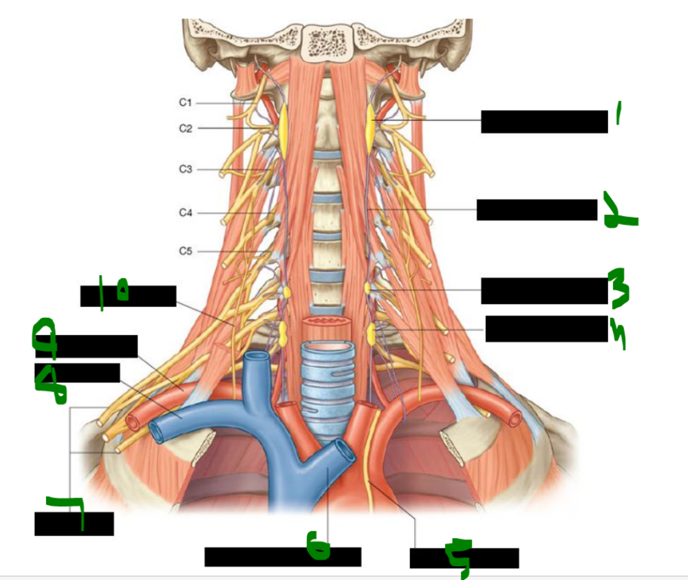
The posterior triangle contains three main arteries: the _____ _____ artery, the _____ cervical artery, and the _____ artery.
dorsal scapular, transverse, suprascapular
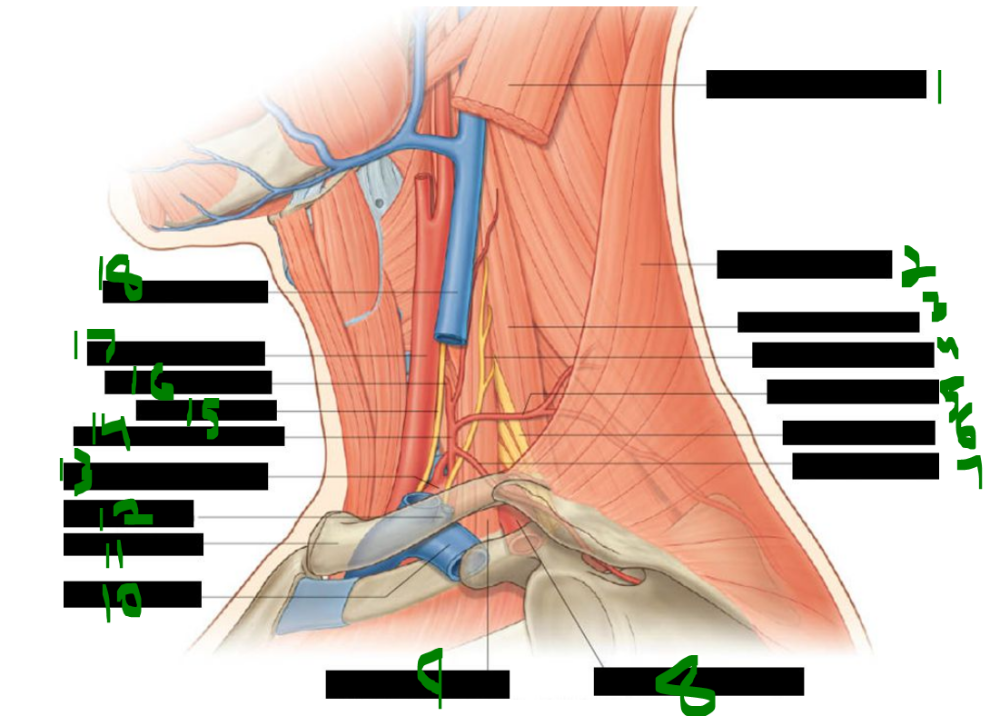
What is 1?
sternocleidomastoid muscle
What is 2?
trapezius muscle
What is 3?
middle scalene muscle
What is 4?
phrenic nerve
What is 5?
transverse cervical artery
What is 6?
brachial plexus
What is 7?
suprascapular artery
What is 8?
3rd part of subclavian artery
What is 9?
anterior scalene muscle
What is 10?
subclavian vein
What is 11?
clavicle
What is 12?
external jugular vein
What is 13?
1st part of subclavian artery
What is 14?
thyrocervical trunk
What is 15?
vagus nerve
What is 16?
inferior thyroid artery
What is 17?
common carotid artery
What is 18?
internal jugular vein
The spinal accessory nerve (CN XI) innervates the _____ and the _____ muscles.
SCM, trapezius
The transverse cervical nerve (C2–C3) provides sensory _____ to the skin over the _____ triangle.
innervation, anterior
The great auricular nerve (C2–C3) innervates the skin over the _____ gland and around the _____.
parotid, auricle
The supraclavicular nerve (C3–C4) pierces the _____ muscle to supply skin over the _____, shoulder, and as far inferiorly as rib _____.
platysma, clavicle, two
The lesser occipital nerve (C2–C3) supplies the skin of the _____ scalp, posterior to the _____.
lateral, auricle
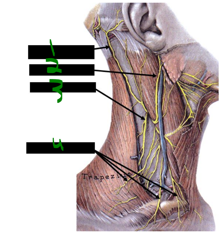
What is 1?
lesser occipital nerve
What is 2?
great auricular nerve
What is 3?
accessory nerve
What is 4?
supraclavicular nerves
the accessory nerve lies on what surface of what muscle?
_____ _____ of _____ _____
anterior surface of levator scauplae
the axillary fascia is a continuation of what fascial layer?
_____ _____, carried by the _____ _____
prevertebral fascia, carried by the brachial plexus
The brachial plexus passes between the _____ and _____ scalene muscles.
anterior, middle
The _____ of the brachial plexus cross the _____ of the posterior triangle.
trunks, base
what branches of the brachial plexus [may] appear in the posterior triangle?
_____ to _____ , _____ _____ nerve, _____ _____ nerve, _____ nerve
-nerve to subclavius, dorsal scapular nerve, long thoracic nerve, suprascapular nerve
(NooDLeS - w/o vowels)
The superior root of the ansa cervicalis is formed by spinal nerves _____ and _____.
C1, C2
The inferior root of the ansa cervicalis arises from spinal nerves _____ and _____.
C2, C3
The transverse cervical and great auricular nerves are both formed from spinal nerves _____ and _____.
C2, C3
The lesser occipital nerve is mainly derived from _____, but also receives contribution from _____.
C2, C3
The supraclavicular nerve arises from spinal nerves _____ and _____.
C3, C4
The phrenic nerve is formed by spinal nerves _____ through _____.
C3, C5
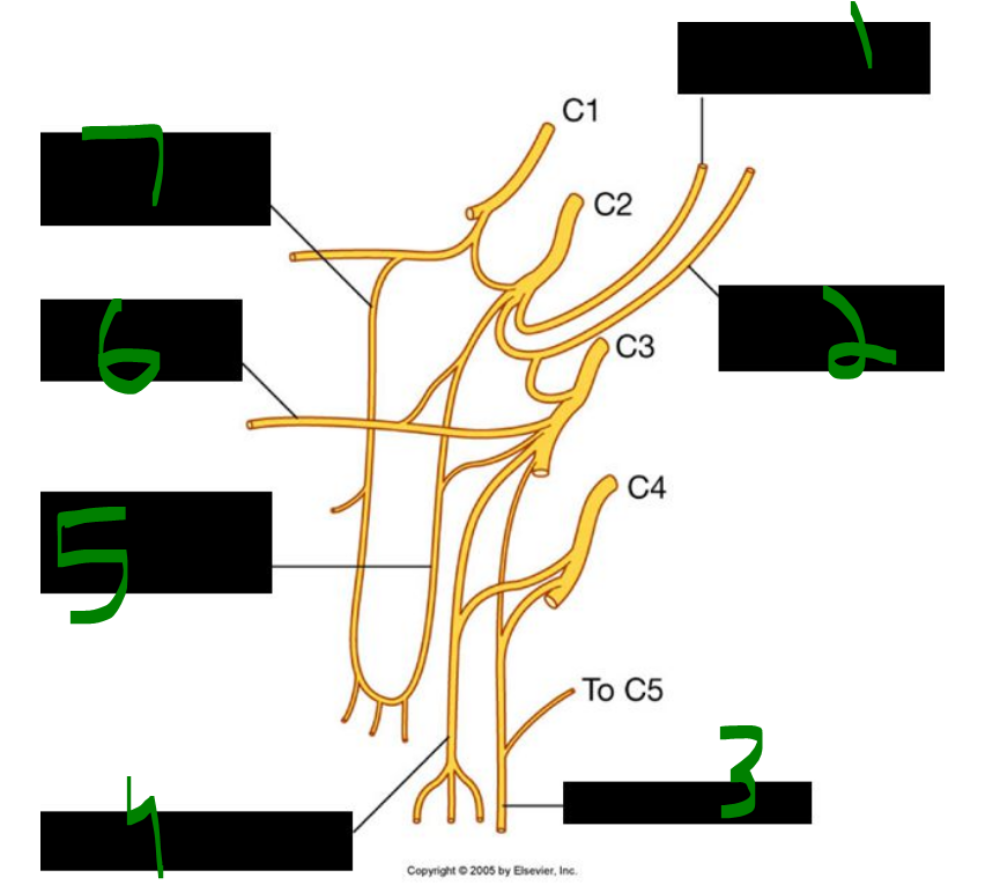
What is 1?
lesser occipital nerve
What is 2?
great auricular nerve
What is 3?
phrenic nerve
What is 4?
supraclavicular nerve
What is 5?
inferior root of ansa cervicalis
What is 6?
transverse cervical nerve
What is 7?
superior root of ansa cervicalis
what muscles are innervated by branches from the ansa cervicalis?
_____, _____, _____
sternohyoid, omohyoid, sternothyroid
What is 1?
C1
C2
C3
What is 2?
inferior root of ansa cervicalis
What is 3?
omohyoid muscle (inferior belly)
What is 4?
sternothyroid muscle
What is 5?
sternohyoid muscle
What is 6?
superior root of ansa cervicalis
What is 7?
omohyoid muscle (superior belly)
What is 8?
thyrohyoid muscle
What is 9?
hypoglossal nerve
Erb’s point, also known as the “nerve point” of the _____, is the site for a cervical _____ nerve block.
neck, plexus
To perform a cervical plexus nerve block at Erb’s point, the anesthetic is injected along the _____ border of the _____, typically near its inferior third.
posterior, SCM
Torticollis is a _____ or _____ of the sternocleidomastoid (SCM) muscle, causing _____ and lateral _____ of the neck and head.
contraction, shortening, rotation, flexion
Congenital torticollis usually appears _____ or shortly after _____. It is most commonly caused by fibrous tissue developing in the _____ before birth.
before, birth, SCM
In some cases of congenital torticollis, tearing of SCM fibers during _____ leads to a _____, which becomes fibrotic and entraps a branch of the _____ nerve (CN XI).
birth, hematoma, spinal accessory
Spasmodic torticollis differs from congenital and typically occurs _____ in _____.
later, life
Sibson’s fascia, also called the _____ membrane, is an extension of the _____ fascia.
suprapleural, endothoracic
Sibson’s fascia provides apical support for the _____ cavity at the root of the _____.
pleural, neck
Sibson’s fascia covers the superior surface of the cervical _____, also known as the _____.
pleura, cupula
Descending into the thoracic cavity, the _____ and _____ nerves lie between the _____ artery and _____ vein.
phrenic, vagus, subclavian, subclavian
what nerve crosses anteriorly to the anterior scalene?
_____ _____
phrenic nerve
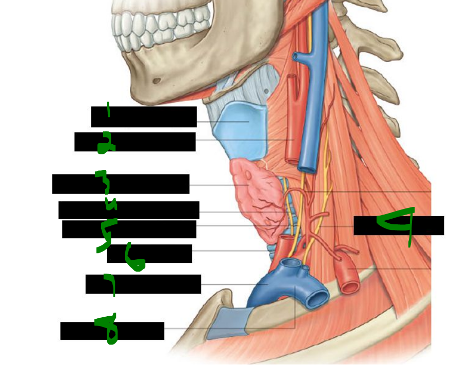
What is 1?
thyroid cartilage
What is 2?
common carotid artery
What is 3?
thyroid gland left lobe (elevated)
What is 4?
left recurrent laryngeal nerve
What is 5?
left vagus nerve (x)
What is 6?
trachea
What is 7?
brachiocephalic vein
What is 8?
subclavian vein
What is 9?
phrenic nerve
The rectus capitis anterior muscle _____ the head, while the rectus capitis lateralis muscle _____ the head _____ .
flexes, flexes, laterally
The longus capitis muscle _____ the head, and the longus colli muscle flexes the neck anteriorly, laterally, and causes slight _____ to the _____ side.
flexes, rotation, opposite
All prevertebral muscles are innervated by the anterior rami of spinal nerves _____ through _____.
C1, C6
The cervical part of the sympathetic trunk lies _____ to the longus colli and longus capitis muscles. The cervical sympathetic trunk lies _____ to the common carotid artery.
anterior
posterior
The cervical sympathetic trunk contains only _____ rami communicantes and does not have any _____ rami communicantes.
gray, white
What is 1?
superior cervical ganglion
What is 2?
sympathetic trunk
What is 3?
middle cervical ganglion
What is 4?
inferior cervical ganglion
What is 5?
left vagus nerve
What is 6?
left brachiocephalic vein
What is 7?
brachial plexus
What is 8?
subclavian vein
What is 9?
subclavian artery
What is 10?
phrenic nerve
The superior cervical ganglion is located anterior to the _____ and _____ vertebrae.
C1, C2
Preganglionic fibers to the superior cervical ganglion originate only from spinal nerve _____.
T1
Postganglionic fibers from the superior cervical ganglion travel via gray rami to spinal nerves _____ through _____.
C1, C4
Postganglionic fibers from the superior cervical ganglion also follow the _____ _____ artery forming the internal carotid plexus.
internal carotid
Other targets of postganglionic fibers from the superior cervical ganglion include the carotid _____ and _____, the _____, and the heart as the superior _____ nerves.
body, sinus, pharynx, cardiac
what structure loops around the subclavian a. to connect the middle & inferior cervical ganglia?
_____ _____
ansa subclavia
The middle cervical ganglion lies anterior to the _____ vertebra.
C6
The middle cervical ganglion sends gray rami communicantes to spinal nerves _____ and _____.
C5, C6
Postganglionic fibers from the middle cervical ganglion _____ to the heart as the _____ _____ _____ .
contribute, middle cardiac nerves
The _____ cervical ganglion is very large and often fused with the _____ thoracic ganglion, forming the _____ ganglion.
inferior, first, cervicothoracic
The inferior cervical ganglion sends gray rami communicantes to spinal nerves _____ and _____.
C7, C8
Branches from the inferior cervical ganglion follow the _____ artery and supply the _____ as the _____ cardiac nerves.
vertebral, heart, inferior
The stellate ganglion is also known as the _____ ganglion.
cervicothoracic
The stellate ganglion refers to the _____ cervical ganglion when fused with the _____ thoracic ganglion.
inferior, first
The stellate ganglion fusion occurs in approximately _____% of people.
80
The costocervical trunk branches from the _____ part of the _____ artery on the left side.
first, subclavian
The costocervical trunk branches from the _____ part of the _____ artery on the right side.
second, subclavian
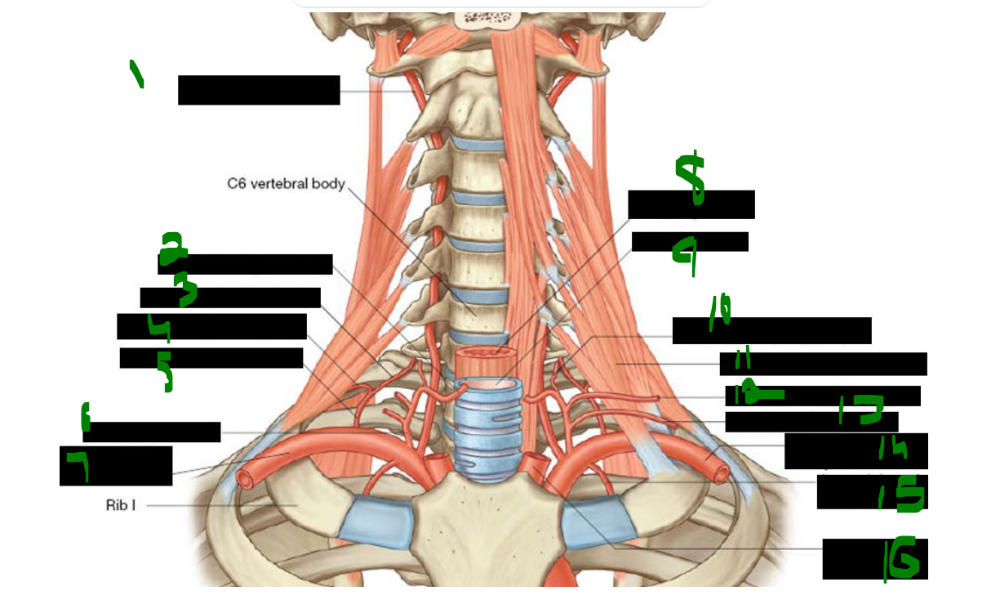
What is 1?
vertebral artery
What is 2?
inferior thryroid artery
What is 3?
deep cervical artery
What is 4?
supreme intercostal artery
What is 5?
costocervical trunk
What is 6?
thyrocervical trunk
What is 7?
right subclavian artery
What is 8?
esophagus
What is 9?
trachea
What is 10?
ascending cervical artery
What is 11?
anterior scalene muscle
What is 12?
transverse cervical artery
What is 13?
suprascapular artery
What is 14?
left subclavian artery
What is 15?
internal thoracic artery
What is 16?
left common carotid artery
The first branch of the subclavian artery is the _____ artery, which ascends and enters the foramina of the transverse processes of vertebrae _____ through _____. It then turns medially, crossing the posterior arch of the _____ (C1), before passing through the _____ _____.
vertebral, C6, C1, atlas, foramen magnum
The ascending cervical artery is a branch of the _____ _____ artery, which arises from the _____ trunk, the _____ branch of the subclavian artery.
inferior thyroid, thyrocervical, second
The third branch of the subclavian artery is the _____ _____ artery, which descends anterior to the _____.
internal thoracic, pleura
The costocervical trunk gives rise to the _____ cervical artery.
The costocervical trunk also gives rise to the _____ intercostal artery, which supplies the _____ and _____ intercostal spaces posteriorly.
deep
supreme, first, second
The supreme intercostal artery branches from the _____ trunk, which arises from the _____ part of the subclavian artery on the left and the _____ part on the right. It supplies blood to the _____ and _____ intercostal spaces posteriorly.
costocervical, first, second, first, second
The main superficial veins draining the neck are the _____ jugular vein and the _____ jugular vein.
external, anterior
The anterior jugular veins are united by the _____ venous arch.
jugular
what structure unites the anterior jugular veins?
_____ _____ _____
jugular venous arch
what does the jugular venous arch unite?
_____ _____ _____
anterior jugular veins
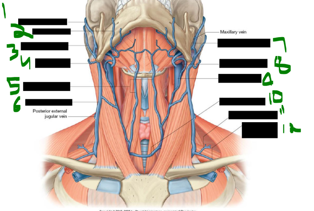
What is 1?
superficial temporal vein
What is 2?
facial vein
What is 3?
posterior auricular vein
What is 4?
occipital vein
What is 5?
anterior jugular vein
What is 6?
external jugular vein
What is 7?
retromandibular vein
What is 8?
common facial vein
What is 9?
internal jugular vein
What is 10?
jugular venous arch
What is 11?
transverse cervical vein
What is 12?
suprascapular vein
The external jugular vein is formed by the union of the posterior division of the _____ vein and the _____ auricular vein.
retromandibular, posterior
The external jugular vein drains into the _____ vein.
subclavian
The tributaries of the external jugular vein located in the posterior triangle include the _____ cervical and _____ arteries.
transverse, suprascapular
The external jugular vein also receives the _____ and _____ jugular veins in the posterior triangle.
anterior, posterior
The occipital and mastoid lymph nodes drain into the _____ _____ _____ _____
superficial cervical lymph nodes.
The occipital and mastoid nodes drain along the _____ jugular vein.
external
The deep cervical lymph nodes receive drainage along the _____ _____ _____ .
internal jugular vein
The pre-auricular, _____, and _____ lymph nodes drain into the deep cervical nodes.
parotid, submandibular
The _____ lymph nodes also drain into the deep cervical nodes.
submental
where does lymph from the tonsils drain?
_____ _____
jugulodigastric node
On the left side, the jugular trunk and subclavian trunk join the _____ duct, which drains into the junction of the _____ jugular and _____ veins.
thoracic, internal, subclavian
On the right side, the jugular trunk and subclavian trunk join the _____ lymphatic duct, which drains into the junction of the _____ jugular and _____ veins.
right, internal, subclavian
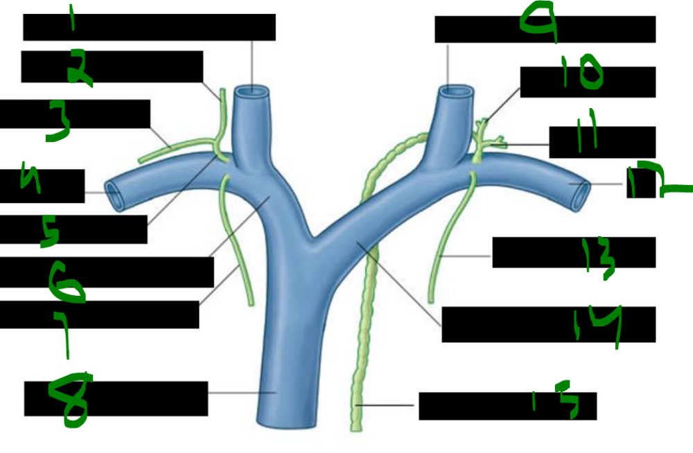
What is 1?
right internal jugular vein
What is 2?
jugular trunk
What is 3?
subclavian trunk
What is 4?
right subclavian vein
What is 5?
right lymphatic duct
What is 6?
right brachiocephalic vein
What is 7?
bronchomediastinal trunk
What is 8?
superior vena cava
What is 9?
left internal jugular vein
What is 10?
jugular trunk
What is 11?
subclavian trunk
What is 12?
left subclavian vein
What is 13?
bronchomediastinal trunk
What is 14?
left brachiocephalic vein
What is 15?
thoracic duct
The thoracic duct enters the root of the neck to the _____ of the esophagus at approximately the _____ vertebral level.
left, C5
The thoracic duct drains into the venous system at the _____ _____ _____.
left venous angle
The internal jugular vein begins at the _____ foramen, travels laterally in the _____ sheath, and passes deep to the _____. It has an inferior _____ and valve that prevent backward flow. It joins with the _____ vein to form the brachiocephalic vein.
jugular, carotid, SCM, bulb, subclavian
The subclavian artery pulse can be palpated in the _____ supraclavicular fossa. Compressing the artery against the _____ rib can prevent blood loss during an upper limb arterial _____.
greater, first, hemorrhage
In a subclavian vein puncture, the needle is inserted inferior to the _____ (at mid-clavicle) and advanced medially toward the tip of the _____ finger placed on the jugular _____.
thumb, index, notch
The subclavian vein puncture targets the _____ venous angle, located posterior to the _____ joint, where the internal jugular and subclavian veins unite to form the _____ vein.
right, sternoclavicular, brachiocephalic
Two major risks of incorrect needle placement during subclavian vein puncture are _____ and accidental entry into the _____ artery.
pneumothorax, subclavian
During subclavian vein puncture, the patient is placed in the _____ position—lying flat with the body elevated above the head by _____ to _____ degrees—to optimize line insertion.
Trendelenburg, 15, 30
The Trendelenburg position is used to insert the needle and _____ during _____ line placement.
catheter, central