Instructions for Side by Side Printing
- Print the notecards
- Fold each page in half along the solid vertical line
- Cut out the notecards by cutting along each horizontal dotted line
- Optional: Glue, tape or staple the ends of each notecard together
Anatomy JV Exam 3: Posterior Triangle
front 1 The posterior triangle is bounded anteriorly by the posterior border of the _____, posteriorly by the anterior border of the _____, and inferiorly by the middle third of the _____. | back 1 SCM, trapezius, clavicle |
front 2 The superior boundary of the posterior triangle is the _____ bone, just posterior to the _____ process. | back 2 occipital, mastoid |
front 3 The roof of the posterior triangle is formed by the _____ layer of the _____ fascia. | back 3 investing, cervical |
front 4 The floor of the posterior triangle is formed by the _____ layer of deep cervical fascia covering the deep _____ muscles. | back 4 prevertebral, neck |
front 5 what are the muscles of the posterior triangle? -_____ _____ , _____ _____ , _____ _____ , _____ _____ , _____ _____ , _____ _____ _____ | back 5 -splenius capitus, levator scapulae, posterior scalene, middle scalene, anterior scalene, omohyoid inferior belly |
front 6 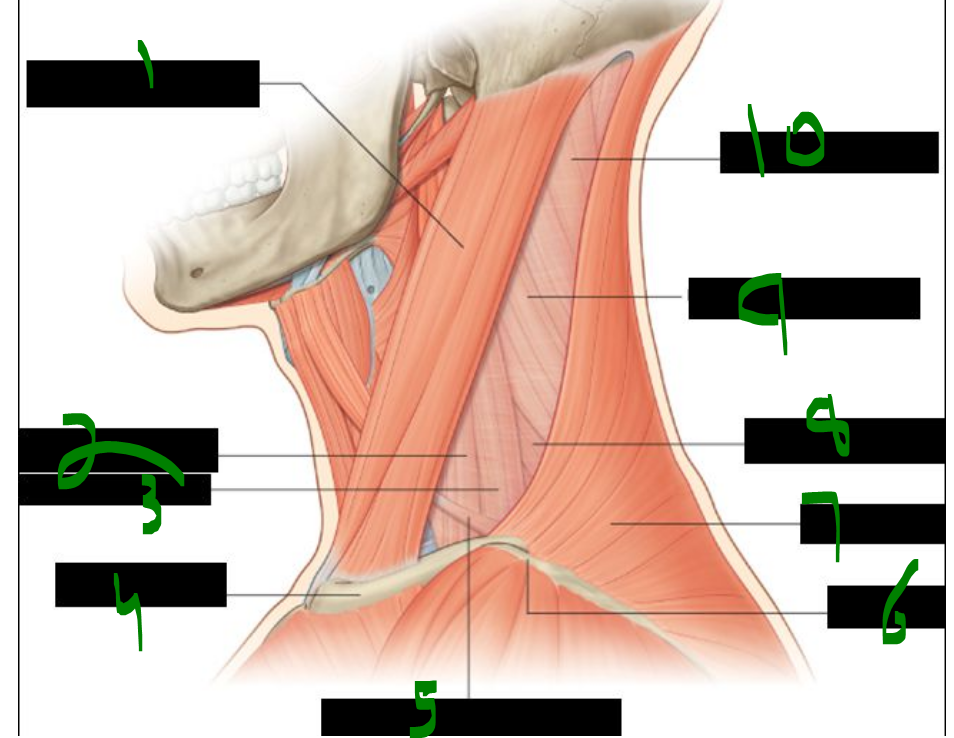 What is 1? | back 6 sternocleidomastoid muscle |
front 7 What is 2? | back 7 anterior scalene muscle |
front 8 What is 3? | back 8 middle scalene muscle |
front 9 What is 4? | back 9 clavicle |
front 10 What is 5? | back 10 inferior belly of omohyoid muscle |
front 11 What is 6? | back 11 acromion |
front 12 What is 7? | back 12 trapezius muscle |
front 13 What is 8? | back 13 posterior scalene muscle |
front 14 What is 9? | back 14 levator scapulae muscle |
front 15 What is 10? | back 15 splenius capitus muscle |
front 16 what fascial layer covers/encloses the muscles of the posterior triangle? ____ ____ | back 16 prevertebral fascia |
front 17 what structure subdivides the posterior triangle & into what? ____ ____ of ____ ____ | back 17 inferior belly of omohyoid muscle |
front 18 The inferior belly of the omohyoid divides the posterior triangle into the _____ triangle and the _____ triangle (also called the omoclavicular or subclavian triangle). | back 18 occipital, supraclavicular |
front 19 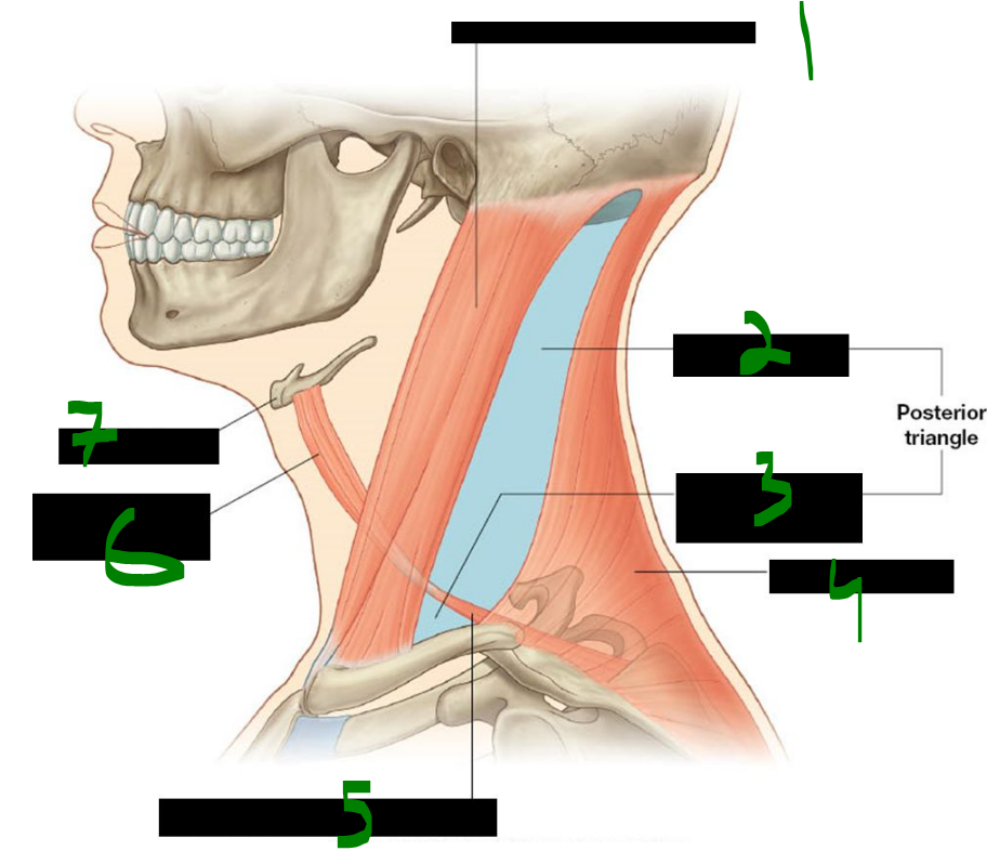 What is 1? | back 19 sternocleidomastoid muscle |
front 20 What is 2? | back 20 occipital triangle |
front 21 What is 3? | back 21 omoclavicular or subclavian triangle |
front 22 What is 4? | back 22 trapezius muscle |
front 23 What is 5? | back 23 inferior belly of omohyoid muscle |
front 24 What is 6? | back 24 superior belly of omohyoid muscle |
front 25 What is 7? | back 25 hyoid bone |
front 26 The omohyoid muscle originates from the superior border of the _____ and inserts onto the inferior border of the _____ bone. | back 26 scapula, hyoid |
front 27 The omohyoid is innervated by the _____ _____, which arises from the anterior rami of spinal nerves _____ to _____. | back 27 ansa cervicalis, C1, C3 |
front 28 The omohyoid muscle functions to _____ and _____ the hyoid bone. | back 28 depress, stabilize |
front 29 The omohyoid has two _____ connected by an intermediate _____ and separated by a fascial _____. | back 29 bellies, tendon, sling |
front 30 The scalene muscles originate from the _____ of the cervical _____ processes. | back 30 tubercles, transverse |
front 31 The anterior and middle scalenes insert on the _____ rib, while the posterior scalene inserts on the _____ rib. | back 31 first, second |
front 32 The scalene muscles are innervated by the anterior rami of spinal nerves _____ through _____. | back 32 C3, C7 |
front 33 The scalene muscles act to _____ the ribs during inspiration and to _____ the neck. | back 33 elevate, flex |
front 34 The subclavian vein lies _____ to the anterior scalene, while the subclavian artery lies _____ to the anterior scalene. | back 34 anterior, posterior |
front 35 The subclavian _____ is more accessible because it is not 'protected' by the anterior scalene muscle, unlike the subclavian _____. | back 35 vein, artery |
front 36 The _____ nerve travels along the _____ surface of the anterior scalene muscle. | back 36 phrenic, anterior |
front 37 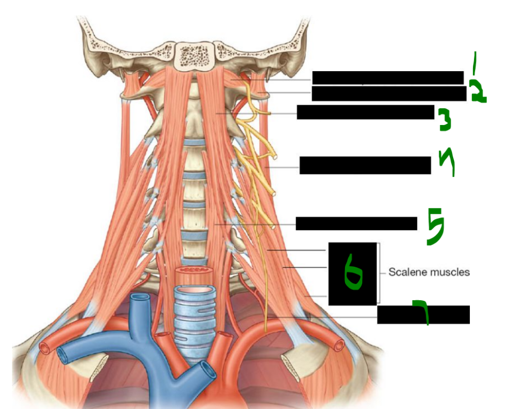 What is 1? | back 37 rectus capitis anterior muscle |
front 38 What is 2? | back 38 rectus capitis lateralis muscle |
front 39 What is 3? | back 39 longus capitis muscle |
front 40 What is 4? | back 40 levator scapulae muscle |
front 41 What is 5? | back 41 longus colli muscle |
front 42 What is 6? | back 42 anterior middle posterior |
front 43 What is 7? | back 43 phrenic nerve |
front 44 The posterior triangle contains three main arteries: the _____ _____ artery, the _____ cervical artery, and the _____ artery. | back 44 dorsal scapular, transverse, suprascapular |
front 45 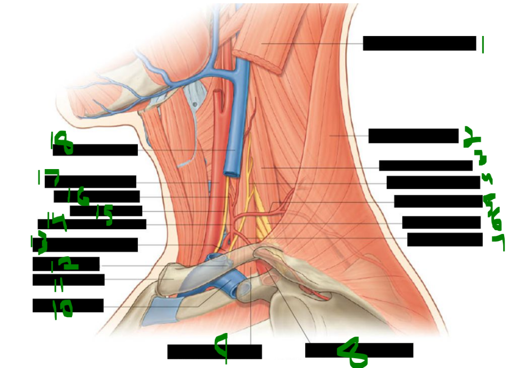 What is 1? | back 45 sternocleidomastoid muscle |
front 46 What is 2? | back 46 trapezius muscle |
front 47 What is 3? | back 47 middle scalene muscle |
front 48 What is 4? | back 48 phrenic nerve |
front 49 What is 5? | back 49 transverse cervical artery |
front 50 What is 6? | back 50 brachial plexus |
front 51 What is 7? | back 51 suprascapular artery |
front 52 What is 8? | back 52 3rd part of subclavian artery |
front 53 What is 9? | back 53 anterior scalene muscle |
front 54 What is 10? | back 54 subclavian vein |
front 55 What is 11? | back 55 clavicle |
front 56 What is 12? | back 56 external jugular vein |
front 57 What is 13? | back 57 1st part of subclavian artery |
front 58 What is 14? | back 58 thyrocervical trunk |
front 59 What is 15? | back 59 vagus nerve |
front 60 What is 16? | back 60 inferior thyroid artery |
front 61 What is 17? | back 61 common carotid artery |
front 62 What is 18? | back 62 internal jugular vein |
front 63 The spinal accessory nerve (CN XI) innervates the _____ and the _____ muscles. | back 63 SCM, trapezius |
front 64 The transverse cervical nerve (C2–C3) provides sensory _____ to the skin over the _____ triangle. | back 64 innervation, anterior |
front 65 The great auricular nerve (C2–C3) innervates the skin over the _____ gland and around the _____. | back 65 parotid, auricle |
front 66 The supraclavicular nerve (C3–C4) pierces the _____ muscle to supply skin over the _____, shoulder, and as far inferiorly as rib _____. | back 66 platysma, clavicle, two |
front 67 The lesser occipital nerve (C2–C3) supplies the skin of the _____ scalp, posterior to the _____. | back 67 lateral, auricle |
front 68 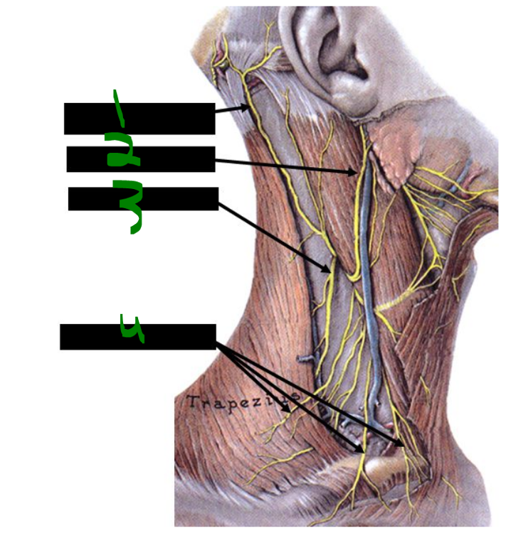 What is 1? | back 68 lesser occipital nerve |
front 69 What is 2? | back 69 great auricular nerve |
front 70 What is 3? | back 70 accessory nerve |
front 71 What is 4? | back 71 supraclavicular nerves |
front 72 the accessory nerve lies on what surface of what muscle? _____ _____ of _____ _____ | back 72 anterior surface of levator scauplae |
front 73 the axillary fascia is a continuation of what fascial layer? _____ _____, carried by the _____ _____ | back 73 prevertebral fascia, carried by the brachial plexus |
front 74 The brachial plexus passes between the _____ and _____ scalene muscles. | back 74 anterior, middle |
front 75 The _____ of the brachial plexus cross the _____ of the posterior triangle. | back 75 trunks, base |
front 76 what branches of the brachial plexus [may] appear in the posterior triangle? _____ to _____ , _____ _____ nerve, _____ _____ nerve, _____ nerve | back 76 -nerve to subclavius, dorsal scapular nerve, long thoracic nerve, suprascapular nerve (NooDLeS - w/o vowels) |
front 77 The superior root of the ansa cervicalis is formed by spinal nerves _____ and _____. | back 77 C1, C2 |
front 78 The inferior root of the ansa cervicalis arises from spinal nerves _____ and _____. | back 78 C2, C3 |
front 79 The transverse cervical and great auricular nerves are both formed from spinal nerves _____ and _____. | back 79 C2, C3 |
front 80 The lesser occipital nerve is mainly derived from _____, but also receives contribution from _____. | back 80 C2, C3 |
front 81 The supraclavicular nerve arises from spinal nerves _____ and _____. | back 81 C3, C4 |
front 82 The phrenic nerve is formed by spinal nerves _____ through _____. | back 82 C3, C5 |
front 83 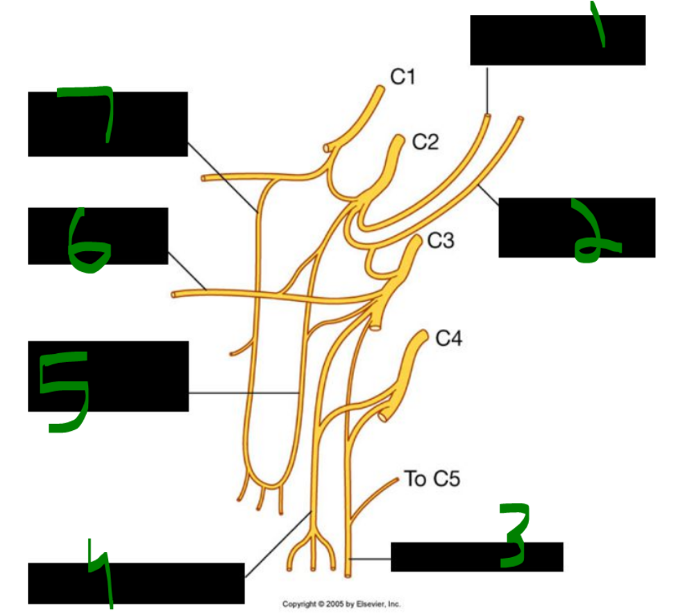 What is 1? | back 83 lesser occipital nerve |
front 84 What is 2? | back 84 great auricular nerve |
front 85 What is 3? | back 85 phrenic nerve |
front 86 What is 4? | back 86 supraclavicular nerve |
front 87 What is 5? | back 87 inferior root of ansa cervicalis |
front 88 What is 6? | back 88 transverse cervical nerve |
front 89 What is 7? | back 89 superior root of ansa cervicalis |
front 90 what muscles are innervated by branches from the ansa cervicalis? _____, _____, _____ | back 90 sternohyoid, omohyoid, sternothyroid |
front 91 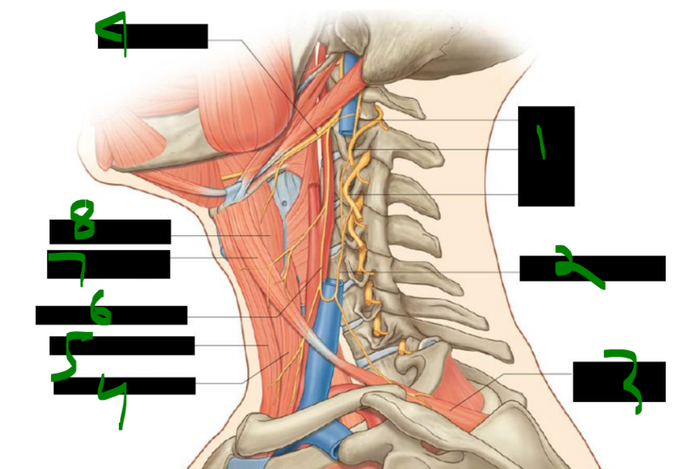 What is 1? | back 91 C1 C2 C3 |
front 92 What is 2? | back 92 inferior root of ansa cervicalis |
front 93 What is 3? | back 93 omohyoid muscle (inferior belly) |
front 94 What is 4? | back 94 sternothyroid muscle |
front 95 What is 5? | back 95 sternohyoid muscle |
front 96 What is 6? | back 96 superior root of ansa cervicalis |
front 97 What is 7? | back 97 omohyoid muscle (superior belly) |
front 98 What is 8? | back 98 thyrohyoid muscle |
front 99 What is 9? | back 99 hypoglossal nerve |
front 100 Erb’s point, also known as the “nerve point” of the _____, is the site for a cervical _____ nerve block. | back 100 neck, plexus |
front 101 To perform a cervical plexus nerve block at Erb’s point, the anesthetic is injected along the _____ border of the _____, typically near its inferior third. | back 101 posterior, SCM |
front 102 Torticollis is a _____ or _____ of the sternocleidomastoid (SCM) muscle, causing _____ and lateral _____ of the neck and head. | back 102 contraction, shortening, rotation, flexion |
front 103 Congenital torticollis usually appears _____ or shortly after _____. It is most commonly caused by fibrous tissue developing in the _____ before birth. | back 103 before, birth, SCM |
front 104 In some cases of congenital torticollis, tearing of SCM fibers during _____ leads to a _____, which becomes fibrotic and entraps a branch of the _____ nerve (CN XI). | back 104 birth, hematoma, spinal accessory |
front 105 Spasmodic torticollis differs from congenital and typically occurs _____ in _____. | back 105 later, life |
front 106 Sibson’s fascia, also called the _____ membrane, is an extension of the _____ fascia. | back 106 suprapleural, endothoracic |
front 107 Sibson’s fascia provides apical support for the _____ cavity at the root of the _____. | back 107 pleural, neck |
front 108 Sibson’s fascia covers the superior surface of the cervical _____, also known as the _____. | back 108 pleura, cupula |
front 109 Descending into the thoracic cavity, the _____ and _____ nerves lie between the _____ artery and _____ vein. | back 109 phrenic, vagus, subclavian, subclavian |
front 110 what nerve crosses anteriorly to the anterior scalene? _____ _____ | back 110 phrenic nerve |
front 111 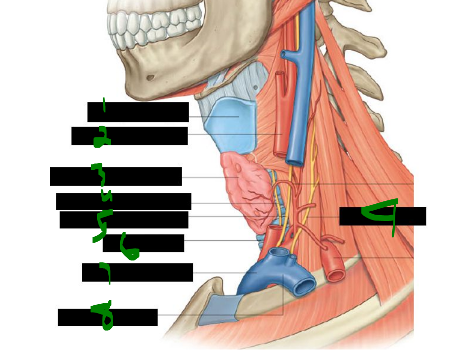 What is 1? | back 111 thyroid cartilage |
front 112 What is 2? | back 112 common carotid artery |
front 113 What is 3? | back 113 thyroid gland left lobe (elevated) |
front 114 What is 4? | back 114 left recurrent laryngeal nerve |
front 115 What is 5? | back 115 left vagus nerve (x) |
front 116 What is 6? | back 116 trachea |
front 117 What is 7? | back 117 brachiocephalic vein |
front 118 What is 8? | back 118 subclavian vein |
front 119 What is 9? | back 119 phrenic nerve |
front 120 The rectus capitis anterior muscle _____ the head, while the rectus capitis lateralis muscle _____ the head _____ . | back 120 flexes, flexes, laterally |
front 121 The longus capitis muscle _____ the head, and the longus colli muscle flexes the neck anteriorly, laterally, and causes slight _____ to the _____ side. | back 121 flexes, rotation, opposite |
front 122 All prevertebral muscles are innervated by the anterior rami of spinal nerves _____ through _____. | back 122 C1, C6 |
front 123 The cervical part of the sympathetic trunk lies _____ to the longus colli and longus capitis muscles. The cervical sympathetic trunk lies _____ to the common carotid artery. | back 123 anterior posterior |
front 124 The cervical sympathetic trunk contains only _____ rami communicantes and does not have any _____ rami communicantes. | back 124 gray, white |
front 125  What is 1? | back 125 superior cervical ganglion |
front 126 What is 2? | back 126 sympathetic trunk |
front 127 What is 3? | back 127 middle cervical ganglion |
front 128 What is 4? | back 128 inferior cervical ganglion |
front 129 What is 5? | back 129 left vagus nerve |
front 130 What is 6? | back 130 left brachiocephalic vein |
front 131 What is 7? | back 131 brachial plexus |
front 132 What is 8? | back 132 subclavian vein |
front 133 What is 9? | back 133 subclavian artery |
front 134 What is 10? | back 134 phrenic nerve |
front 135 The superior cervical ganglion is located anterior to the _____ and _____ vertebrae. | back 135 C1, C2 |
front 136 Preganglionic fibers to the superior cervical ganglion originate only from spinal nerve _____. | back 136 T1 |
front 137 Postganglionic fibers from the superior cervical ganglion travel via gray rami to spinal nerves _____ through _____. | back 137 C1, C4 |
front 138 Postganglionic fibers from the superior cervical ganglion also follow the _____ _____ artery forming the internal carotid plexus. | back 138 internal carotid |
front 139 Other targets of postganglionic fibers from the superior cervical ganglion include the carotid _____ and _____, the _____, and the heart as the superior _____ nerves. | back 139 body, sinus, pharynx, cardiac |
front 140 what structure loops around the subclavian a. to connect the middle & inferior cervical ganglia? _____ _____ | back 140 ansa subclavia |
front 141 The middle cervical ganglion lies anterior to the _____ vertebra. | back 141 C6 |
front 142 The middle cervical ganglion sends gray rami communicantes to spinal nerves _____ and _____. | back 142 C5, C6 |
front 143 Postganglionic fibers from the middle cervical ganglion _____ to the heart as the _____ _____ _____ . | back 143 contribute, middle cardiac nerves |
front 144 The _____ cervical ganglion is very large and often fused with the _____ thoracic ganglion, forming the _____ ganglion. | back 144 inferior, first, cervicothoracic |
front 145 The inferior cervical ganglion sends gray rami communicantes to spinal nerves _____ and _____. | back 145 C7, C8 |
front 146 Branches from the inferior cervical ganglion follow the _____ artery and supply the _____ as the _____ cardiac nerves. | back 146 vertebral, heart, inferior |
front 147 The stellate ganglion is also known as the _____ ganglion. | back 147 cervicothoracic |
front 148 The stellate ganglion refers to the _____ cervical ganglion when fused with the _____ thoracic ganglion. | back 148 inferior, first |
front 149 The stellate ganglion fusion occurs in approximately _____% of people. | back 149 80 |
front 150 The costocervical trunk branches from the _____ part of the _____ artery on the left side. | back 150 first, subclavian |
front 151 The costocervical trunk branches from the _____ part of the _____ artery on the right side. | back 151 second, subclavian |
front 152 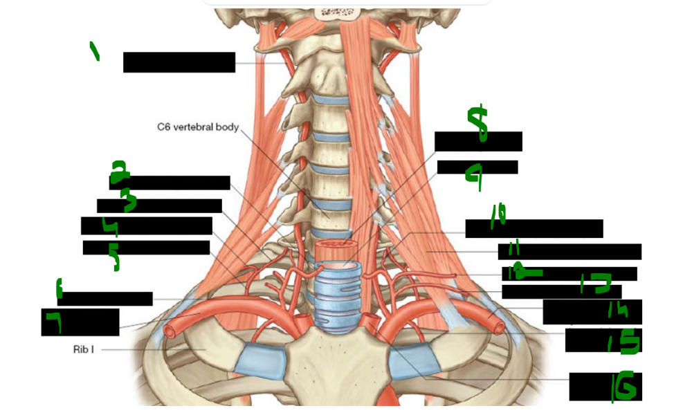 What is 1? | back 152 vertebral artery |
front 153 What is 2? | back 153 inferior thryroid artery |
front 154 What is 3? | back 154 deep cervical artery |
front 155 What is 4? | back 155 supreme intercostal artery |
front 156 What is 5? | back 156 costocervical trunk |
front 157 What is 6? | back 157 thyrocervical trunk |
front 158 What is 7? | back 158 right subclavian artery |
front 159 What is 8? | back 159 esophagus |
front 160 What is 9? | back 160 trachea |
front 161 What is 10? | back 161 ascending cervical artery |
front 162 What is 11? | back 162 anterior scalene muscle |
front 163 What is 12? | back 163 transverse cervical artery |
front 164 What is 13? | back 164 suprascapular artery |
front 165 What is 14? | back 165 left subclavian artery |
front 166 What is 15? | back 166 internal thoracic artery |
front 167 What is 16? | back 167 left common carotid artery |
front 168 The first branch of the subclavian artery is the _____ artery, which ascends and enters the foramina of the transverse processes of vertebrae _____ through _____. It then turns medially, crossing the posterior arch of the _____ (C1), before passing through the _____ _____. | back 168 vertebral, C6, C1, atlas, foramen magnum |
front 169 The ascending cervical artery is a branch of the _____ _____ artery, which arises from the _____ trunk, the _____ branch of the subclavian artery. | back 169 inferior thyroid, thyrocervical, second |
front 170 The third branch of the subclavian artery is the _____ _____ artery, which descends anterior to the _____. | back 170 internal thoracic, pleura |
front 171 The costocervical trunk gives rise to the _____ cervical artery. The costocervical trunk also gives rise to the _____ intercostal artery, which supplies the _____ and _____ intercostal spaces posteriorly. | back 171 deep supreme, first, second |
front 172 The supreme intercostal artery branches from the _____ trunk, which arises from the _____ part of the subclavian artery on the left and the _____ part on the right. It supplies blood to the _____ and _____ intercostal spaces posteriorly. | back 172 costocervical, first, second, first, second |
front 173 The main superficial veins draining the neck are the _____ jugular vein and the _____ jugular vein. | back 173 external, anterior |
front 174 The anterior jugular veins are united by the _____ venous arch. | back 174 jugular |
front 175 what structure unites the anterior jugular veins? _____ _____ _____ | back 175 jugular venous arch |
front 176 what does the jugular venous arch unite? _____ _____ _____ | back 176 anterior jugular veins |
front 177 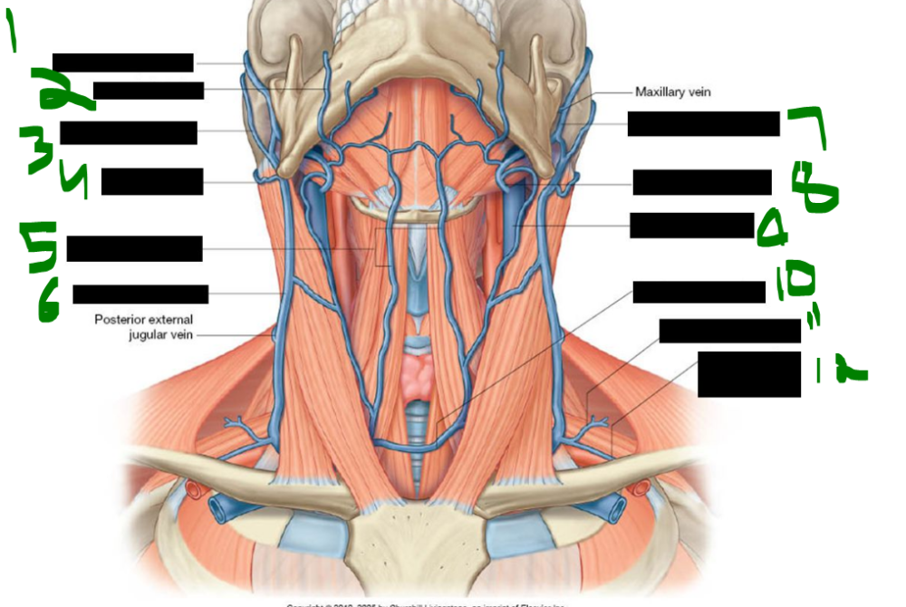 What is 1? | back 177 superficial temporal vein |
front 178 What is 2? | back 178 facial vein |
front 179 What is 3? | back 179 posterior auricular vein |
front 180 What is 4? | back 180 occipital vein |
front 181 What is 5? | back 181 anterior jugular vein |
front 182 What is 6? | back 182 external jugular vein |
front 183 What is 7? | back 183 retromandibular vein |
front 184 What is 8? | back 184 common facial vein |
front 185 What is 9? | back 185 internal jugular vein |
front 186 What is 10? | back 186 jugular venous arch |
front 187 What is 11? | back 187 transverse cervical vein |
front 188 What is 12? | back 188 suprascapular vein |
front 189 The external jugular vein is formed by the union of the posterior division of the _____ vein and the _____ auricular vein. | back 189 retromandibular, posterior |
front 190 The external jugular vein drains into the _____ vein. | back 190 subclavian |
front 191 The tributaries of the external jugular vein located in the posterior triangle include the _____ cervical and _____ arteries. | back 191 transverse, suprascapular |
front 192 The external jugular vein also receives the _____ and _____ jugular veins in the posterior triangle. | back 192 anterior, posterior |
front 193 The occipital and mastoid lymph nodes drain into the _____ _____ _____ _____ | back 193 superficial cervical lymph nodes. |
front 194 The occipital and mastoid nodes drain along the _____ jugular vein. | back 194 external |
front 195 The deep cervical lymph nodes receive drainage along the _____ _____ _____ . | back 195 internal jugular vein |
front 196 The pre-auricular, _____, and _____ lymph nodes drain into the deep cervical nodes. | back 196 parotid, submandibular |
front 197 The _____ lymph nodes also drain into the deep cervical nodes. | back 197 submental |
front 198 where does lymph from the tonsils drain? _____ _____ | back 198 jugulodigastric node |
front 199 On the left side, the jugular trunk and subclavian trunk join the _____ duct, which drains into the junction of the _____ jugular and _____ veins. | back 199 thoracic, internal, subclavian |
front 200 On the right side, the jugular trunk and subclavian trunk join the _____ lymphatic duct, which drains into the junction of the _____ jugular and _____ veins. | back 200 right, internal, subclavian |
front 201 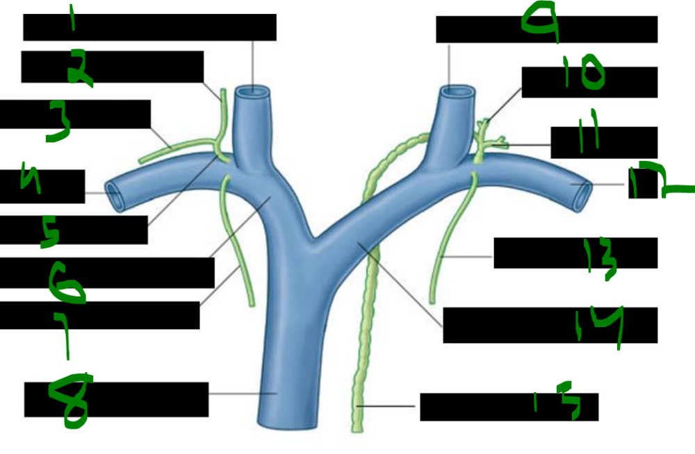 What is 1? | back 201 right internal jugular vein |
front 202 What is 2? | back 202 jugular trunk |
front 203 What is 3? | back 203 subclavian trunk |
front 204 What is 4? | back 204 right subclavian vein |
front 205 What is 5? | back 205 right lymphatic duct |
front 206 What is 6? | back 206 right brachiocephalic vein |
front 207 What is 7? | back 207 bronchomediastinal trunk |
front 208 What is 8? | back 208 superior vena cava |
front 209 What is 9? | back 209 left internal jugular vein |
front 210 What is 10? | back 210 jugular trunk |
front 211 What is 11? | back 211 subclavian trunk |
front 212 What is 12? | back 212 left subclavian vein |
front 213 What is 13? | back 213 bronchomediastinal trunk |
front 214 What is 14? | back 214 left brachiocephalic vein |
front 215 What is 15? | back 215 thoracic duct |
front 216 The thoracic duct enters the root of the neck to the _____ of the esophagus at approximately the _____ vertebral level. | back 216 left, C5 |
front 217 The thoracic duct drains into the venous system at the _____ _____ _____. | back 217 left venous angle |
front 218 The internal jugular vein begins at the _____ foramen, travels laterally in the _____ sheath, and passes deep to the _____. It has an inferior _____ and valve that prevent backward flow. It joins with the _____ vein to form the brachiocephalic vein. | back 218 jugular, carotid, SCM, bulb, subclavian |
front 219 The subclavian artery pulse can be palpated in the _____ supraclavicular fossa. Compressing the artery against the _____ rib can prevent blood loss during an upper limb arterial _____. | back 219 greater, first, hemorrhage |
front 220 In a subclavian vein puncture, the needle is inserted inferior to the _____ (at mid-clavicle) and advanced medially toward the tip of the _____ finger placed on the jugular _____. | back 220 thumb, index, notch |
front 221 The subclavian vein puncture targets the _____ venous angle, located posterior to the _____ joint, where the internal jugular and subclavian veins unite to form the _____ vein. | back 221 right, sternoclavicular, brachiocephalic |
front 222 Two major risks of incorrect needle placement during subclavian vein puncture are _____ and accidental entry into the _____ artery. | back 222 pneumothorax, subclavian |
front 223 During subclavian vein puncture, the patient is placed in the _____ position—lying flat with the body elevated above the head by _____ to _____ degrees—to optimize line insertion. | back 223 Trendelenburg, 15, 30 |
front 224 The Trendelenburg position is used to insert the needle and _____ during _____ line placement. | back 224 catheter, central |