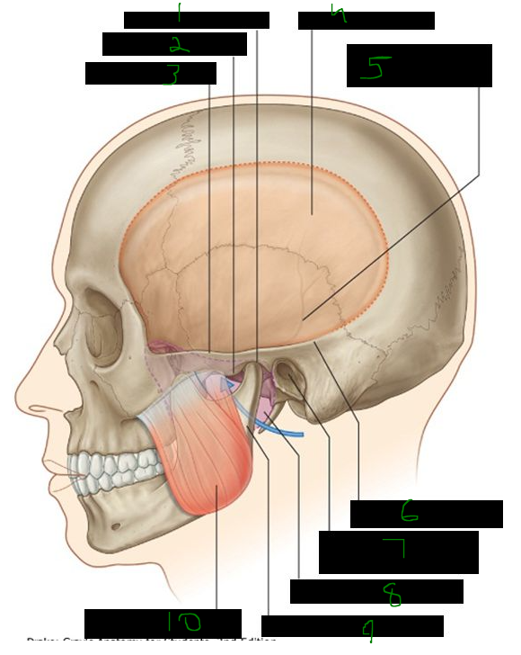
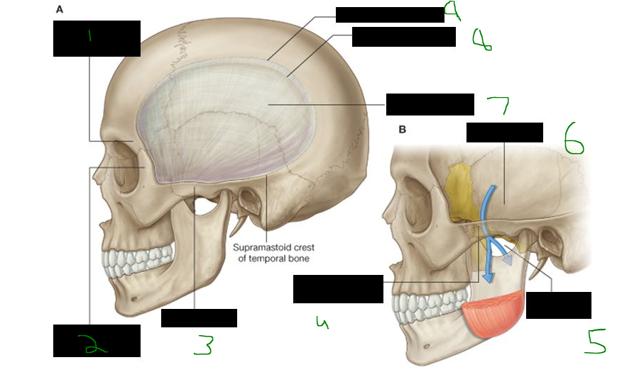
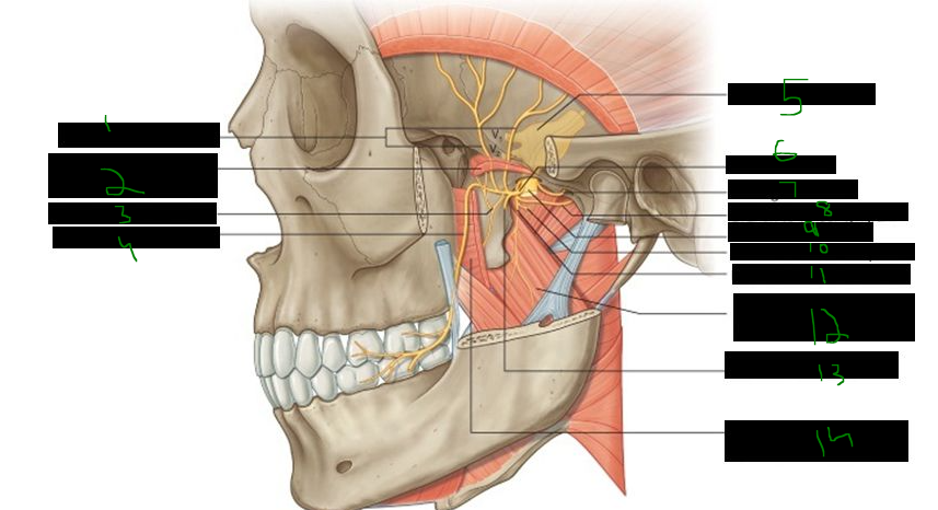
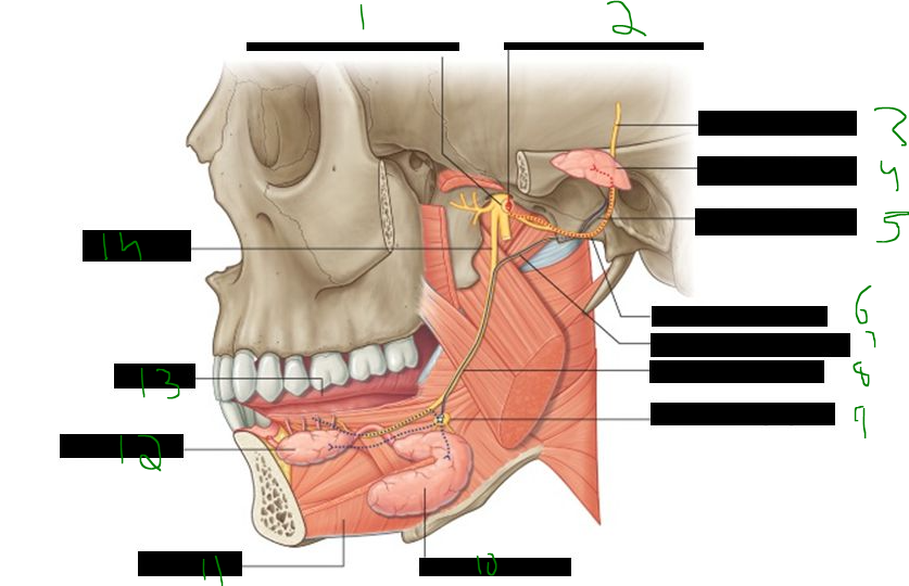
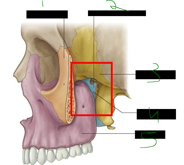
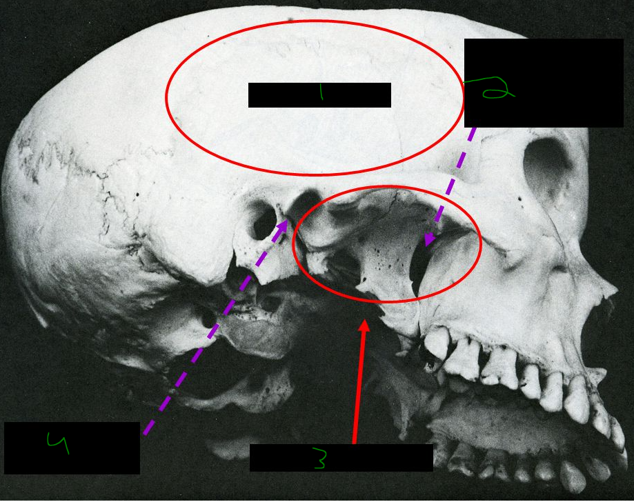
What is 1?
mandibular fossa
What is 2?
articular tubercle
What is 3?
zygomatic arch
What is 4?
temporal fossa
What is 5?
groove for middle temporal artery
What is 6?
supramastoid crest
What is 7?
external auditory meatus
What is 8?
infratemporal fossa
What is 9?
ramus of mandible
What is 10?
masseter mucle
The ______ ______ separates the temporal from the infratemporal fossae, which are ______ with each other; the temporal fossa lies ______ to the infratemporal fossa.
zygomatic arch, continuous, superior
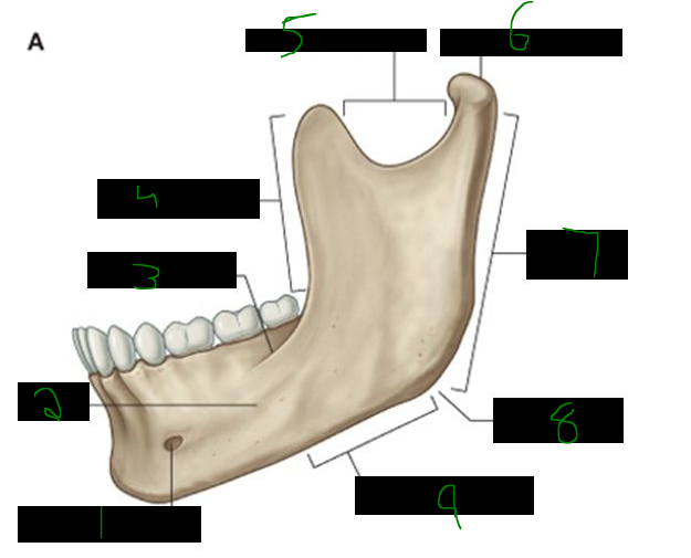
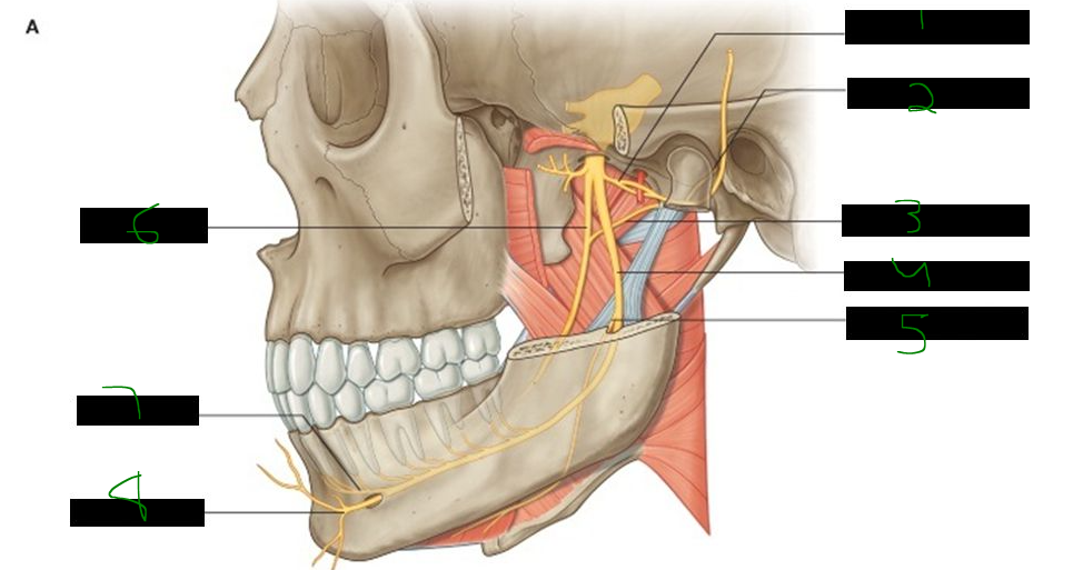
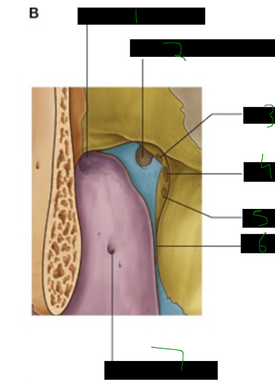
What is 1?
mental foramen
What is 2?
body
What is 3?
oblique line
What is 4?
anterior border
What is 5?
superior border
What is 6?
condylar process
What is 7?
ramus
What is 8?
angle
What is 9?
inferior border
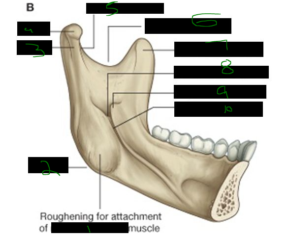
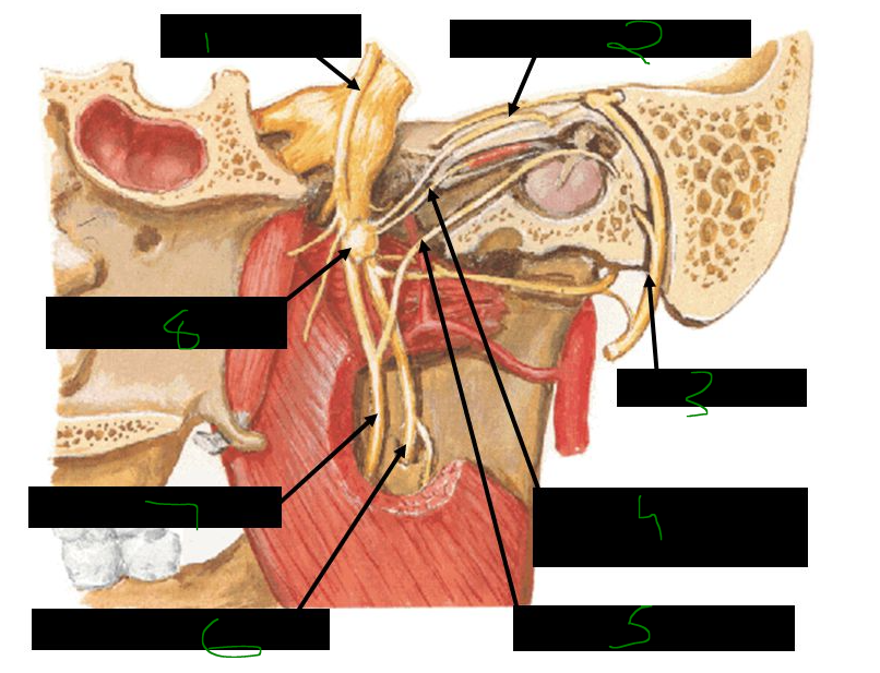
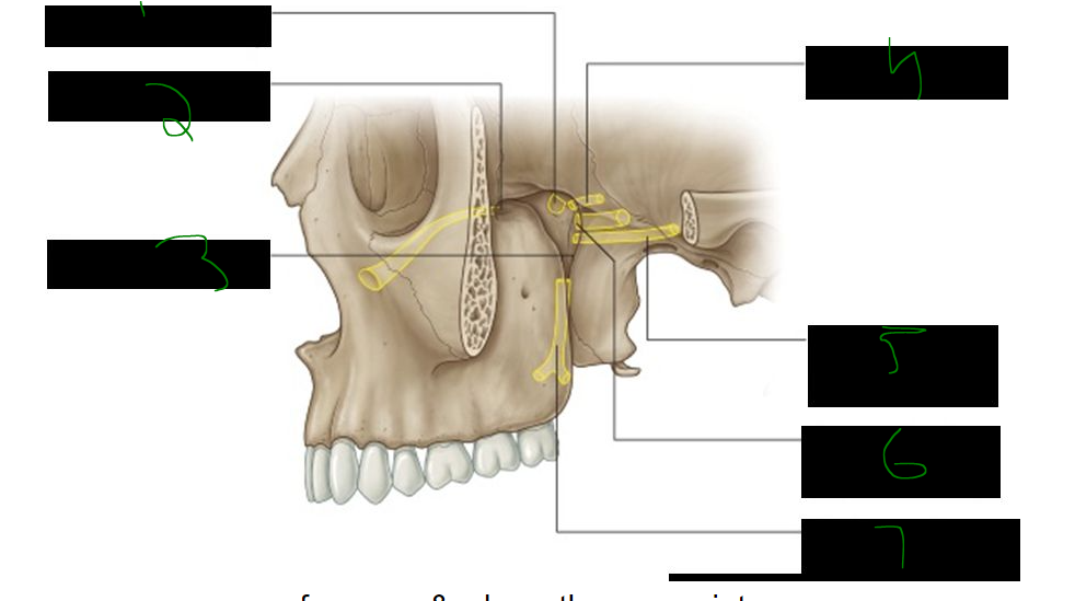
What is 1?
medial pterygoid muscle
What is 2?
angle
What is 3?
neck
What is 4?
head
What is 5?
pterygoid fovea
What is 6?
mandibular notch
What is 7?
coronoid process
What is 8?
mandibular foramen
What is 9?
lingula
What is 10?
mylohyoid groove
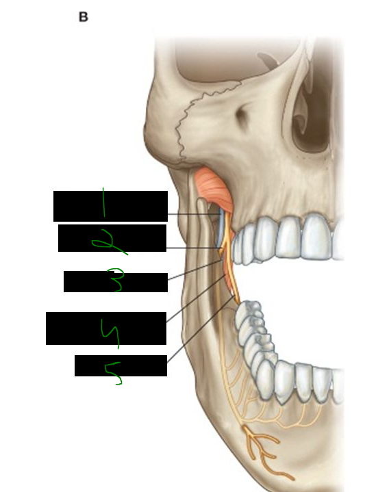
The infratemporal fossa is located deep to the ______ muscle and the ______ of the ______.
masseter, ramus, mandible
The lingula is located on the ______ part of the ______, where the ______ ligament attaches.
medial, mandible, sphenomandibular
where does the sphenomandibular ligament attach?
______ of ______ (______ surface)
lingula of mandible (medial surface)
Protrusion of the mandible at the TMJ is primarily performed by the ______ pterygoid and assisted by the ______ pterygoid.
lateral, medial
Retraction of the mandible involves the posterior fibers of the ______, the deep part of the ______, and the ______ and digastric muscles.
temporalis, masseter, geniohyoid
Elevation of the mandible is performed by the ______, ______, and ______ pterygoid muscles.
temporalis, masseter, medial
Depression of the mandible is assisted by ______, the ______ muscle, the ______ muscle, and the mylohyoid.
gravity, digastric, geniohyoid
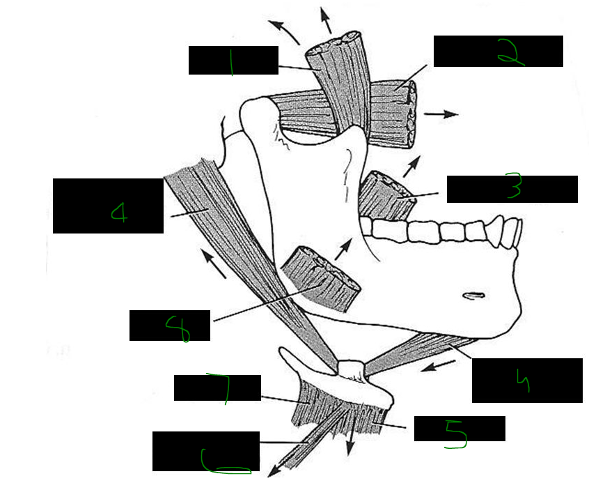
What is 1?
temporalis
What is 2?
lateral pterygoid
What is 3?
medial pterygoid
What is 4?
digastric (anterior belly)
What is 5?
sternohyoid
What is 6?
omohyoid (superior belly)
What is 7?
thyrohyoid
What is 8?
masseter
What is 9?
digastric (posterior belly)
At the TMJ, forward movement of the mandible and articular disc occurs in the ______ portion of the joint, while ______ movement between the condylar process and articular disc occurs in the ______ portion.
upper, hinge, lower
what is the main muscle that allows protrusion of the jaw?
______ ______ (assisted by ______ pterygoid)
lateral pterygoid (assisted by medial pterygoid)
opening of the jaw is a composite of what actions?
mandibular ______ & ______
mandibular protrusion & depression
what muscle inserts into the TMJ joint capsule in the region of the articular disc?
______ ______
lateral pterygoid
Jaw dislocation most often occurs when the mandible is fully ______, making the joint ______ and susceptible to dislocation from minor ______.
depressed, unstable, trauma
To reduce a dislocated jaw, apply downward pressure on the ______ ______ and push the mandible ______.
lower molars, backward
Traumatic dislocation of the jaw can injure the ______ nerve, a branch of the mandibular division of cranial nerve ______.
auriculotemporal, V3
during surgical procedures involving the TMJ, what nerve(s) are susceptible to damage?
______ & ______
facial & auriculotemporal
Jaw clicking and popping is caused by delayed movement of the ______ ______ followed by its sudden movement during ______ and ______ of the mandible.
articular disc, opening, closing
The sphenomandibular ligament runs from the ______ of the sphenoid to the ______ of the ______ of the mandible.
spine, lingula, ramus
The stylomandibular ligament extends from the ______ process of the ______ bone to the ______ of the mandible.
styloid, temporal, angle
The lateral ligament connects the margin of the ______ ______ to the ______ of the mandible.
articular tubercle, neck
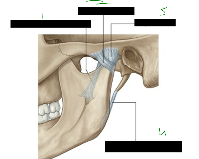
What is 1?
sphenomandibular ligament
What is 2?
lateral ligament
What is 3?
capsule
What is 4?
stylomandibular ligament
The masseter muscle originates from the ______ ______ and the maxillary process of the ______ bone.
zygomatic arch, zygomatic
The insertion of the masseter is at the ______ and ______ surface of the ______.
angle, lateral, mandible
The masseter is innervated by the ______ nerve, a branch of the mandibular division of cranial nerve ______.
masseteric, V3
The masseter functions in powerful ______ of the mandible and contributes slightly to ______.
elevation, protrusion
The blood supply to the masseter muscle comes from the ______ branch of the ______ artery.
masseteric, maxillary
What is 1?
zygomatic process of frontal bone
What is 2?
frontal process of zygomatic bone
What is 3?
zygomatic arch
What is 4?
infratemporal crest of sphenoid
What is 5?
infratemporal fossa
What is 6?
temporal fossa
What is 7?
temporal fascia
What is 8?
inferior temporal line
What is 9?
superior temporal line
The temporalis muscle originates from the ______ ______, with anterior fibers oriented ______ and posterior fibers more ______.
temporal fossa, vertically, horizontal
The temporalis inserts on the anterior surface of the ______ process and the ______ of the mandible.
coronoid, ramus
The temporalis is a powerful ______ of the mandible, assists in ______, and also produces ______ movements.
elevator, retraction, side to side
Innervation to the temporalis muscle comes from the ______ ______ nerves, which are branches of cranial nerve ______.
deep temporal, V3
Blood supply to the temporalis comes from the ______ ______ arteries (from the maxillary artery) and the ______ ______ artery (from the superficial temporal artery).
deep temporal, middle temporal
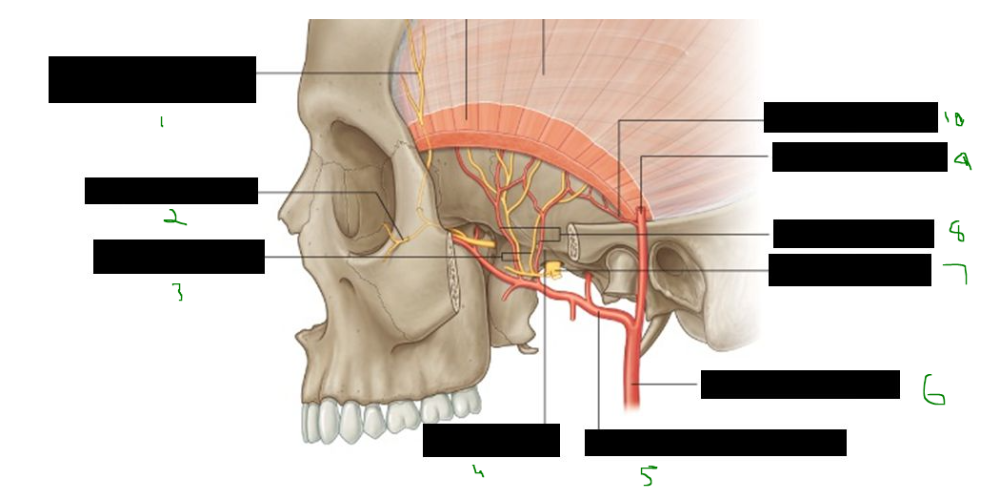
What is 1?
zygomaticotemporal nerve (branch of maxillary nerve v2)
What is 2?
zygomaticofacial nerve
What is 3?
deep temporal nerves
What is 4?
infratemporal crest
What is 5?
maxillary artery in infratemporal fossa
What is 6?
external carotid artery
What is 7?
mandibular nerve (v3)
What is 8?
deep temporal arteries
What is 9?
superficial temporal artery
What is 10?
middle temporal artery
Contents of the temporal fossa include the ______ muscle, deep ______ nerves and vessels, and the ______ branches of the maxillary nerve (V2).
temporalis, temporal, zygomaticotemporal
Openings into or out of the roof of the infratemporal fossa include the ______ fossa, foramen ______, foramen ______, and the ______ fissure.
temporal, ovale, spinosum, petrotympanic
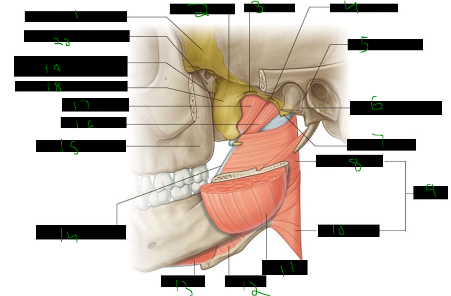
What is 1?
greater wing of sphenoid bone
What is 2?
infratemporal crest
What is 3?
foramen ovale
What is 4?
foramen spinosum
What is 5?
petrotympanic fissure
What is 6?
head and neck of mandible
What is 7?
spine of sphenoid
What is 8?
superior constrictor
What is 9?
pharynx
What is 10?
middle constrictor
What is 11?
masseter
What is 12?
hyoglossus
What is 13?
mylohyoid
What is 14?
pterygomandibular raphe
What is 15?
posterior surface of maxilla
What is 16?
levator veli palatini
What is 17?
tensor veli palatini
What is 18?
lateral plate of pterygoid process
What is 19?
pterygomaxillary fissure
What is 20?
pterygopalatine fossa
Openings in the anterior part of the infratemporal fossa include small foramina in the ______ for the ______ ______ alveolar blood vessels and nerves supplying the ______.
maxilla, posterior superior, teeth
The medial part of the infratemporal fossa has the ______ ______, which leads into the ______ ______.
pterygomaxillary fissure, pterygopalatine fossa
The medial pterygoid muscle has two heads: the deep head originates from the ______ plate of the ______ process, and the superficial head originates from the ______ of the maxilla.
lateral, pterygoid, tuberosity
Both heads of the medial pterygoid muscle insert on the ______ surface of the mandible near the ______, passing deep to the ______ ligament.
medial, angle, sphenomandibular
The medial pterygoid muscle functions to ______ the mandible and assist with ______.
elevate, protrusion
Innervation to the medial pterygoid muscle is via the ______ to medial pterygoid nerve, a branch of cranial nerve ______.
nerve, V3
The lateral pterygoid muscle has two heads: the upper head originates from the ______ bone, and the lower head originates from the ______ plate of the ______ process.
sphenoid, lateral, pterygoid
The lateral pterygoid inserts on the neck of the mandible at the ______ ______ and the capsule of the TMJ at the ______ ______.
pterygoid fovea, articular disc
The lateral pterygoid functions to ______ the mandible and pull the ______ ______ anteriorly; most fibers are ______ oriented.
protrude, articular disc, horizontally
Innervation of the lateral pterygoid comes from the ______ to lateral pterygoid nerve, a branch of cranial nerve ______.
nerve, V3
When the lateral and medial pterygoid muscles contract on one side, the chin moves to the ______ side, helping to ______ food by the ______.
opposite, grind, molars
The four muscles of mastication are ______, lateral ______, medial ______, and ______, all innervated by branches of the mandibular nerve (CN ______).
temporalis, pterygoid, pterygoid, masseter, V3
The buccinator assists in mastication by holding food between the ______ and ______ teeth, and is innervated by the ______ nerve (buccal branch).
upper, lower, facial
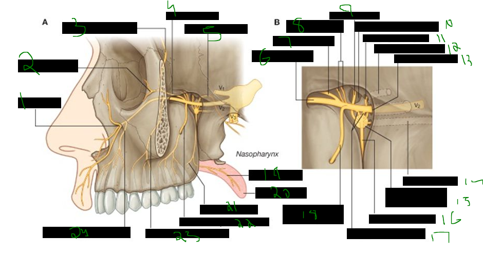
The mandibular nerve (V3) provides sensation from the teeth and gingivae of the ______, anterior two-thirds of the ______, mucosa on the floor of the ______ cavity, lower lip, skin over the ______, lower ______, and part of the cranial dura mater.
mandible, tongue, oral, temple, face
Motor functions of the mandibular nerve include innervating the muscles of ______, the ______ tympani, and the tensor ______ ______ of the soft palate.
mastication, tensor, veli palatini
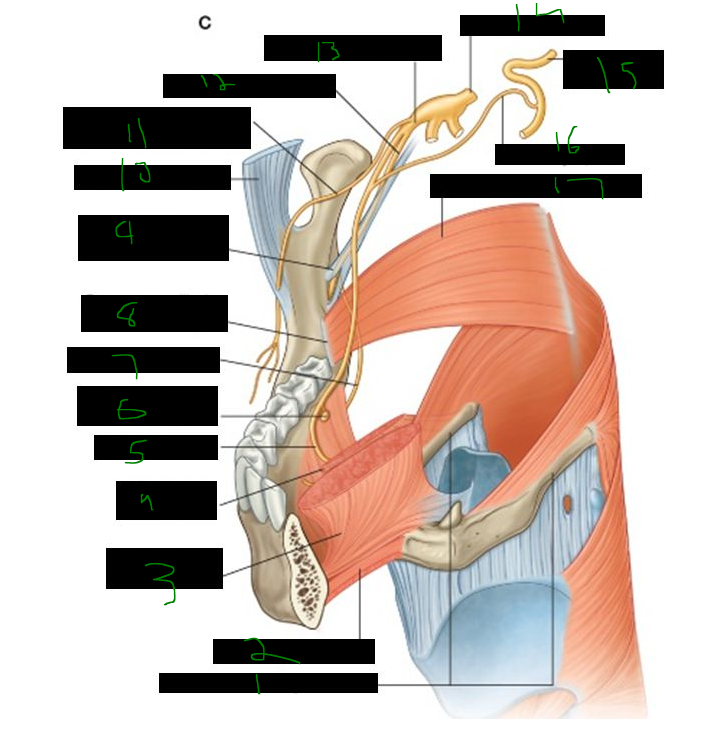
All branches of the mandibular nerve (V3) originate in the ______ ______.
infratemporal fossa
The nine branches of the mandibular nerve (V3) are ______, nerve to ______ pterygoid, nerve to ______ pterygoid (which branches to ______ veli palatini and ______ tympani), ______ temporal, ______, ______, ______ temporal, ______ alveolar (which branches to ______ and anterior belly of ______), and ______.
masseteric, lateral, medial, tensor, tensor, deep, meningeal, buccal, auriculotemporal, inferior, mylohyoid, digastric, lingual
Just distal to the foramen ovale, the mandibular nerve gives off the ______ branch, which enters the foramen ______, and the nerve to ______ ______.
meningeal, spinosum, medial, pterygoid
What is 1?
deep temporal nerves
What is 2?
upper head lateral pterygoid (cut)
What is 3?
nerve to lateral pterygoid
What is 4?
buccal nerve
What is 5?
trigeminal nerve
What is 6?
anterior trunk
What is 7?
meningeal nerve
What is 8?
branch to tensor tympani
What is 9?
posterior trunk
What is 10?
branch to tensor veli palatini
What is 11?
nerve to medial pterygoid
What is 12?
deep head medial pterygoid
What is 13?
nerve to masseter
What is 14?
lower head lateral pterygoid (cut)
The four branches of the anterior trunk of V3 are ______ (sensory), ______, ______, and nerve to ______ pterygoid (all motor).
buccal, masseteric, deep temporal, lateral
The three nerves of the posterior trunk of V3 are ______, ______, and ______ alveolar, which gives motor branches to ______ and anterior belly of ______ muscles before entering the mandibular foramen.
auriculotemporal, lingual, inferior, mylohyoid, digastric
What is 1?
auriculotemporal nerve
What is 2?
petrotympanic fissure
What is 3?
chorda tympani nerve
What is 4?
inferior alveolar nerve
What is 5?
nerve to mylohyoid
What is 6?
lingual nerve
What is 7?
incisive nerve
What is 8?
mental nerve
Motor branches from the inferior alveolar nerve to ______ and the anterior belly of ______ branch off before the nerve enters the ______ foramen.
mylohyoid, digastric, mandibular
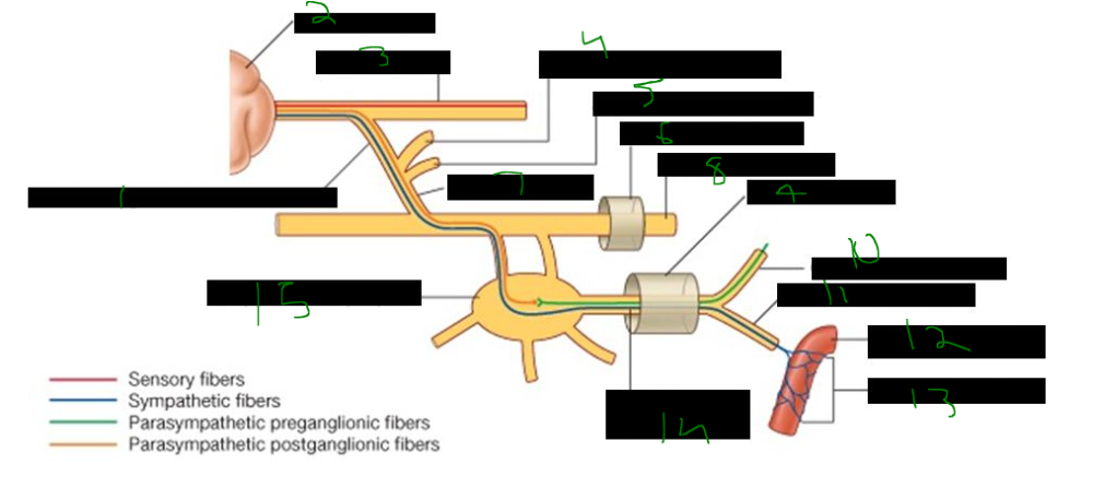
The ______ nerve carries parasympathetic axons from the otic ganglion to the ______ gland and is a branch of the mandibular nerve (CN ______).
auriculotemporal, parotid, V3
The ______ nerve supplies general sensation to the anterior two-thirds of the tongue, while ______ ______ (from CN ______) provides taste to the same region and carries parasympathetics to the sublingual and submandibular glands.
lingual, chorda tympani, VII
The ______ alveolar nerve (branch of ______) passes through the mandibular foramen, gives sensation to the lower ______, and exits the mental foramen as the ______ nerve to supply the ______.
inferior, V3, teeth, mental, chin
What is 1?
sphenomandibular ligament
What is 2?
inferior alveolar nerve
What is 3?
lingula
What is 4?
medial pterygoid muscle
What is 5?
lingual nerve
What is 1?
greater horns of hyoid bone
What is 2?
geniohyoid muscle
What is 3?
genioglossus muscle
What is 4?
hyoglossus muscle
What is 5?
lingual nerve
What is 6?
submandibular ganglion
What is 7?
nerve to mylohyoid
What is 8?
pterygomandibular raphe
What is 9?
sphenomandibular ligament
What is 10?
temporalis tendon
What is 11?
buccal nerve (branch of anterior trunk)
What is 12?
inferior alveolar nerve
What is 13?
mandibular nerve (v2)
What is 14?
trigeminal nerve (V)
What is 15?
facial nerve (7)
What is 16?
chorda tympani
What is 17?
superior constrictor muscle
What is 1?
otic ganglion (medial to V3)
What is 2?
lesser petrosal nerve (IX)
What is 3?
auriculotemporal nerve
What is 4?
top of parotid gland
What is 5?
petrotympanic fissure
What is 6?
auriculotemporal nerve
What is 7?
chroda tympani nerve of VII
What is 8?
lingual nerve
What is 9?
submandibular ganglion
What is 10?
submandibular gland
What is 11?
mylohyoid
What is 12?
sublingual gland
What is 13?
tongue
What is 14?
lingual nerve
What is 1?
motor root
What is 2?
lesser petrosal nerve
What is 3?
VII
What is 4?
nerve to tensor tympani
What is 5?
chorda tympani
What is 6?
inferior alveolar
What is 7?
lingual nerve
What is 8?
otic ganglion
The maxillary artery has three parts:
- First part gives off ______ meningeal and ______ alveolar arteries.
- Second part lies near the ______ pterygoid muscle and gives ______ branches (deep temporal, masseteric, buccal, pterygoid).
- Third part enters the ______ fossa and gives off terminal branches.
middle, inferior, lateral, muscular, pterygopalatine
What is 1?
branches of middle meningeal in cranail artery
What is 2?
maxillary artery
What is 3?
superficial temporal artery
What is 4?
middle meningeal artery
What is 5?
pterygoid artery
What is 6?
artery to masseter
What is 7?
inferior alveolar artery
What is 8?
external carotid
What is 9?
mental artery
What is 10?
buccal artery
What is 11?
lower head of pterygoid (cut)
What is 12?
pterygopalatine fossa
What is 13?
upper head of lateral pterygoid (cut)
What is 14?
deep temporal arteries
The maxillary artery is a branch of the ______ carotid artery, arises within the ______ gland, and passes between the ______ and the sphenomandibular ligament.
external, parotid, mandible
what artery passes between the mandible & sphenomandibular ligament?
______ ______
maxillary a.
what veins connect the pterygoid plexus to the cavernous sinus?
______ ______
emissary veins
What is 1?
facial vein
What is 2?
deep facial vein
What is 3?
inferior ophthalmic vein
What is 4?
emissary veins
What is 5?
superficial temporal vein
What is 6?
maxillary vein
What is 7?
inferior alveolar vein
What is 8?
retromandibular vein
What is 9?
external jugular vein
What is 10?
internal jugular vein
The contents of the infratemporal fossa include the ______ nerve (V3), ______ petrosal nerve and ______ ganglion, ______ tympani nerve, ______ plexus of veins, medial and lateral ______ muscles, the ______ artery, and the sphenomandibular ligament.
mandibular, lesser, otic, chorda, pterygoid, pterygoid, maxillary
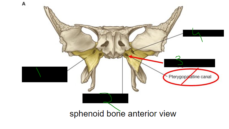
borders of the pterygopalatine fossa?
anterior wall - ______ surface of ______
medial wall - ______ surface of ______ bone
posterior wall & roof - ______ of ______ bone
anterior wall - posterior surface of maxilla
medial wall - lateral surface of palatine bone
posterior wall & roof - parts of sphenoid bone
What is 1?
zygomatic bone
What is 2?
pterygopalatine fossa
What is 3?
sphenoid bone
What is 4?
palatine bone
What is 5?
maxilla
The openings into the pterygopalatine fossa include the ______ orbital fissure, ______ foramen, ______ canal (which opens to the pharynx), ______ rotundum, ______ canal, and ______ canal.
inferior, sphenopalatine, palatovaginal, foramen, pterygoid, palatine
What is 1?
inferior orbital fissure
What is 2?
sphenopalatine foramen
What is 3?
palatovaginal canal
What is 4?
foramen rotundum
What is 5?
pterygoid canal
What is 6?
palatine canal
What is 7?
alveolar foramen
What is 1?
sphenopalatine foramen
What is 2?
inferior orbital fissure
What is 3?
pterygomaxillary fissure
What is 4?
palatovaginal canal
What is 5?
pterygoid canal
What is 6?
foramen rotundum
What is 7?
palatine canal

What is 1?
infra-orbital
What is 2?
zygomaticofacial
What is 3?
zygomaticotemporal
What is 4?
zygomatic
What is 5?
pharyngeal nerve
What is 6?
infra-orbital nerve
What is 7?
zygomatic nerve
What is 8?
orbital branches
What is 9?
nasal nerves
What is 10?
sphlenopalatine foramen
What is 11?
pharyngeal nerve
What is 12?
palatovaginal canal
What is 13?
foramen rotundum
What is 14?
pterygoid canal
What is 15?
pterygopalatine ganglion
What is 16?
palatine nerves
What is 17?
ganglionic branches
What is 18?
posterior superior alveolar
What is 19?
lesser palatine
What is 20?
soft palate
What is 21?
greater palatine
What is 22?
posterior superior alveolar
What is 23?
middle superior alveolar
What is 24?
antioer superior alveolar
What is 1?
surface related to pterygopalatine fossa
What is 2?
palatovagianl groove
What is 3?
pterygoid canal
What is 4?
foramen rotundum
What is 1?
cartilage filling foramen lacerum
What is 2?
greater petrosal nerve of VII
What is 3?
maxillary nerve (v2)
What is 4?
internal carotid artery
What is 5?
superior orbital fissure
What is 6?
lesser wing
What is 7?
greater wing
What is 8?
foramen rotundum
What is 9?
pterygoid process
What is 10?
posterior opening of bony part of pterygoid canal
What is 1?
pterygoid canal
What is 2?
greater petrosal nerve
What is 3?
deep petrosal nerve
What is 4?
pterygopalatine ganglion
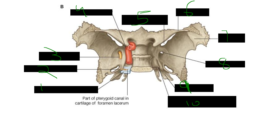
The nerve of the pterygoid canal is formed by the ______ petrosal nerve (from CN ______) and the ______ petrosal nerve (from the internal carotid plexus), and it carries preganglionic parasympathetics and postganglionic sympathetics to the pterygopalatine ganglion.
greater, VII, deep
What is 1?
branch of zygomaticotemporal nerve
What is 2?
lacrimal gland
What is 3?
lacrimal nerve
What is 4?
zygomaticotemporal nerve
What is 5?
zygomaticofacial nerve
What is 6?
foramen rotundum
What is 7?
zygomatic nerve
What is 8?
maxillary nerve (v2)
What is 9?
pterygoid canal
What is 10?
greater petrosal nerve
What is 11?
deep petrosal nerve
What is 12?
internal carotid artery
What is 13?
sympathetic plexus
What is 14?
nerve of pterygoid canal
What is 15?
pterygopalatine ganglion
The six terminal branches of the maxillary artery are the ______ artery (which gives rise to the anterior alveolar artery), ______ superior alveolar artery, ______ palatine artery, ______ artery, ______ artery, and the artery of the ______ canal.
infraorbital, posterior, greater, sphenopalatine, pharyngeal, pterygoid
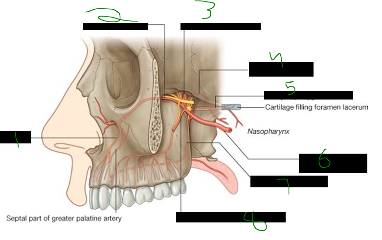
What is 1?
anterior alveolar artery
What is 2?
infraorbital artery
What is 3?
sphenopalatine artery
What is 4?
pharyngeal artery
What is 5?
artery of pterygoid canal
What is 6?
maxillary artery in infratemporal fossa
What is 7?
greater palatine artery
What is 8?
posterior superior alveolar artery
Veins from the pterygopalatine fossa pass out through the ______ fissure into the ______ fossa and drain into the ______ plexus of veins.
pterygomaxillary, infratemporal, pterygoid
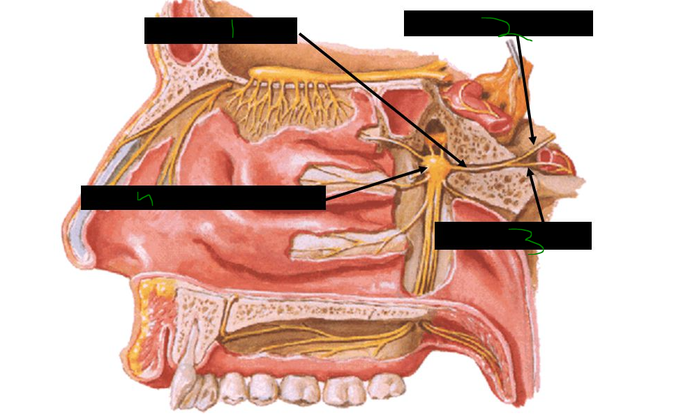
The pterygopalatine fossa contains the ______ ganglion, the ______ nerve (V2), preganglionic parasympathetics in the ______ petrosal nerve (from CN VII), and postganglionic parasympathetics that supply mucous glands of the nasal cavity, oral cavity, nasopharynx, and the ______ gland.
pterygopalatine, maxillary, greater, lacrimal
What is 1?
temporal fossa
What is 2?
pterygomaxillary fissue leading to pterygopalatine fossa
What is 3?
infratemporal fossa
What is 4?
petrotympanic fissure
what fissure leads to the pterygopalatine fossa?
___ ___
pterygomaxillary fissure
The nerve to the lateral pterygoid muscle branches from the ______ trunk of the mandibular nerve (V3), while the nerve to the medial pterygoid branches directly from V3 just distal to the ______ ______, along with the ______ branch.
anterior, foramen ovale, meningeal
In the infratemporal fossa, the chorda tympani from CN ______ joins the ______ nerve (V3), and the lesser petrosal nerve from CN ______ carries preganglionic parasympathetics to the otic ganglion.
VII, lingual, IX
Otic ganglion is small or large?
small
otic ganglion is located _______ to mandibular n. V3
medial
where do all branches of the mandibular nerve (V3) originate?
_______ _______
infratemporal fossa