Paget's disease (osteodystrophia deformans)
chronic progressive disorder of excessive breakdown and formation of bone typically seen in pelvis, femur, and lumbar vertebrae.
Age > 50
insidious onset with slow progession
lab tests include hypercalcimia (active state) and elevated phosphate levels increase risk of Paget's
appears "fluffy" on imaging
presentation of Paget's disease (osteodystrophia deformans)
bone pain, headache, diziness, lightheadedness, impingement on spine, vertigo, INCREASED CARDIAC OUTPUT (cardiopulmonary)
Paget's disease (osteodystrophia deformans) treatment
promote bone health, pharmacologic management, NSAID for pain, surgical intervention
Osteogenesis imperfecta (brittle bone disease)
collagen synthesis disorder (congenital), does not present until child ambulates, LE>UE, bruise easily, increased ligamentous laxity, thin skin, hearing impairments, CV issues, motor skill delays
Osteogenesis imperfecta (brittle bone disease) diagnosis
skin biopsy examining collagen, bone scan/radiograph
Osteogenesis imperfecta (brittle bone disease) treatment
orthopedic management, biophosphates to inhibit osteoclast activity, bone marrow transplant
Marfan's syndrome
mutation of FBN1 gene - structural disorder of connective tissue
Marfan's syndrome presentation
tall stature with elongation of arms and legs, severe myopia, aortic imperfections, px in abdomen, weakness in LE
diagnosed by systemic feature scoring table
Marfan's syndrome treatment
aortic repair, beta blockers, ocular care, moderate aerobic exercise
Ethers-Danlos syndrome (stretchy skin)
genetic CT disorder, 13 different types, mutation in COL5A1 or COL5A2 gene
Ethers-Danlos syndrome (stretchy skin) diagnosis
skin biopsy, collagen typing, genetic testing, echocardiogram
Ethers-Danlos syndrome (stretchy skin) treatment
activity modification
signs of Ethers-Danlos syndrome (stretchy skin)
joint hypermobility, thin stretchy velvety skin, abnormal scar formation, system involvement, chronic pain
Osteochondrosis
an interruption of blood supply of a bone, abnormal bone growth at ossification centers of epiphysis, developmental, injury/overuse related
most common site of osteochondrosis in Osgood-Schlatter's disease
knee
most common site of osteochondrosis in Sever's disease
ankle
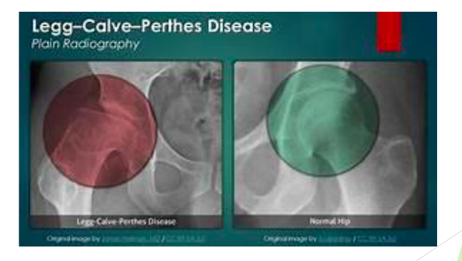
most common site of osteochondrosis in Legg-Calve-Perthes disease
hip
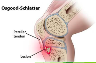
Osgood-Schlatter's disease
mechanical issue with extensor of the knee, force of the quadriceps muscle and repetitive stress on tibial tubercle
Osgood-Schlatter's disease risk factors
genu valgum, excessive pronation, adolescent males (predominant) 10-15 years old
Osgood-Schlatter's disease signs
constant ache, pain at site, tender to palpation, decreased flexiblity, pain with jumping activities
Osgood-Schlatter's disease diagnosis
radiograph
Osgood-Schlatter's disease treatment
rest, activity modification, NSAID, ice, THEREX
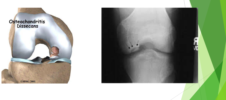
Osteochondritis dessicans
disorder of ossification site with localized subchondral necrosis followed by reossification of bone
Osteochondritis dessicans cause
repetitive trauma resulting in ischemia and disruption of subchondral growth causing articular cartilage softening and fragmentation, cartilage injured and forms a crater
Osteochondritis dessicans presentation
pain with specific activities, swelling, giving way
Osteochondritis dessicans diagnosis
radiograph, MRI
Osteochondritis dessicans treatment
varies on severity of lesion... immobilize, activity modification, possible surgical intervention/debridement-tissue implementation
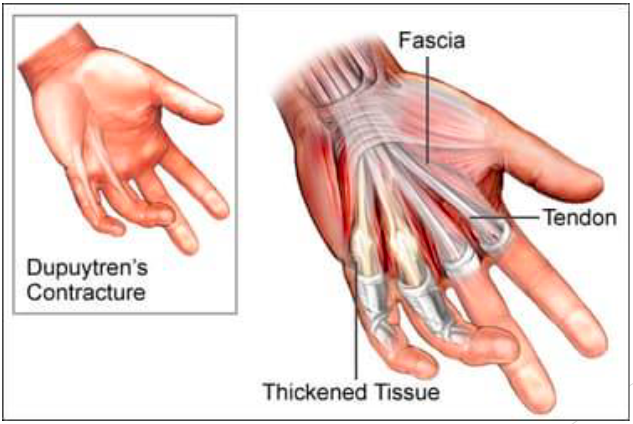
Dyputren's Contracture (soft tissue)
palmer fibromatosis, flexion contracture, palmar nodules, thickened cord, 3rd & 4th digits in patients with DC, 4th & 5th in general population, etiology unknown.. thought to be associated through alcohol or Northern European/Scandinavian descent