how many total vertebrae are there?
33 CTLSC 7,12,5,5,4
distinctive feature of cervical vertebrae
transverse foramina, have bifoid spinous processes, and superior articular facets face up
distinctive feature of thoracic vertebrae
have costal facets, heart shaped bodies, superior articular facets face POSTERIOR, inferior articular facets face ANTERIOR
distinctive feature of lumbar vertebrae
massive bodies, superior articular facets face MEDIAL, inferior articular facets face LATERAL
distinctive feature of sacrum
fused, 4 pairs of foramina, transmits weight from spine to pelvis
distinctive feature of coxxyx
tailbone, no weightbearing function, attachment point for muscles and ligament
what are the 3 main joints of the vertebral column?
anterior intervertebral joints, zygapophyseal joints (facet joints), craniovertebral joints
Anterior intervertebral joints
between adjacent vertebral bodies
zygapophyseal joints (facet joints)
between superior and inferior articular processes
cranioertebral joints
atlanto-occipital and atalanto-axial
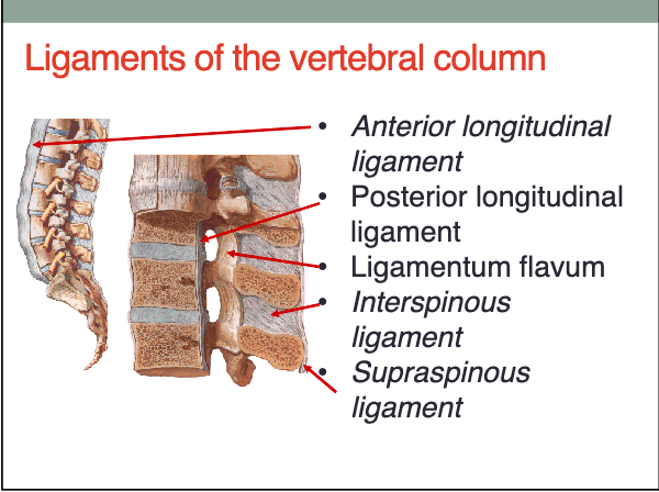
ligaments of the vertebral column
anterior longitudinal ligament, posterior longitudinal ligament, ligamentum flavum, interspinous ligament, supraspinous ligament... ligamentum nuchae as well
what are the motions of intervertebral joints?
rocking, gliding, rotation
what are the motions of zygapophyseal joints?
gliding
where is flexion motion greatest?
cervical spine
where is extension greatest?
lumbar spine
where is lateral flexion greatest?
cervical and lumbar spine
where is rotation greatest?
thoracic spine
Concave forward
kyphosis
Where is kyphosis typically seen?
thoracic and sacral spine
Concave rearward
lordosis
Where is lordosis typically seen?
cervical and lumbar regions of the spine
Superior boundary of the abdomen
xiphoid process, costal rib margin, costal cartilage ribs 7-10
Inferior boundary of the abdomen
line between ASIS and pubic syphysis (inguinal ligament)
Lateral boundary of the abdomen
vertical line from ASIS to costal margin
Anterior boundary of the abdomen
umbillicus
linea semilunaris
half moon shaped, lateral aspect of rectus abdominis
camper fascia
superficial fatty layer
scarpa fascia
deep membraneous layer
layers of fat in order
skin, camper fascia, scarpa fascia, investing fascia, external oblique, internal oblique, transverse abdominal muscle, endoabdominal muscle, parietal peritoneum
rectus sheath above acruate line.. ANTERIOR
aponerousis of external oblique and half of aponeurosis of internal oblique
rectus sheath above acruate line... POSTERIOR
half of aponeurosis of internal oblique and aponeurosis of transverse abdominal
rectus sheath below acruate line... ANTERIOR
aponeuroses of external and internal obliques and transverse abdominal all pass in front of the rectus abdominis
rectus sheath below acruate line.. POSTERIOR
no aponeuroses behind rectus abdominis, thin transversalis fascia only
function of inguinal region
structures enter and exit abdominal cavity, 75% of all abdominal hernias
where is the inguinal canal located?
lies parallel and superior to the medial half of the inguinal ligament
inguinal canal anterior wall
aponeurosis of external oblique
inguinal canal posterior wall
transversalis fascia & conjoint tendon
inguinal canal roof
fibers of internal oblique and transverse abdominis
inguinal canal floor
fold under of external oblique (inguinal ligament)
inguinal rings (superficial)
inferomedial (location of inguinal hernias)
inguinal rings (deep)
superolateral
contents of inguinal canal in men
spermatic cord
contents of inguinal canal in women
round ligament of uterus
inguinal hernia (direct)
occurs due to weakness of anterior abdominal wall; accounts for 1/3 - 1/4 of hernias, rare in women
inguinal hernia (indirect)
congenital, occurs due to processus vaginalis remaining in part prior to birth, most common in men
diaphragm attachment (sternal part)
2 slips that attach to posterior xiphoid
diaphragm attachment (costal part)
slips attach to internal aspect of inferior 6 costal cartilages and adjoining ribs, forming right and left domes
diaphragm attachment (lumbar part)
musculotendinous bundles attaching to anterior aspect of bodies L1-L3 ascent to central tendon
diaphragm attachment (central tendon)
top and center of the hood, where all parts attach