Types of prokaryotic cell division
binary fission
conjugation
Binary fission
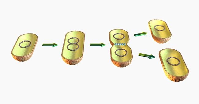
when prokaryotes, replicate the DNA and then split in half.
Forms two identical cells
Asexual reproduction
Conjugation
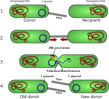
process that occurs in bacteria
one cell transfers a copy of extra chromosomal DNA (plasmid)
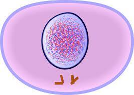
chromatin
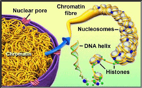
DNA wrapped around histone proteins
in this stage during interphase and cytokinesis
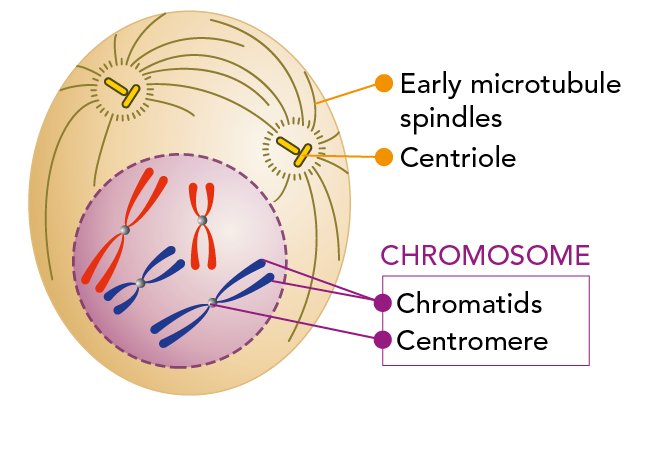
chromosome
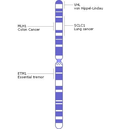
Chromatin that condenses and supercoils before cell division
sister chromatids
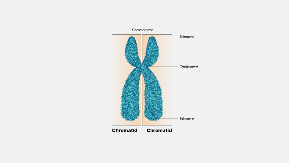
a chromosome paired with its identical copy
held together by centromere
Cell Cycle
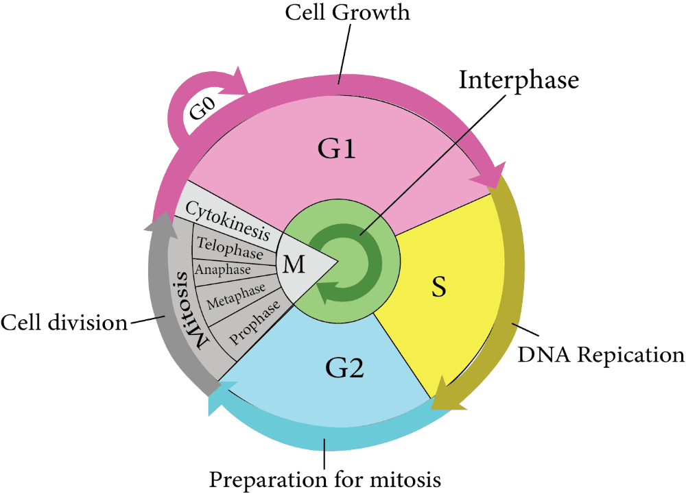
3 stages in the life of a cell
Interphase
Mitosis
Cytokinesis
Interphase
the time in between cell divisions
where a cell spends most of it life
3 steps: G1, S, G2
G1
First growth in interphase
Gap 1 phase
time where cell does its job
eats, gets rid of waste, makes proteins
S phase
synthesis stage
Time when DNA replication occurs during interphase
G2
2nd Growth phase
Gap 2 phase
Cell produces any extra materials or organelles needed for cell division
centromere
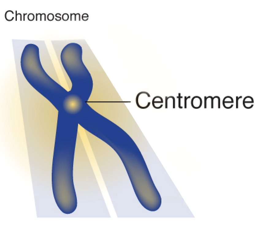
structure that holds sister chromatids together
centrioles
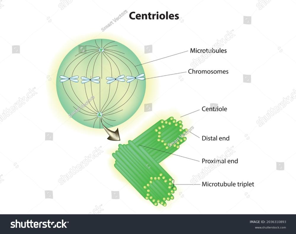
organelles in animal cells that release the spindle fibers
spindle fibers
rope like structures that attach to the centromeres and help separate the sister chromatids
Mitosis
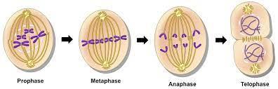
division of the nucleus
prophase
first stage of mitosis
sister chromatids appear held by a centromere
nuclear envelope and nucleolus start to break down
centrioles move to opposite poles
metaphase
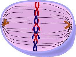
sister chromatids line up along the midline or equator of the cell
spindle fibers attach to the centromeres
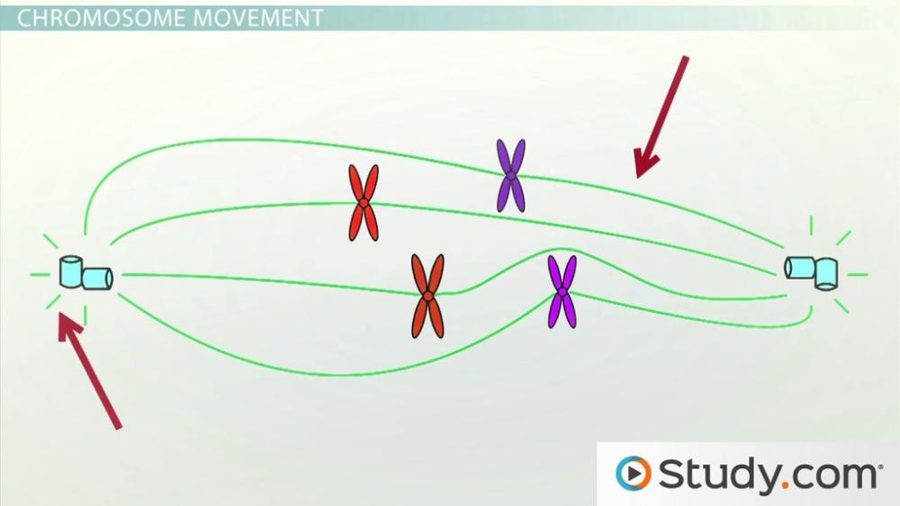
anaphase
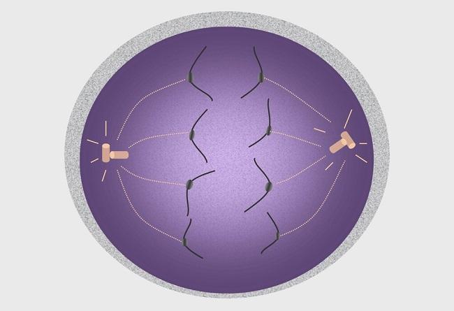
third stage of mitosis
spindle fibers retract and pull sister chromatids apart
individual chromosomes move towards opposite poles
telophase
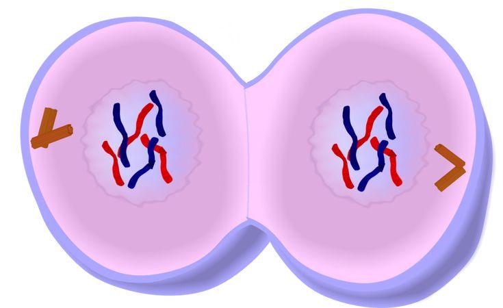
end of mitosis
chromosomes reach opposite ends of cells
cell is elongated
new nuclei and nucleoli begin to form
cytokinesis
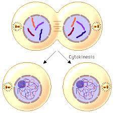
cell splits into two identical daughter cells
animal cells have a cleavage furrow (pinching of cell membrane )
plant cells have a cell plate that forms between the 2 cells and separates them
cancer cells
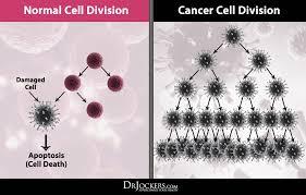
cells that divide more often
do not spend time in interphase