The major opposing forces that influence tooth position originate from the (...).
surrounding musculature
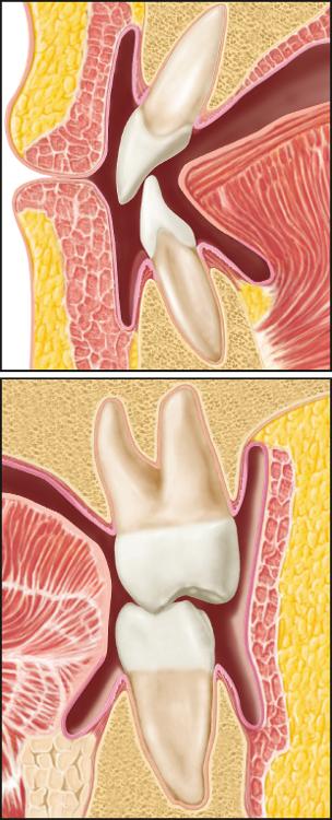
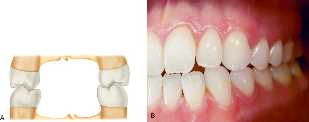
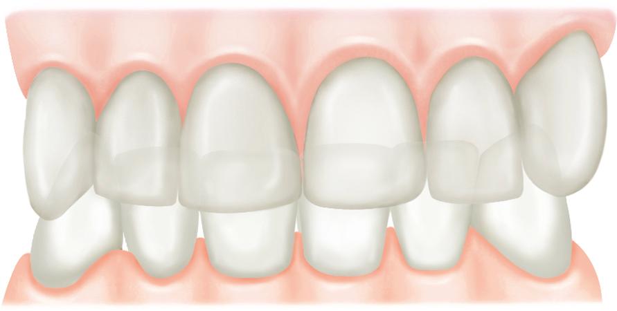
If the tongue is either unusually active or large, it can displace the neutral space (...), resulting in (...) of the anterior teeth which presents clinically as (...).
labially; labial flaring; anterior openbite
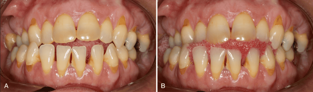
This patient presents with labial flaring of the anterior teeth due to excessive labially directed forces produced by the tongue when swallowing.
- Both the statement and reason are true.
- Both the statement and reason are false.
- The statement is true, but the reason is false.
- The statement is false, but the reason is true.
- The statement and the reason are both true, but unrelated.
(c) The statement is true, but the reason is false.
The anterior teeth are displaced labially by the constant resting or posturing position of the tongue, not the actual activity of swallowing; the tongue thrusting forward during a swallow is more likely associated with the patient’s attempt to seal the mouth, which is necessary for efficient swallowing.
The tooth position in the oral cavity where the labiolingual and buccolingual forces are equal.
neutral space
(...) between adjacent teeth helps maintain the teeth in normal alignment.
proximal contact
If proximal contact is lost, the adjacent tooth will (...) to restore it.

drift mesially
Another important factor that helps to stabilize tooth alignment is (...), which prevents the extrusion or supereruption of teeth.
occlusal contact
If occlusal contact is lost, the opposing tooth will (...) to restore it.
super erupt
Refers to the relationship of the teeth to each other within the dental arch.
intraarch tooth alignment
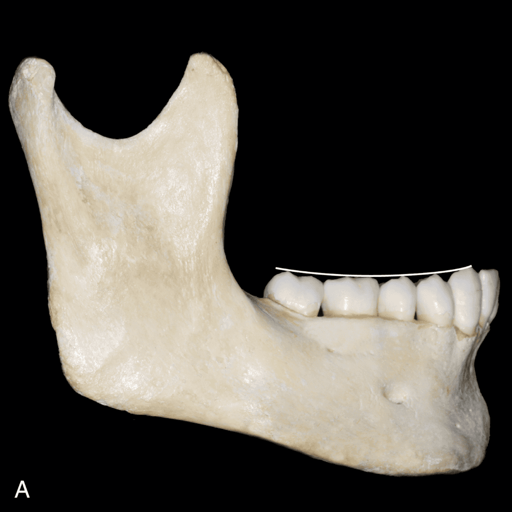
The plane that would be established if a line were drawn through all the buccal cusp tips, lingual cusp tips, and incisal edges of the mandibular teeth.
plane of occlusion
The occlusal planes of the dental arches are curved in a manner that permits (...) during function.
maximum utilization of tooth contacts
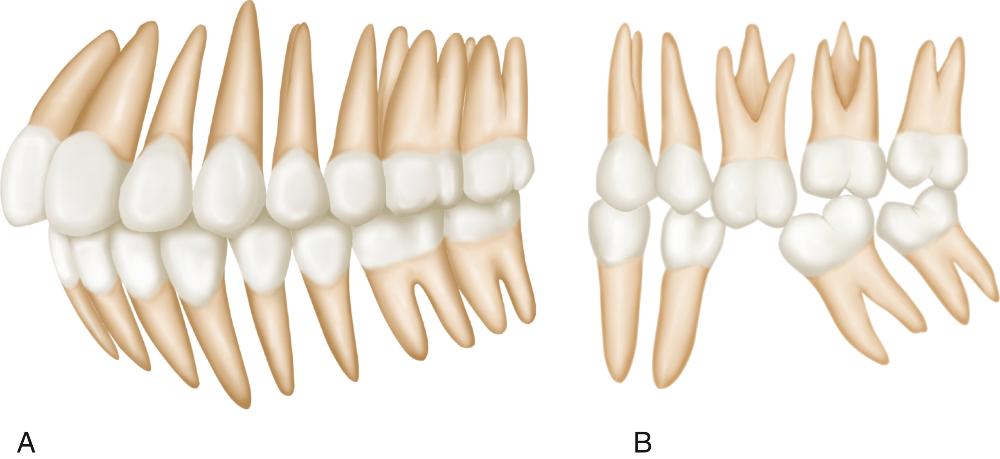
When examining the arches from the lateral view, the mesiodistal axial relationship can be seen. In the mandibular arch both the anterior and the posterior teeth are inclined (...).
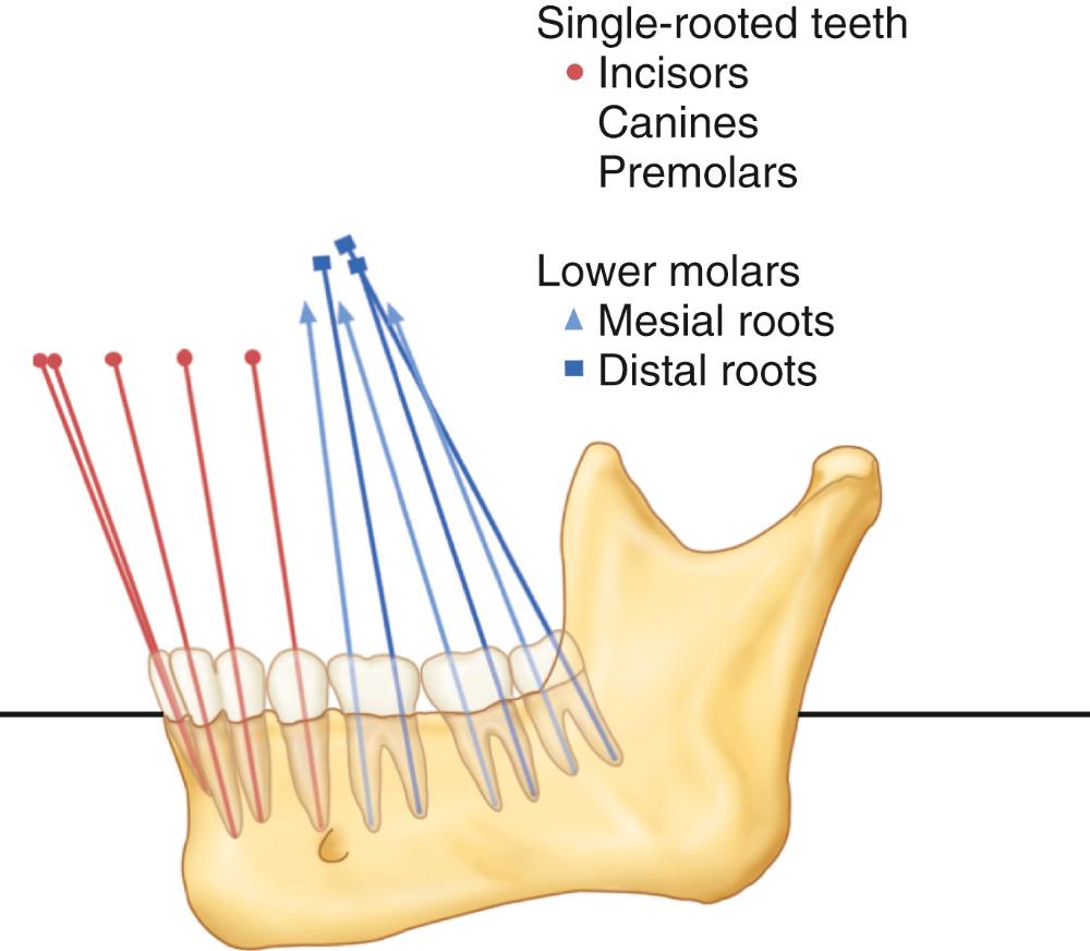
mesially
When examining the arches from the lateral view, the mesiodistal axial relationship can be seen. In the maxillary arch, the anterior teeth are inclined (...), while the most posterior molars are inclined (...).
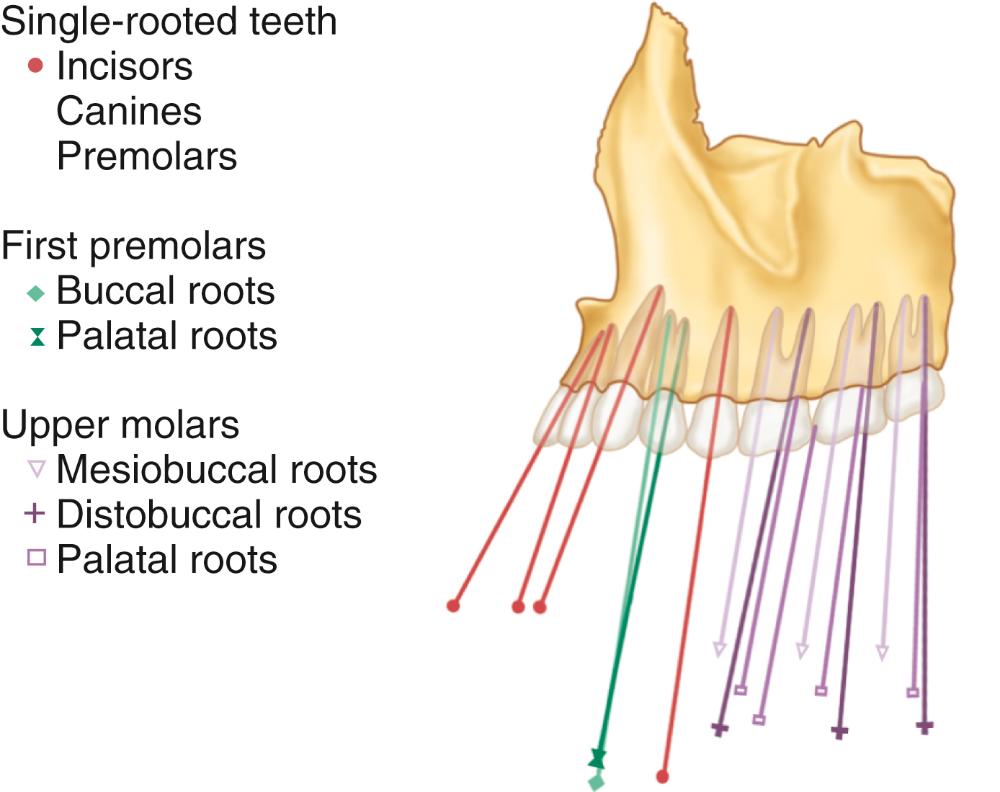
mesially; distally
The curved line following the plane of occlusion from the lateral view, which is convex for the maxillary arch and concave for the mandibular arch.
curve of Spee
When observing the dental arches from the frontal view, the buccolingual axial relationship can be seen. In the maxillary arch, the posterior teeth have a slight (...) inclination.
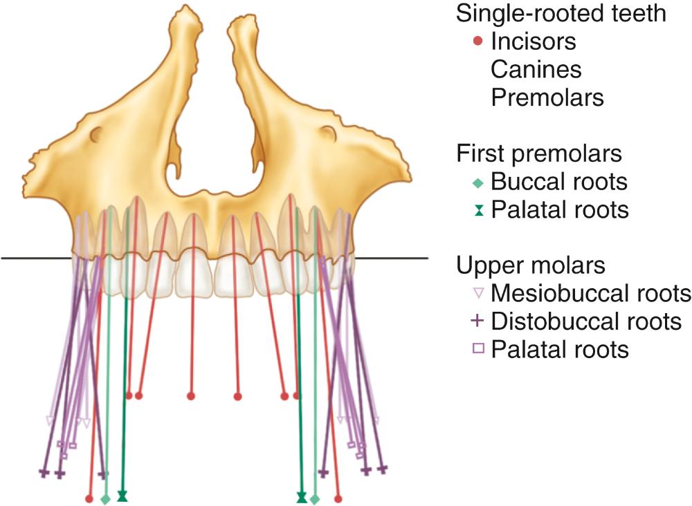
buccal
When observing the dental arches from the frontal view, the buccolingual axial relationship can be seen. In the mandibular arch, the posterior teeth have a slight (...) inclination.
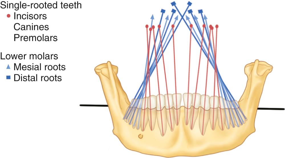
lingual
The curved line following the plane of occlusion from the frontal view, which is convex for the maxillary arch and concave for the mandibular arch.
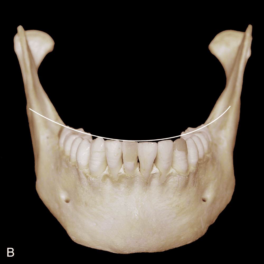
curve of Wilson
Bonwill, one of the first to describe the dental arches, noted that an equilateral triangle with 4-inch sides existed between the (...) and the (...).
centers of the condyles; mesial contact areas of the mandibular central incisors
In 1932, Monson utilized Bonwill’s triangle and proposed a theory that a sphere existed with a radius of 4 inches whose center was an equal distance from the (...) and from the (...).
occlusal surfaces of the posterior teeth; centers of the condyles
The area of the tooth between the buccal and lingual cusp tips of the posterior teeth, where the major forces of mastication are applied.
occlusal table
The occlusal table represents approximately (...)% of the total buccolingual dimension of the posterior tooth and is positioned over the (...).
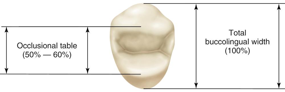
50% to 60%; long axis of the root structure
The occlusal table is considered the (...) of the tooth, since it falls between the cusp tips. Likewise, the occlusal area outside the cusp tips is called the (...).
inner aspect; outer aspects
The inner aspects of the tooth that extend from the cusp tips to the central fossa areas are called the (...), while the outer aspects of the tooth that extend from the cusp tips to height of the contour are called the (...).
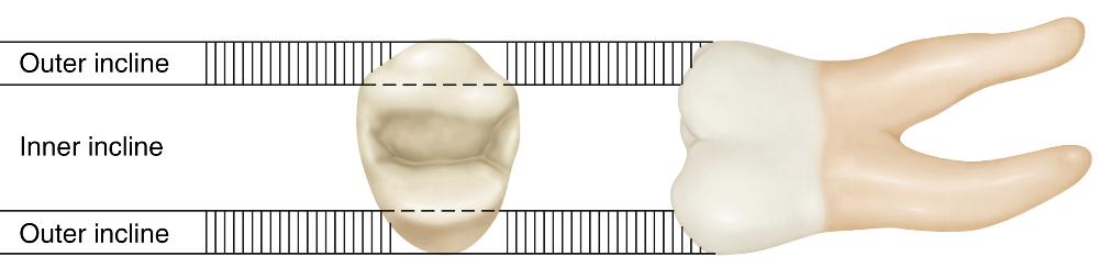
inner inclines; outer inclines
Tooth inclines are also identified with respect to the surface toward which they are directed. (...) inclines are those that face the mesial portion of the tooth, while (...) inclines are those that face the distal portion.
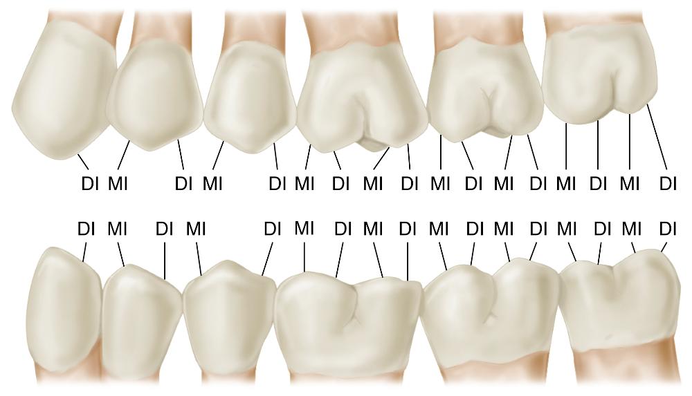
mesial; distal
Refers to the relationship of the teeth in one arch to those in the other.
interarch tooth alignment
The distance of a line that begins at the distal surface of the third molar, extends mesially through all of the proximal contact areas around the entire arch, and ends at the distal surface of the opposite.
arch length
Both arches have approximately the same length, with the (...) arch being only slightly smaller, as a result of the narrower mesiodistal distance of the incisors.
mandibular
The distance across the arch.
arch width
The width of the (...) arch is slightly less than that of the (...) arch; thus when the arches occlude, each maxillary tooth is positioned more (...) than the occluding mandibular tooth.
mandibular; maxillary; facially
In the normal occlusal relationship of the posterior teeth, the mandibular (...) cusps to occlude along the central fossa areas of the maxillary teeth. Likewise, the maxillary (...) cusps occlude along the central fossa areas of the mandibular teeth
buccal; lingual
Occasionally, because of discrepancies in skeletal arch size or eruption patterns, the teeth occlude in such a manner that the maxillary buccal cusps contact in the central fossa area of the mandibular teeth. This relationship is referred to as (...).
cross-bite
The buccal cusps of the mandibular posterior teeth and the lingual cusps of the maxillary posterior teeth are called the (...). cusps.
centric (or supporting)
The centric cusps are responsible for maintaining the (...). These cusps also play a major role in (...) since contact occurs on both the inner and the outer aspects.
vertical dimension of occlusion (VDO); mastication
The centric cusps are broad and rounded. When viewed from the occlusal, their tips are located approximately (...) the distance into the total buccolingual width of the tooth.
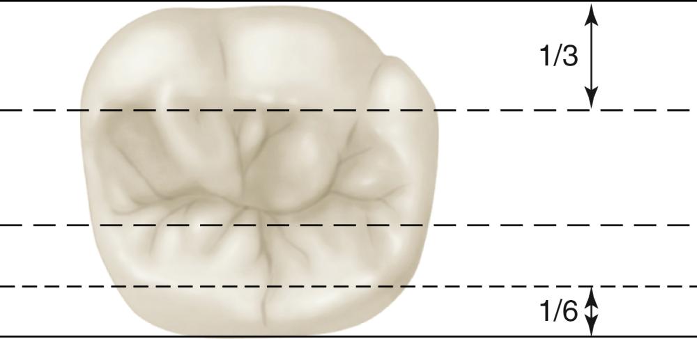
one-third
The buccal cusps of the maxillary posterior teeth and the lingual cusps of the mandibular posterior teeth are called the (...) cusps.
noncentric (or guiding)
The noncentric cusps are relatively sharp, with definite tips located approximately (...) the distance into the total buccolingual width of the tooth.
one-sixth
The small area of the centric cusp (about 1 mm) that functions against the inner incline of the noncentric cusp is called the (...).
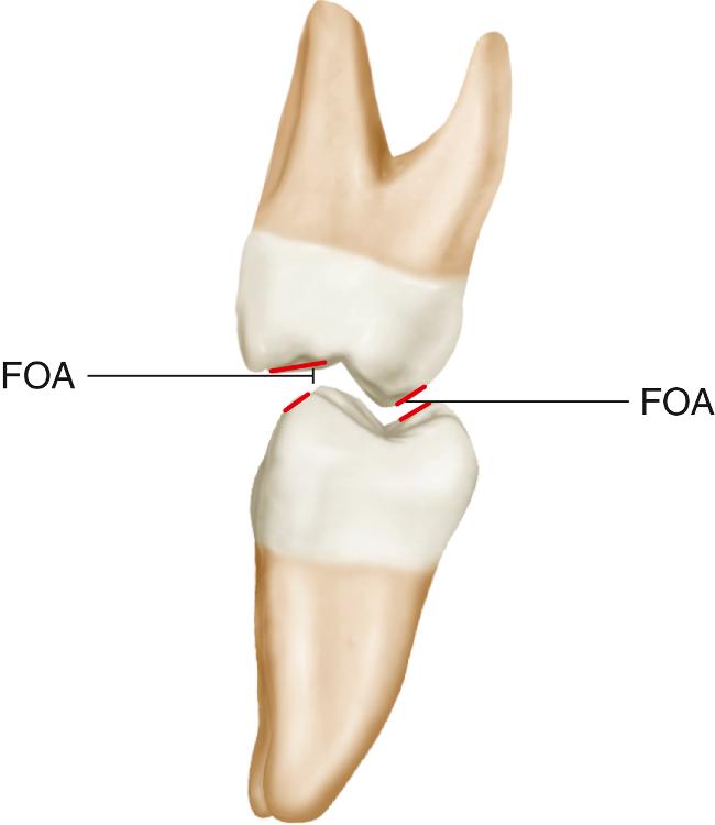
functional outer aspect
Since the contact between the noncentric cusps and the functional outer aspect assists in cutting food during mastication, the noncentric cusps have also been called the (...) cusps.
shearing
The major role of the noncentric cusps is to minimize (...), to maintain the bolus of food on the occlusal table for mastication, and to guide the mandible into (...).
tissue impingement; maximum intercuspal position (MICP)
The relationship of the teeth in full occlusion, so that tight definite occlusal relationship results.
maximum intercuspal position (MICP)
An imaginary line that extends through all the buccal cusp tips of the mandibular posterior teeth.
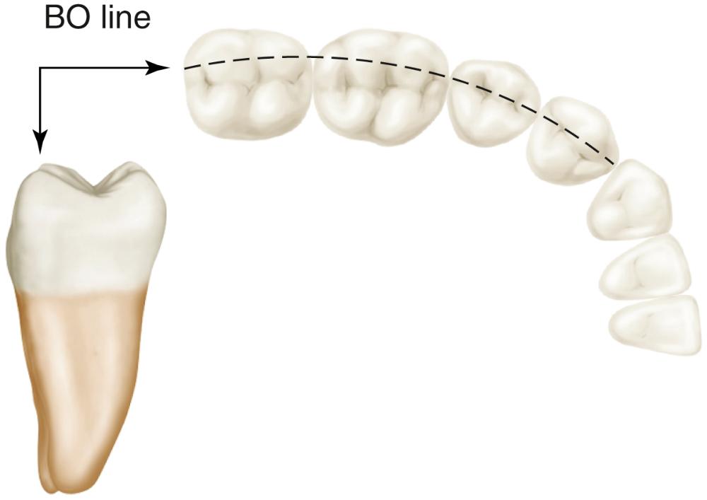
buccoocclusal (B-O) line
An imaginary line that extends through all the lingual cusp tips of the maxillary posterior teeth.
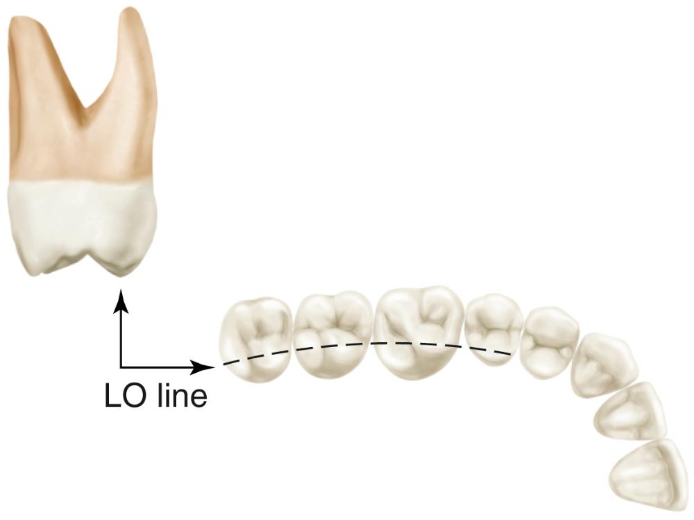
linguoocclusal (L-O) line
An imaginary line that extends through the central developmental grooves of the maxillary and mandibular posterior teeth.
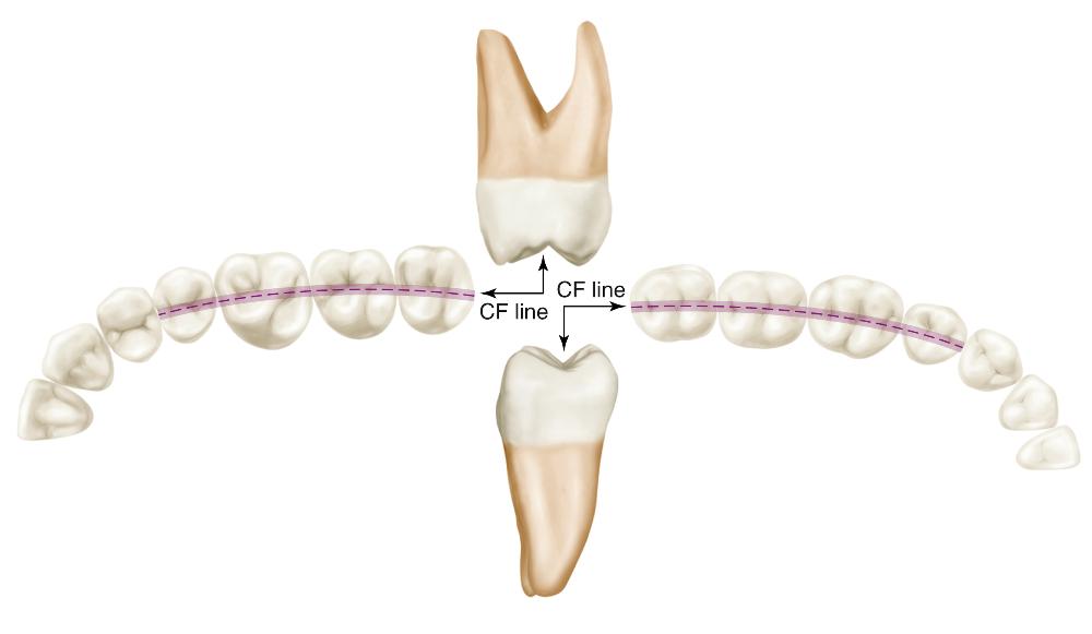
central fossa (C-F) line
The proximal contact areas are generally located slightly (...) to the C-F line. This allows for a greater (...) embrasure area and a smaller (...) embrasure area, which provides a spillway for food and improves the efficiency of mastication.
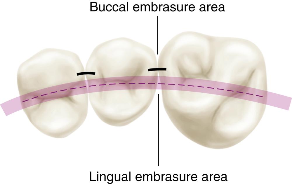
buccal; lingual; buccal
The (...) line of the mandibular teeth occludes with the C-F line of the maxillary teeth.
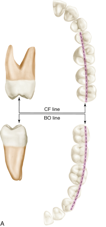
B-O
The (...) line of the maxillary teeth occludes with the C-F line of the mandibular teeth.
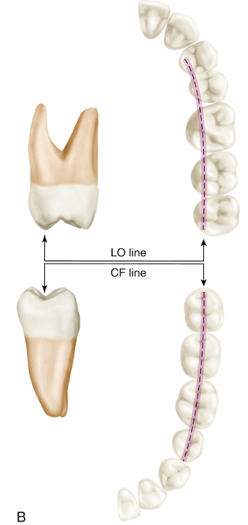
L-O
Occlusal contacts occur when the centric cusps contact the opposing central fossa line. Viewed from the facial, these cusps typically contact in one of which two areas?
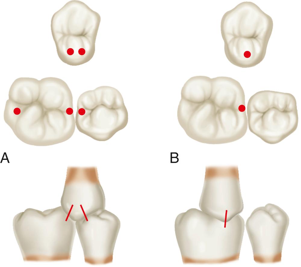
(1) central fossa area or (2) marginal ridge and embrasure area
When the normal interarch tooth relationship is viewed from the lateral, it can be seen that each tooth occludes with two opposing teeth, except for the (...) and the (...), which occlude with only one opposing tooth.
mandibular central incisors; maxillary third molars
Throughout the arch, any given tooth is found to occlude with its (...) in the opposing arch plus (...), which helps distribute occlusal forces.
namesake; an adjacent tooth
In the normal relationship, the mandibular teeth are positioned slightly (...) to their counterparts. This is true of both the posterior and the anterior teeth.
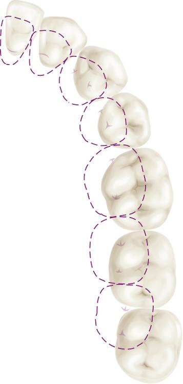
mesiolingual
In examining the occlusal relationships of the posterior teeth, much attention is centered around the first molar. The mandibular first molar is normally situated slightly (...) to the maxillary first molar.
mesial
The variation in molar relationship was first described by (...) and is therefore called an (...) molar relationship.
Angle; Angle Class I, II, or III
In an Angle Class I molar relationship:
- The mesiobuccal cusp of the mandibular first molar occludes in the (...).
- The mesiobuccal cusp of the maxillary first molar is aligned over the (...).
- The mesiolingual cusp of the maxillary first molar is situated in the (...).
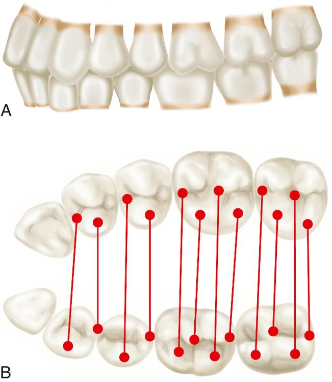
- embrasure area between the maxillary second premolar and first molar
- buccal groove of the mandibular first molar
- central fossa area of the mandibular first molar
In an Angle Class II molar relationship:
- The mesiobuccal cusp of the mandibular first molar occludes in the (...).
- The mesiobuccal cusp of the mandibular first molar is aligned with the (...).
- The distolingual cusp of the maxillary first molar occludes in the (...).
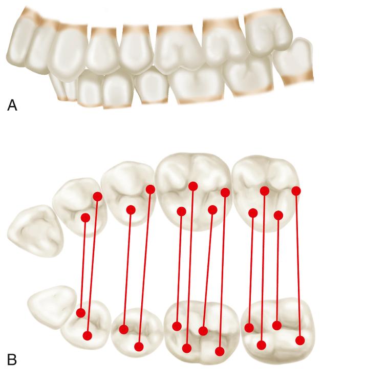
- central fossa area of the maxillary first molar
- buccal groove of the maxillary first molar
- central fossa area of the mandibular first molar
When compared to the Class I relationship, each occlusal contact pair in a Class II relationship is situated to (...) by approximately the mesiodistal width of (...).
distally; a premolar
In an Angle Class III molar relationship:
- The distobuccal cusp of the mandibular first molar is situated in the (...).
- The mesiobuccal cusp of the maxillary first molar is situated over the (...).
- The mesiolingual cusp of the maxillary first molar is situated in the (...).
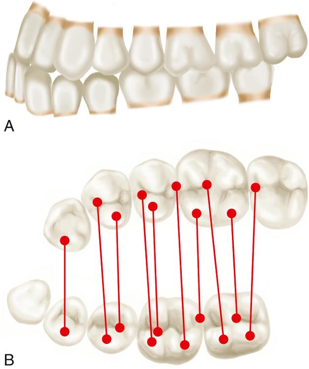
- embrasure between the maxillary second premolar and first molar
- embrasure between the mandibular first and second molar
- mesial pit of the mandibular second molar
When compared to the Class I relationship, each occlusal contact pair in a Class III relationship is situated to (...) by approximately the mesiodistal width of (...).
mesially; a premolar
What is the most commonly found molar relationship?
Class I
A Class II or III (...) describes a condition that is not Class I, yet is not extreme enough to satisfy the description of Class II or III.
tendency
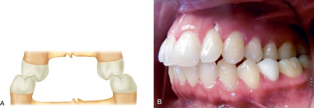
Like the maxillary posterior teeth, the maxillary anterior teeth are normally positioned labial to the mandibular anterior teeth. Unlike the posterior teeth, both maxillary and mandibular anteriors are inclined to the (...).
labial by 12 to 28° from a vertical reference line
Although a great amount of variation occurs, the normal relationship will find the (...) of the mandibular incisors contacting the (...) of the maxillary incisors.
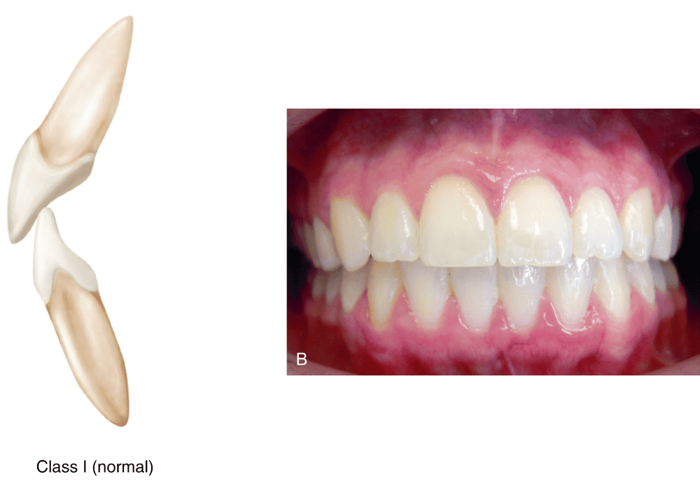
incisal edges; lingual surfaces
Anterior contacts commonly occur in the lingual fossae of the maxillary incisors approximately (...) mm gingival to the incisal edges.
4 mm (i.e. 3 to 5 mm of the mandibular incisors are hidden)
The anterior tooth contacts that provide guidance of the mandible through the various lateral movements are called (...).
anterior guidance
The distance between the labial incisal edge of the maxillary incisor and the labial surface of the mandibular incisor in the intercuspal position.
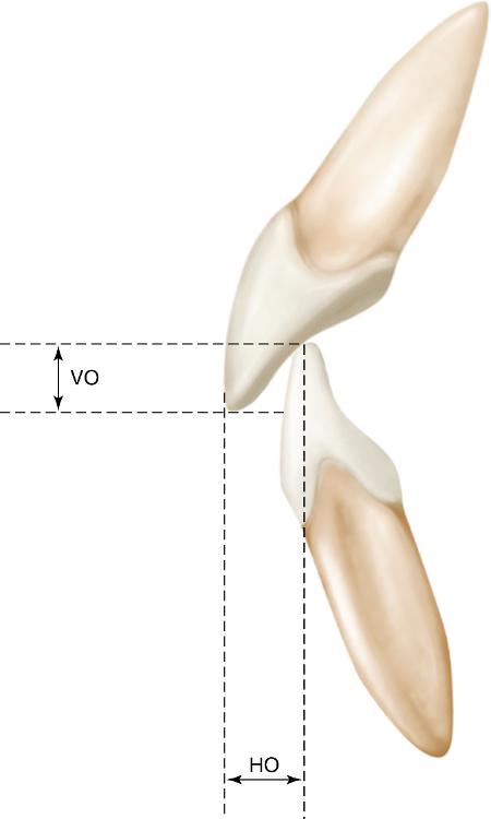
horizontal overlap (sometimes called overjet)
The distance between the incisal edges of the opposing anterior teeth, which is approximately 3 to 5 mm in normal occlusion.
vertical overlap (sometimes called overbite)
When a person has an underdeveloped mandible (Class II molar relationship), the mandibular anterior teeth often contact at the gingival third of the lingual surfaces of the maxillary teeth. This anterior relationship is termed a (...).
deep-bite (or deep overbite)
If in an anterior Class II relationship the maxillary central and laterals are at a normal labial inclination, it is considered to be (...),
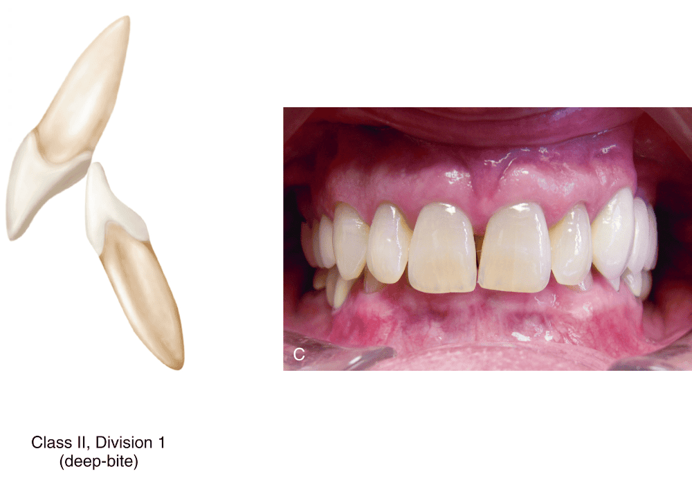
Class II, division 1
If in an anterior Class II relationship the maxillary incisors are lingually inclined, the anterior relationship is termed (...).
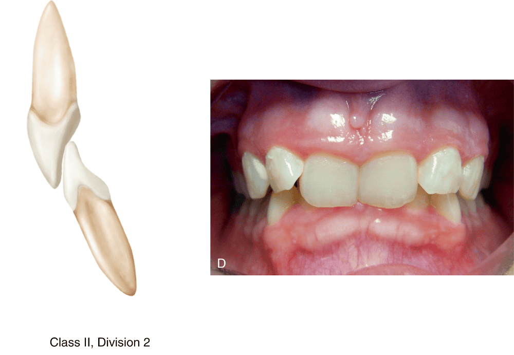
Class II, division 2
In people with pronounced mandibular growth, the mandibular anterior teeth are often positioned forward and contact with the incisal edges of the maxillary anterior teeth (molar Class III relationship). This is termed an (...) relationship.
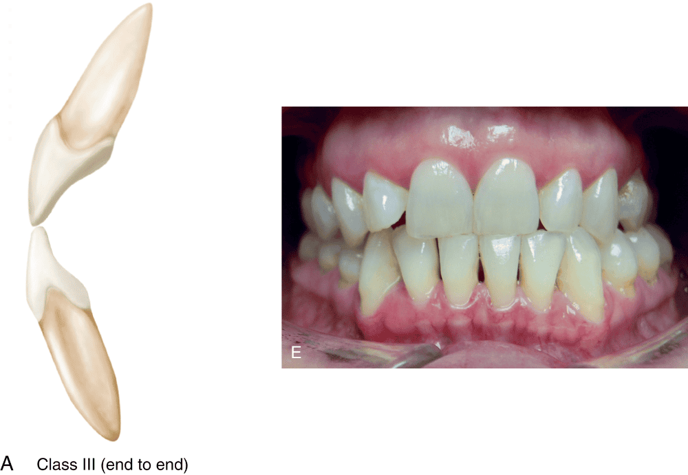
end-to-end (or edge-to-edge)
In extreme cases of (...) occlusion, the mandibular anterior teeth can be positioned so far forward that no contact occurs in the intercuspal position.
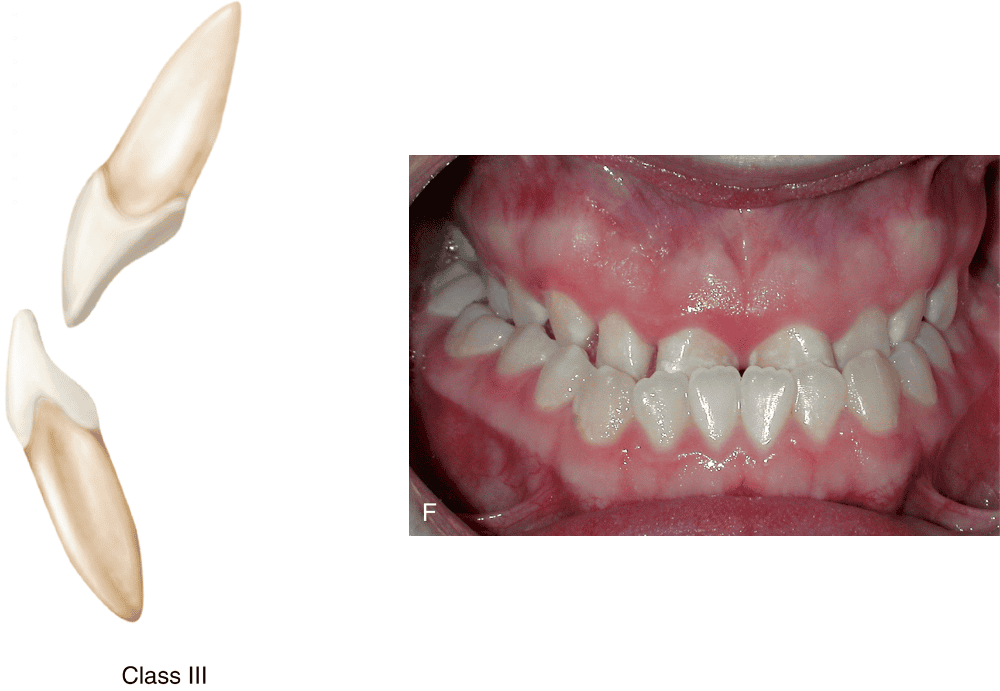
Class III
When the posterior teeth in maximum intercuspation and the opposing anterior teeth do not overlap or even contact each other, there is negative vertical overlap. This anterior relationship is termed an (...).
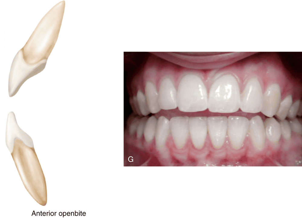
anterior openbite
The temporomandibular joints and associated musculature permit the mandible to move in which three planes?
sagittal, horizontal, and frontal
The term (...) has been used to describe any movement of the mandible from the intercuspal position that results in tooth contact.
eccentric
What are the three basic eccentric movements of the mandible?
protrusive, laterotrusive, and retrusive
A (...) mandibular movement occurs when the mandible moves forward from the intercuspal position.
protrusive
Any area of a tooth that contacts an opposing tooth during protrusive movement is considered to be (...).
protrusive contact
In a normal occlusal relationship, the predominant protrusive contacts occur on the anterior teeth, between the (...) of the mandibular incisors against the (...) of the maxillary incisors.
incisal and labial edges; lingual fossa areas and incisal edges
The (...) of the teeth are the surfaces responsible for the characteristics of anterior guidance.
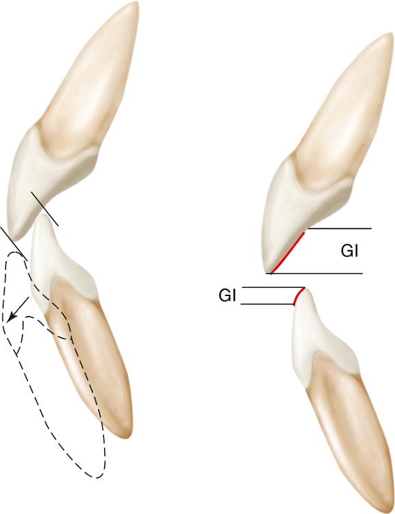
guiding inclines (GI)
Posterior protrusive contacts can occur between the (...) of maxillary cusps and the (...) of mandibular cusps.
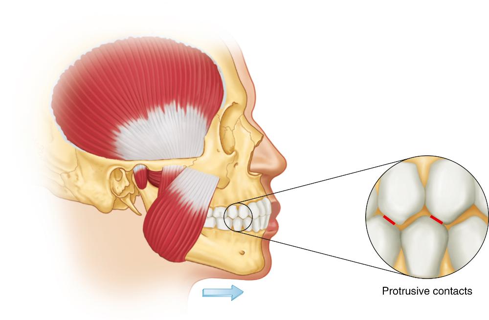
distal inclines; mesial inclines
During (...) mandibular movement, the right and left mandibular posterior teeth move across their opposing teeth in different directions.
laterotrusive
Any area of a tooth that contacts an opposing tooth on the working side during laterotrusive movement is considered to be (...).
laterotrusive (or working-side) contact
Laterotrusive contacts can occur:
- between the (...) of the maxillary buccal cusps and the (...) of the mandibular buccal cusps, and
- between the (...) of the maxillary lingual cusps and the (...) of the mandibular lingual cusps.
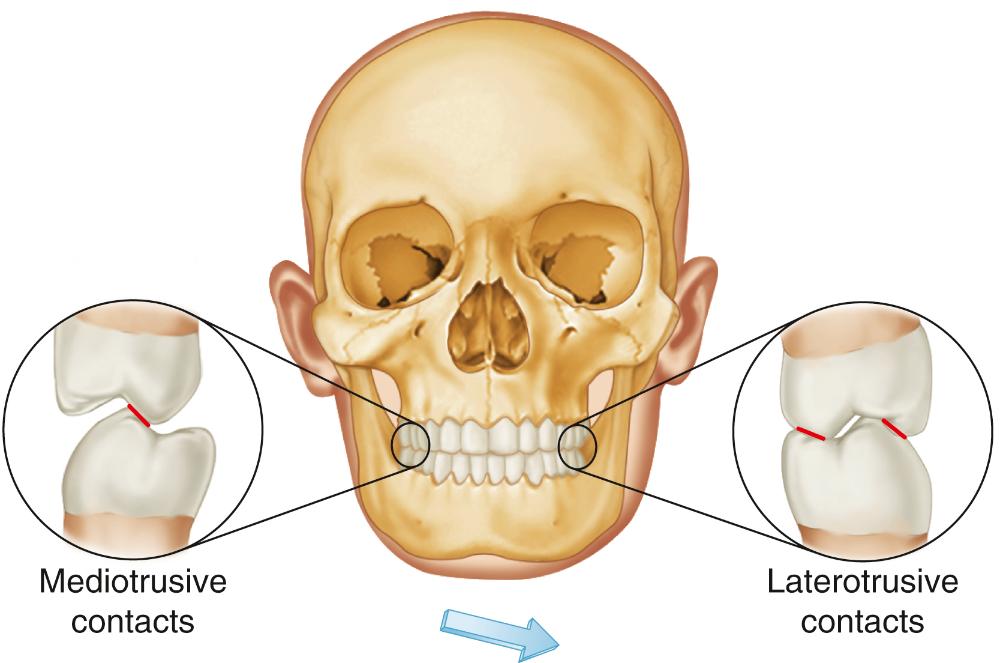
inner inclines ; outer inclines; outer inclines; inner inclines
Any area of a tooth that contacts an opposing tooth on the non-working side during laterotrusive movement is considered to be (...).
mediotrusive (or non-working-side) contacts
Mediotrusive contacts can only occur between the (...) of the maxillary lingual cusps and the (...) of the mandibular buccal cusps.
inner inclines; inner inclines
In earlier literature, mediotrusive (non-working-side) contacts were also called (...).
balancing contacts
A (...) movement occurs when the mandible moves posteriorly from the intercuspal position.
retrusive
Compared to the other movements, retrusive movement is quite small (1 or 2 mm), because it is (...).
restricted by the ligamentous structures
Posterior retrusive contacts can occur between the (...) of the maxillary cusps and the (...) of the mandibular cusps.
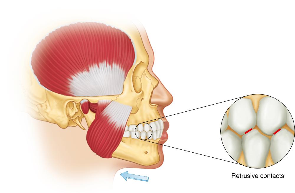
mesial inclines; distal inclines
Each centric cusp can potentially provide eccentric contact with the opposing tooth on (...) of its inclines. Each noncentric cusps can contact an opposing tooth on (...) of its inclines.
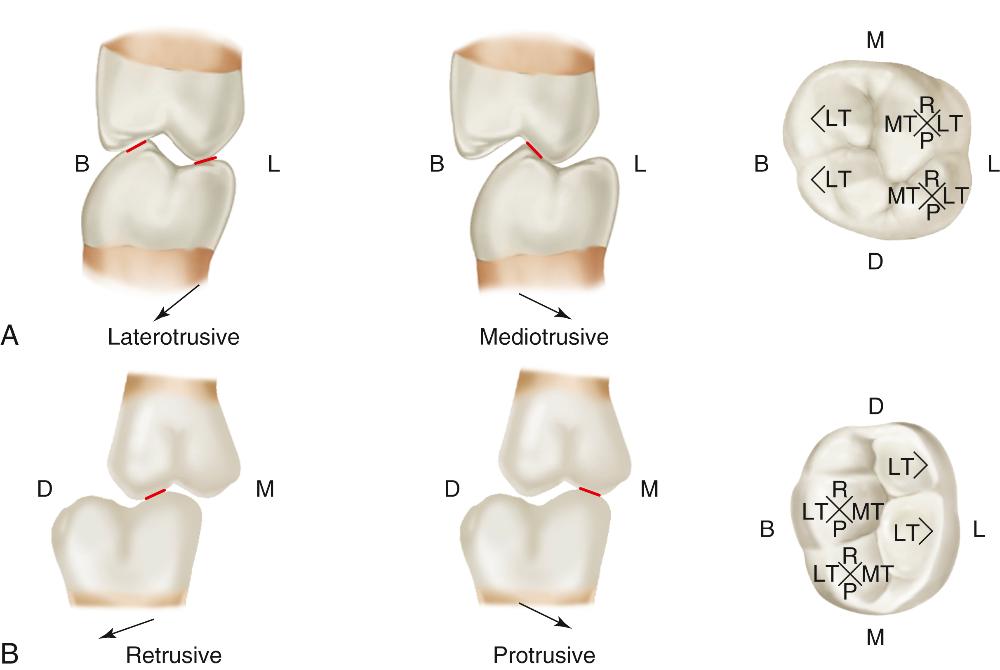
all four; only one (inner)
In a normal occlusal relationship, the first anterior teeth from the midline to participate in laterotrusive movements are the (...).

canines