Chapter 12: The CNS (Brain and Spinal Cord)
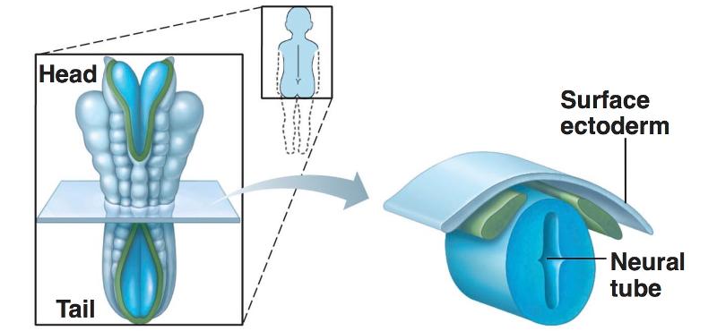
Development of the Neural Tube from Embryonic Ectoderm
4 steps:
1-neural plate forms from surface of ectoderm
2-the neural plate invaginates (folds), forming neural groove with neural folds
3-neural fold cells migrate to form neural crest, which forms much of the PNS and many other structures
4-the neural groove becomes the neural tube, which will form CNS structures
Embryonic Ectoderm-
Neural tube development: Step 1
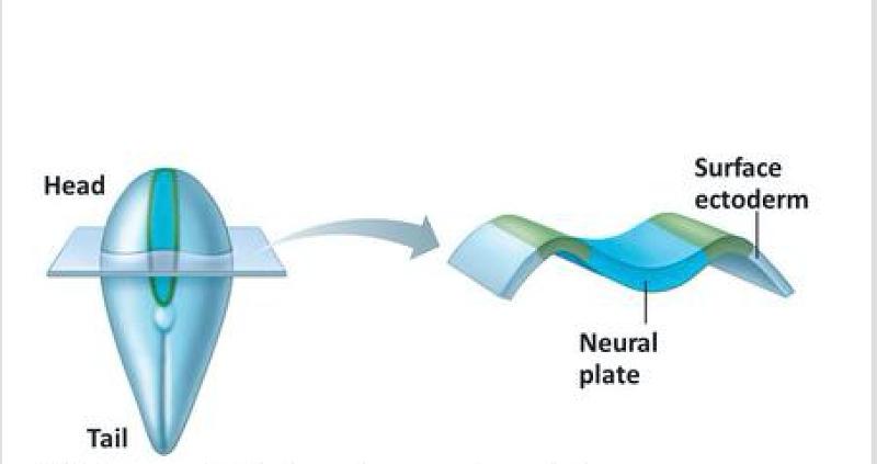
Embryonic Ectoderm-
Neural tube development: Step 2
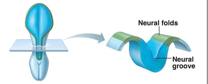
Embryonic Ectoderm-
Neural tube development: Step 3
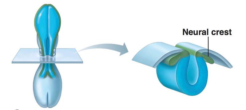
Embryonic Ectoderm-
Neural tube development: Step 4
Embryonic Development of Human Brain
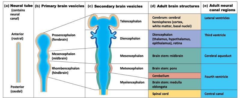
Neural Tube contains what important thing?
a neural canal
has two sides: anterior and posterior
Primary Brain Vesicles
Prosencephalon (forebrain)
Mesencephalon (midbrain)
Rhombencephalon (hindbrain)
Secondary Brain Vesicles
1-Telencephalon (end brain)
2-Diencephalon (interbrain)
3-Mesencephalon (midbrain)
4-Metencephalon (after brain)
5-Myelencephalon (spinal brain)
Telencephalon's Adult Brain Structures
Cerebrum: cerebral hemispheres
(cortex, white matter, basal nuclei)
Telencephalon's Adult Neural Region
Lateral Ventricles
Diencephalon's Adult Brain Structures
Diencephalon (thalamus, hypothalamus, epithalamus), retina
Diencephalon's Adult Neural Canal Region
Third Ventricle
Mesencephalon's Adult Brain Structures
Brain stem: midbrain
Mesencephalon's Adult Neural Canal Region
Cerebral Aquaduct
Metancephalon's Adult Brain Structures
Brain Stem: pons
Cerebellum
Metencephalon and Myelencephalon share what neural canal region?
Fourth Ventricle
Myelencephalon's Adult Brain Structure
Brain Stem: Medulla Oblongata
Spinal Cord has what neural canal region?
Central Canal
Effect of Space Restriction on Brain Development: Because the brain grows quicker than the skull...

2 flexures develop...the midbrain flexure and the cervical flexure
both move the forebrain toward the brain stem
Effect of Space Restriction on Brain Development: Because the cerebral hemispheres are forced to take a horseshoe-shaped course and grow posteriorly and laterally...

they grow back over and almost completely envelop the diencephalon and midbrain
Effect of Space Restriction on Brain Development: By week 26, the further growth of cerebral hemispheres causes their surfaces to crease and fold, producing convolutions and increasing surface area to allow...
more neurons to occupy the limited space
Ventricles in the brain
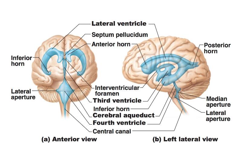
4 of them!
Lateral Ventricle
Third Ventricle
Cerebral Aquaduct
Fourth Ventricle
Ventricles are lined with...
Ependymal Cells
2 Lateral apertures and a single median aperture in the 4th ventricle are the openings that connect the 4 ventricles to the...
subarachnoid space
The fluid that surrounds the brain, spinal cord, and ventricles is...
cerebral spinal fluid (CSF)
The Cerebral Hemispheres take up...
85% of total brain mass
Ridges/folds of the hemispheres are called...
Gyri
"shallow grooves" are called...
sulci
Sulci divide the brain into 5 hemispheres. What are they?
Frontal
Parietal
Temporal
Occipital
Insula
"deep grooves" are called...
fissures
What jobs do fissures have?
separate large regions of brain
The median longitudinal fissure separates...
the brain into right and left hemispheres
The transverse cerebral fissure separates...
the cerebral hemisphere from the cerebellum
Lobes and Fissures of the Cerebral Hemispheres
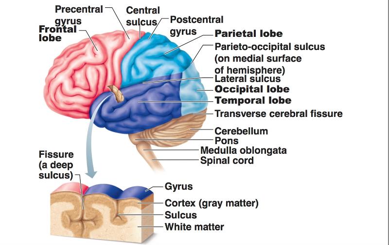
Cerebral Cortex Composition
outer layer of gray matter
neuron cell bodies
dendrites
neuroglia cells
blood vessels
NO axon or fiber tracts!!!
**billions of neurons arranged in 6 layers..(40% of brain mass)
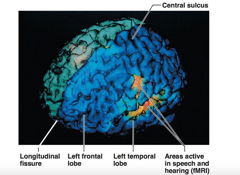
MRI of the cerebral cortex
Brain Cortex: 3 kinds of Functional Areas
Motor areas
Sensory areas
Association areas
All the neurons of the Brain Cortex are...
interneurons
More Facts of Brain Cortex Function
each hemisphere controls the sensory and motor functions of the opposite side of the body
some functions of the brain are mostly found in one side of the brain
no functional area of the cortex acts alone
conscious behavior involves the entire cortex in one way or another
Functional and Structural Areas of the Cerebral Cortex
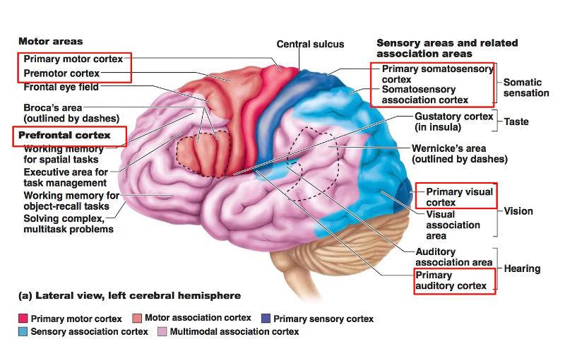
Cool map to help remember the important areas for structure and function
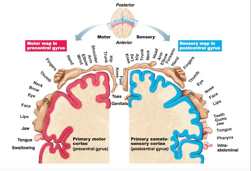
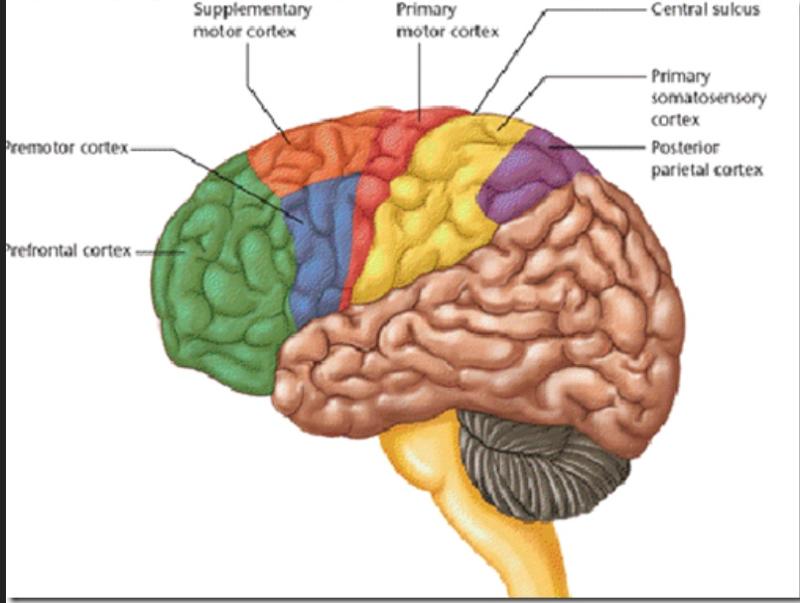
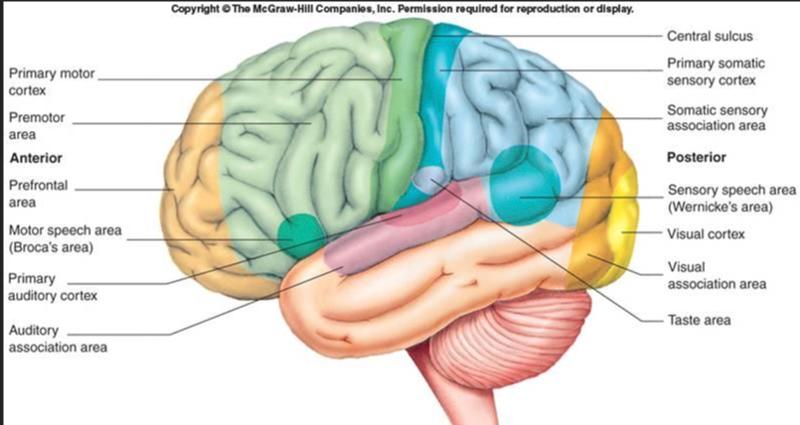
Primary Motor Cortex Location
pre central gyrus of frontal lobe
The Primary Motor Cortex has...
large neurons called pyramidal cells
Pyramidal cells allow...
conscious control of muscles
Pyramidal cells' long axons form...
motor tracts called pyramidal tracts (corticospinal tracts) in the spinal cord where innervation is contralateral (as in taking place on opposite sides)
The kind of mapping where pyramidal cells controlling specific body parts are grouped together...

somatotopy
Somatotopy's way of distorting the size of a figure to show how much gyrus is devoted to the body part is...
motor homunculi
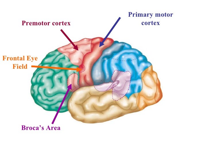
Premotor Cortex Location
just anterior to the precentral gyrus in the frontal lobe
Premotor Cortex Job
Controls learned motor skills of a repetitious or patterned nature (ex: typing, piano, driving)
Broca's Area Location
anterior to the inferior region of the premotor area
present only in the left hemisphere
Broca's Area Job
special motor speech
Frontal Eye Field Location
partially in and anterior to the premotor cortex and superior to the Broca's Area
Frontal Eye Field Job
controls voluntary movement of eyes
Stroke Symptoms by Stroke Location:
stroke to primary motor cortex
loss of voluntary control (paralysis) of muscle
reflexes intact
Stroke Symptoms by Stroke Location:
stroke to premotor cortex
loss of muscle motor memory
retain voluntary muscle control, but have to learn activities all over again
reflexes intact
Primary Somatosensory Cortex Location
in the post central gyrus of parietal lobe
2 Groups of neurons that supply the Primary Somatosensory Cortex
sensory skin receptors (touch)
proprioceptors (position sense receptors)
**give a sense of spatial discrimination
and represented graphically by the somatosensory homunculus
Somatosensory Association Cortex Location
just posterior to the primary somatosensory cortex
Somatosensory Association Cortex Job
integrate sensory inputs
as in temperature, pressure, texture, size, memory
used when you pick up an object in your pocket without looking at it/feeling for something in the dark
Visual Areas of the Cortex Location
on the extreme posterior tip of the occipital lobe
buried deep in the calcarine sulcus
Visual Areas of the Cortex Job
receives visual info from the retinas
Visual Association Area Location
surrounds primary visual cortex and covers much of the occipital lobe
Visual Association Area Job
uses past visual experiences to interpret visual stimuli
enables us to recognize and appreciate what we see
Primary Auditory Cortex Location
each one is located in the superior margin of the temporal lobe abutting the lateral sulcus
Primary Auditory Cortex Job
interprets pitch, loudness, location
Auditory Association Area Job
permits perception of sound stimulus:
understanding what we hear
storing memories of sound
Wernick's Area includes parts of the Auditory Cortex
Primary Olfactory (smell) Cortex Location
on the medial aspect of the temporal lobe
Primary Olfactory Cortex Job
makes us aware of different odors
Gustatory Cortex Location
in the insula, deep in the temporal lobe
Gustatory Cortex Job
involved in the perception of taste
Visceral Sensory Area Location
in cortex of insula, posterior to gustatory cortex
Visceral Sensory Area Job
perception of visceral sensations:
upset stomach, full bladder, constipation..
Vestibular (Equilibrium) Cortex Location
posterior insula, deep in the Temporal Lobe
Vestibular (Equilibrium) Cortex Job
helps with conscious awareness of balance, head position
Multimodal Association Areas: sensory information flow in the brain (direction)
1st-to the primary sensory cortex (you can see the words on the test)
2nd-to the sensory association cortex (you recognize the words as something you read)
3rd-to the multimodal association cortex (the words you read have meaning, based on memory)
3 Parts of the Multimodal Association Cortex
Anterior Association Area (prefrontal cortex)
Posterior Association Area
Limbic Association Area
Anterior Association Area (prefrontal cortex)
Location
frontal lobe
Anterior Association Area (prefrontal cortex)
Job
intellect, complex learning, recall, personality, working memory, abstract ideas, judgment, reasoning, persistence, planning
heavily dependent on positive and negative feedback from one's social environment
Posterior Association Area
Location
temporal, parietal, and occipital lobes
Posterior Association Area
Job
role in recognizing patterns, faces, localization, binding sensory inputs, understanding written and spoken language
way to remember: mmm..look at that guy's posterior!
Limbic Association Area
Location
cingulate gyrus, hippocampus areas
Limbic Association Area
Job
provides the emotional impact of a scene
Lateralization means that...
each hemisphere has some unique abilities
in 90%, the left hemisphere is dominant for...
language, math, logic
in 90%, the right hemisphere is dominant for...
visual-spacial, intuition, emotion, art, music
90% of people with left hemisphere dominance tend to be...
right-handed
In 10% of people, the roles of the hemispheres...
are reversed or share functions equally
Cerebral White Matter consists largely of...
myelinated fibers bundled into large tracts
Tract Name Classification is determined by...
the direction in which they run
The 3 Tract Classification Names
Commissural Tracts
Association Fibers
Projection Fibers
Commissural Tract Example
corpus callosum--it connects the right and left hemispheres
Association Fibers Job
Connect different parts of the same hemisphere
Projection Fibers Job
connect the cerebral hemispheres to the lower brain and spinal cord
Basal Nuclei are...
spots deep in the brain where large groups of neuron cell bodies are located
The 3 groups of basal nuclei/neuron cell bodies
Caudate nucleus
Putamen
Globus Pallidus
Basal Nuclei Job
important in starting, stopping, and monitoring the intensity of movements executed by the cortex, especially if they are slow or stereotyped
ex: swinging arms when walking
AND inhibit unwanted movements
ex: absent in Huntington's Chorea and Parkinson's Dz--Chorea (dance)
The Thalamus is...
one of the Diencephalon brain structures
composed of multiple separate nuclei
2 egg-shaped collections of nuclei in the center of the brain with a small space between them called the 3rd ventricle
3rd Ventricle
a space between 2 egg-shaped collections of nuclei inside the thalamus
Interthalamic adhesion (intermediate mass) is a..
small connection between the 2 egg-shaped collections of nuclei inside the thalamus
Thalamus Jobs
sorting and editing information
processing touch, pressure, pain
directing information to appropriate locations
Hypothalamus is...
below the thalamus
Hypothalamus composition
mammillary bodies
infundibulum
Mammillary bodies are...
relay stations in the olfactory pathway
The infundibulum is...
the connecting stalk of pituitary
Hypothalamus Jobs (multiple)
autonomic control center
center for emotional response
body temp regulation
regulation of food intake
regulation of water balance and thirst
regulation of sleep-wake cycles
control of endocrine system functioning
Epithalamus Location
most dorsal part of the diencephalon
forms the roof of the third ventricle
pineal gland is located here
Pineal Gland (body) Jobs
secretes the hormone melatonin to regulate the sleep-wake cycle
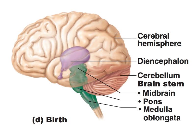
Brain Stem Composition
midbrain
pons
medulla oblongata
10 of the 12 cranial nerves come out of the...
Brain Stem
Midbrain Location
between the diencephalon and the pons
Cerebral Aquaduct Location
in the middle of the midbrain joining the 3rd and 4th ventricles
surrounding the Cerebral Aqueduct is the...
periaquaductal gray matter
periaquaductal gray matter jobs
involved in pain suppression and serves as a link for the fight-flight response
The 3rd and 4th cranial nerves are located here...
Midbrain
Pons Location
in between the midbrain and medulla oblangata
Behind the pons is the...
4th ventricle
Nuclei for CN V, CN VI, and CN VII are here...
Pons
Medulla Oblongata Location
between the pons and the spinal cord
Pyramids are located at the lowest part of the...
Medulla Oblangata
Pyramids are...
a collection of longitudinal fibers
Decussation of the pyramids
fibers that cross from left to right and vice versa (the cross-over point)
CN nuclei located in the medulla oblongata are...
CN VIII, CN IX, CN X, CN XI, CN XII
Medulla Oblongata Jobs
Cardiovascular center (for heart rate)
Respiratory center (for rate of breathing)
vomiting, hiccuping, swallowing, sneezing, coughing (reflexes)
Cerebellum means
"small brain"
Cerebellum Location
behind the pons and medulla
below the transverse cerebral fissure
Cerebellum Jobs
precise contraction of skeletal muscles for smooth, coordinated movements (as in driving, typing, sports)
all of these functions are subconscious
Limbic System Jobs
emotional or affective feelings
helps recognize facial expressions
helps assess danger
elicits fear response
Psychosomatic Illness
a connection with an emotion that can induce an illness (like anxiety)
Reticular Formation Location
extends through the core of the medulla oblongata, pons, and midbrain
Reticular Activating System Job
sends continuous stream of impulses to the cerebral cortex to keep it alert and enhance excitability
can filter out familiar or weak signals so you can focus on new things
Why can you ignore background noise while doing something?
The Reticular Activating System*
Meninges Location
layers around the brain
3 Layers of Meninges
Dura mater
Arachnoid mater
Pia mater
Falx Cerebri
Falx Cerebelli
Tentorium Cerebelli
are partitions that subdivide the cranial cavity of the dura mater
Falx Cerebri location
between right and left hemispheres
Falx Cerebelli location
between right and left cerebellum
Tentorium Cerebelli location
separates the cerebrum and cerebellum
Arachnoid mater contains the...
subdural space and subarachnoid space
Subarachnoid space is between...and is filled with...
the arachnoid mater and pia mater
CSF
The Pia mater is...
directly attached to the brain
Dural Septa and Dural Venous Sinuses
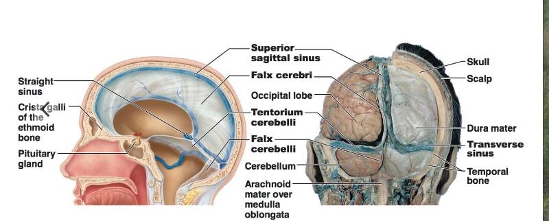
ex: falx cerebri, falx cerebelli, tentorium cerebelli
Cerebrospinal Fluid Location
in and around spinal cord and brain
CSF Jobs
protects brain and spinal cord
Choroid Plexus is found...
It is...
in the roof of each ventricle
a bunch of capillaries where CSF is released
CSF is constantly...
being formed and reabsorbed
replaced every 8 hours
about 500 ml is formed per day
Circulation of CSF
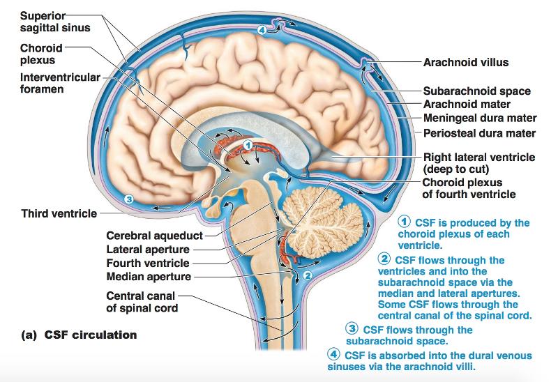
lateral ventricles --> foramen of Monro/third ventricle --> aqueduct of Sylvius --> fourth ventricle --> foramina of Magendie and Luschka --> subarachnoid space over brain and spinal cord --> reabsorption into venous sinus blood via arachnoid granulations
Hydrocephalus
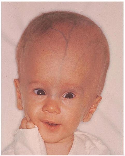
CSF accumulation
The Spinal Cord is protected by the...
spinal dura mater and CSF and an epidural space is exterior to the spinal dura mater
Spinal Cord tip ends at about
L1 or L2
Cone-shaped end of the spinal cord is the...
conus medullaris
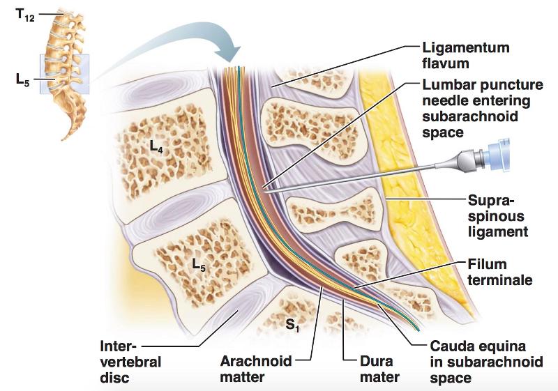
Cauda equina is...
a bunch of sacral nerve fibers that come out of the conus medullaris like a horse tail
What is the best area to do a lumbar spinal tap?
the cauda equina
Spinal cord Cross-Sectional Anatomy
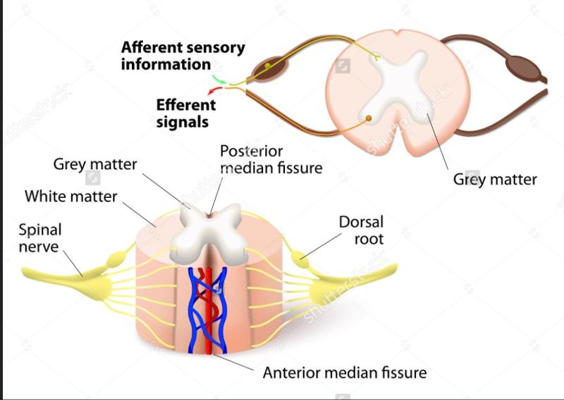
The gray matter of the spinal cord looks like the letter "H" or like a "butterfly"
central canal
The middle of the spinal cord is the...
central canal
2 posterior projections of the spinal cord's gray matter are...
the dorsal horns
Dorsal Horns
Jobs

where afferent fibers bring info into the spinal cord
where a dorsal root ganglion is part of the entering nerve
ex: nerve fiber stretched by reflex hammer
2 anterior projections of the spinal cord's gray matter are...
the ventral horns
Ventral Horns
Jobs
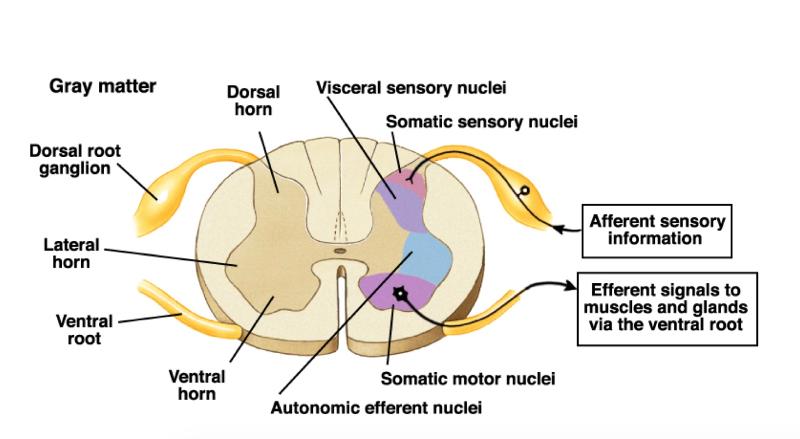
where efferent fibers project information out of the spinal cord
ex: muscle is stimulated by nerve to contract
Lumbar Tap Puncture
Organization of the Gray Matter of the Spinal Cord

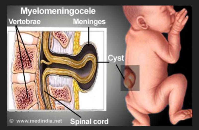
Lumbar Myelomeningocele/Spina Bifita