Athletic Training Special Tests
Cranial Nerve Assessment
Spurling's Test
- Patient's head extended and rotated toward the painful shoulder.
- Examiner applies an axial load to the cervical spine
(+) Pain
-Indication: Cervical Nerve Root Impingement usually caused by disc herniation
Stork's Test
- Patient is standing on one leg and performs backwards bending
- Examiner applies an axial load to the cervical spine
(+) test: Pain
Indication: Spondylolysis/ Spondylosisthesis
Slump Test
Test for Lumbar Spine
- Patient is sitting
- Ask the patient t perform the following motions.
- Cervical Spine Flexion
- Knee Extension
- Ankle Dorsiflexion
- Release Neck Flexion
- Bilateral Knee Extension
(+) Test: Increased radiating pain as sequential changes in posture occur
Indication: Increased neural tension caused by disc herniation or nerve root impingement
Straight Leg Raise Test
- Patient is supine and slowly lifts a leg while keeping the knee extended
- At the end of motion (Max Flexion) dorsiflex the ankle
(+) test: Pain
Indication:
- Pain at 30 degrees indicated hip problem or inflamed nerve
- Pain from 30-60 degrees indicates sciatic nerve impingement
- Pain from 70-90 degrees indicates sacroiliac joint problem
- Pain with dorsiflexion at the end of motion indicates nerve root impingement or sciatic nerve irritation
Phalen's Test
Position:
- Patient flexes the wrists and puts the back of the hands together
- The position is held for approx 1 minute
(+) Test:
- Pain & Numbness
Indication:
- Carpal Tunnel Syndrome
Finklestein's Test
Position:
- Athlete makes a fist with the thumb tucked on the inside
- Wrist is ulnarly deviated
(+) Test:
- Pain
Indication:
- DeQuarvain's Syndrome
Valgus or Varus Stress Test for Elbow
Valgus (Ulnar)
- Position:
- Patient’s elbow flexed to 25°
- One hand supports the lateral elbow with the fingers reaching behind the joint to palpate the medial joint line
- The opposite grasps the distal forearm
- Examiner applies valgus torque to the elbow
- (+) Test:
- Increased laxity compared to the opposite
side, and/or
pain
- Increased laxity compared to the opposite
side, and/or
- Indication:
- Injury to the ulnar collateral ligament
Valgus or Varus Stress Test for Knee
Test for Lateral Epicondylitis
Test for Medial Epicondylitis
Murphy's Sign
Position:
- Athlete is sitting or standing
- Athlete makes a fist and the examiner observes the position of the third metacarpal
(+) Test:
- Third metacarpal is at level with the second and the fourth metacarpals
Indications:
- Dislocated lunate
Neer's Impingement Sign
Position:
- Patient's shoulder is placed in internal rotation
- While maintaining shoulder internal rotation, examiner passively flexes the shoulder to end range
(+) test:
- Pain in subacrominal space between 90-180 deg of flexion
Indication:
- Subacromial impingement
Hawkins - Kennedy Test
Position:
- Patient's shoulder is placed in 90 deg of flexon with elbow flexed to 90 deg
- While maintaining shoulder flexion, examiner passively internally rotates the shoulder
(+) test:
- Pain in subacromial space in end range
Indication:
- Subacrominal Impingement
Roo's Test
Position:
- Patient abducts both arms and flexes elbows to 90 deg flexion
- Patient opens and closes the hands for 3 minutes
(+) Test:
- Inability to maintain testing position or replication of motor/sensory symptoms
Indication:
- Thoracic Outlet Syndrome
Allen's Test
Position:
- Examiner palpates the radial pulse
- Examiner abducts the patients shoulder and flex the elbow to 90 deg and externally rotates the arm while asking the patient to rotate the head away from injured side
(+) test:
- Diminished or disappearance of radial pulse
Indication:
- Thoracic Outlet Syndrome
Speed's Test
Position:
• Patient sitting with elbow extended and palm facing forward
•
Examiner resists shoulder flexion while palpating for tenderness over
the bicipital groove.
(+) Test:
• Pain over LH biceps tendon
Indication:
• LH biceps tendinopathy
Empty Can Test
Position:
• Patient’s shoulders elevated 90° in a scapular plane
(30° anterior to the frontal plane)
• With patient’s thumb
pointing down (i.e. empting the can), examiner applies downward
pressure at the wrist
(+) Test:
• Weakness or pain
accompanying the movement
Indication:
• Supraspinatus
pathology (ex. impingement, RC tear, tendinopathy)
O'Brien's Test
Position:
• Patient’s shoulder in 90° flexion and 15° horizontal
adduction
• With patient’s thumb pointing down ( = shoulder IR),
examiner applies downward pressure at the wrist
• Repeats this
with palm facing up ( = shoulder ER)
(+) Test:
• Pain or
clicking within the GH joint experienced with the arm
internally rotated but is decreased during external
rotation
Indication:
• Labral pathology
*Note that pain at AC joint may indicate AC joint injury
Anterior Drawer Test of the Knee
Position:
- Patient is supine with knee flexed to 90°, muscles relaxed
- Examiner stabilizes the foot (can sit on the foot), and pulls the proximal tibia anterior
(+) Test:
- Forward movement of the tibia that is greater than the normal side
Indication:
- Damage to the Anterior Cruciate Ligament
Lachman's Test
Position:
- Patient supine with knee flexed to 30°, muscles relaxed
- Examiner grasps the patient's femur with 1 hand, and proximal tibia with another, and pulls the proximal tibia anterior while stabilizing the femur
(+) Test:
- Forward movement of the tibia that is greater than the normal side
Indication:
- Damage to the Anterior Cruciate Ligament
McMurray's Test
Position:
- Examiner places one hand on top of the knee with thumb over one joint line and index and middle finger on the other joint line
- From full flexion, move the knee into extension while:
- Maintaining tibial external rotation and applying varus torque (stresses medial meniscus)
- Maintaining tibial internal rotation and applying valgus torque (stresses lateral meniscus)
(+) Test:
- Popping, clicking, or locking of the knee
Indication:
- Possible meniscal tear
Posterior Sag Test
Position:
- Patient is supine of a treatment table
- Examiner supports the patient's legs in a 90/90 position
(+) Test:
- Posterior sagging of the tibia
Indication:
- Damage to the posterior cruciate ligament
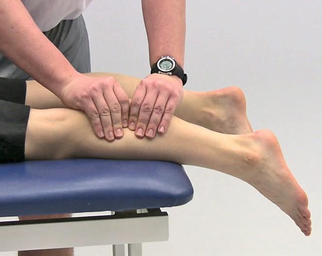
Thompson Test
Leg Special Test
Position:
- Examiner squeezes the patient's calf muscles, while the foot is extended off the table
(+) Test:
- Absence of foot movement (plantar flexion)
Indication:
- Achilles tendon rupture
Inversion Talor Tilt
Position:
- Patient's ankle in 90 degree dorsiflexion
- Examiner cups a hand around the calcaneous and inverts the foot while palpating the lateral ligaments]
(+) test:
- Excessive motion and/or clunking sound as it reaches the end point
Indication:
- Damage to the calcaneofibular (CF) ligament (primarily) and other lateral structures
Eversion Talor Tilt
Position:
- Patient's ankle in 90° dorsiflexion
- Examiner cups a hand around the calcaneous and everts the foot while palpating the deltoid ligaments
(+) Test:
- Excessive motion and/or clunking sound as it reaches the end point
Indication:
- Damage to the deltoid ligaments
Klieger's Test
Position:
- Examiner stabilizes the distal leg, while grasping the patient's foot and passively moving the ankle into dorsiflexion and external rotation
(+) Test:
- Pain
Indication:
- Damage to the deltoid ligament, distal ankle syndesmosis, anterior/posterior distal tibiofibular ligaments. and interosseous membrane
Yeargason's Test
Position:
- Patient sitting with elbow flexed 90° and forearm pronated
- Examiner resists elbow flexion and supination
- (+) Test:
• Pain or snapping in the bicipital groove - Indication:
• LH biceps tendinopathy
Appley's Compression Test (Pulling upwards)
Position:
- Patient is prone with knee flexed 90°
- Examiner applies axial loading to the leg while rotating tibia (foot) into internal and external rotation
(+) Test:
- Pain
Indication:
- Possible meniscus injury
Appley's Distraction Test (Pushing downwards)
Position:
- Patient is prone with knee flexed 90°
- Examiner applies distraction to the leg while rotating tibia (foot) into internal and external rotation
(+) Test:
- Pain
Indication:
- Possible injury to the ligament or joint capsule
- No pain if meniscal injury is present