Print Options

Card layout: ?

← Back to notecard set|Easy Notecards home page

Instructions for Side by Side Printing
  1. Print the notecards
  2. Fold each page in half along the solid vertical line
  3. Cut out the notecards by cutting along each horizontal dotted line
  4. Optional: Glue, tape or staple the ends of each notecard together
  1. Verify Front of pages is selected for Viewing and print the front of the notecards
  2. Select Back of pages for Viewing and print the back of the notecards
    NOTE: Since the back of the pages are printed in reverse order (last page is printed first), keep the pages in the same order as they were after Step 1. Also, be sure to feed the pages in the same direction as you did in Step 1.
  3. Cut out the notecards by cutting along each horizontal and vertical dotted line
To print: Ctrl+PPrint as a list

71 notecards = 18 pages (4 cards per page)

Viewing:

The Urinary System

front 1

Which is not a major function of the kidney?

  1. regulation of blood ionic composition
  2. regulation of blood cell size
  3. regulation of blood volume
  4. regulation of blood pressure
  5. regulation of blood pH

back 1

B

front 2

This is the formation of a new glucose molecule.

  1. glycolysis
  2. gluconeogenesis
  3. glucosamine
  4. glucose
  5. calcitriol

back 2

B

front 3

Which of the following is a waste product normally excreted by the kidneys?

  1. urea
  2. glucose
  3. insulin
  4. cholesterol
  5. carbon dioxide

back 3

A

front 4

This is smooth dense irregular connective tissue that is continuous with the outer coat of the ureter.

  1. adipose capsule
  2. renal capsule
  3. renal hilus
  4. renal cortex
  5. renal medulla

back 4

B

front 5

The portion of the kidney that extends between the renal pyramids is called the

  1. renal columns
  2. renal medulla
  3. renal pelvis
  4. calyces
  5. renal papilla

back 5

A

front 6

Which is the correct order of blood flow?

  1. renal artery-segmental artery-interlobular artery-peritubular capillaries- afferent arterioles
  2. interlobar arteries-arcuate arteries-glomerular capillaries-arcuate veins
  3. arcuate veins-arcuate arteries- glomerular capillaries- renal vein
  4. renal vein-segmental arteries-interlobar arteries- efferent arterioles
  5. interlobar veins- afferent arterioles- efferent arterioles- glomerular capillaries

back 6

B

front 7

Which is the correct order of filtrate flow?

  1. glomerular capsule, Proximal Convoluted tubule (PCT), Loop of Henle, Distal Convoluted tubule (DCT), Collecting duct
  2. Loop of Henle, glomerular capsule, PCT, DCT, Collecting duct
  3. Ascending limb of Loop, PCT, DCT, Collecting duct
  4. Collecting duct, DCT, PCT, Collecting duct, glomerular capsule
  5. PCT, glomerular capsule, DCT, Collecting duct, Loop of Henle

back 7

A

front 8

Which structure of the nephron reabsorbs the most substances?

  1. glomerular capsule
  2. Loop of Henle
  3. Ascending limb
  4. Collecting duct
  5. Proximal convoluted tubule

back 8

E

front 9

This is the structure of the nephron that filters blood.

  1. glomerular capsule
  2. Loop of Henle
  3. Ascending limb
  4. Collecting duct
  5. Renal corpuscle

back 9

A

front 10

This term means the return of substances into the blood stream from the filtrate.

  1. reabsorption
  2. filtration
  3. secretion
  4. excretion
  5. none of the above

back 10

A

front 11

This is a nephron process that results in a substance in blood entering the already formed filtrate.

  1. reabsorption
  2. filtration
  3. secretion
  4. excretion
  5. none of the above

back 11

C

front 12

This layer of filtration membrane is composed of collagen fibers and proteoglycans in a glycoprotein matrix.

  1. glomerular endothelial cells
  2. basal lamina
  3. pedicels
  4. filtration slites
  5. slit membrane

back 12

B

front 13

This occurs when stretching triggers contraction of smooth muscle walls in afferent arterioles.

  1. glomerular filtration rate
  2. tubulomerular feedback
  3. myogenic mechanism
  4. renal autoregulation
  5. capsular hydrostatic pressure

back 13

C

front 14

This occurs when a substance passes from the fluid in the tubular lumen through the apical membrane, across the cytosol, and then into the interstitial fluid.

  1. paracellular reabsoprtion
  2. transcellular reabsoprtion
  3. apical reasborption
  4. basolateral reabsorption
  5. active transport

back 14

B

front 15

Once fluid enters the proximal convoluted tubule

  1. it is less dense
  2. it has a higher K+ concentration
  3. it is called tubular fluid
  4. all the Na+ is removed
  5. it is headed to the ascending loop

back 15

C

front 16

The proximal convoluted tubules reabsorb what percentage of filtered water?

  1. 25%
  2. 50%
  3. 65%
  4. 80%
  5. 99%

back 16

C

front 17

Which of the following is NOT a way angiotensin II affects the kidneys?

  1. It increases GFR
  2. It can decrease GFR
  3. It enhances reabsorption of certain ions
  4. It stimulates the release of aldosterone
  5. None of the above

back 17

A

front 18

Urea recycling can cause a buildup of urea in the

  1. Renal capsule
  2. Loop of Henle
  3. Ascending tubule
  4. Renal medulla
  5. Renal pelvis

back 18

D

front 19

Increased secretion of Hydrogen ions would result in a ______________ of blood ____________?

  1. increase, pressure
  2. decrease, volume
  3. increase, sodium levels
  4. decrease, pH
  5. increase, urea

back 19

D

front 20

Increased secretion of Aldosterone would result in a ______________ of blood ____________?

  1. increase, potassium
  2. decrease, volume
  3. increase, calcium levels
  4. decrease, pH
  5. increase, sodium

back 20

E

front 21

The ascending loop of Henle is impermeable to

  1. urea
  2. water
  3. albumin
  4. sodium
  5. chloride

back 21

B

front 22

An analysis of the physical, chemical and microscopic properties of urine is called

  1. Urinalysis
  2. Filtration study
  3. Concentration study
  4. Diuretic
  5. Osmolarity

back 22

A

front 23

Water accounts for what percentage of the total volume of urine?

  1. 25%
  2. 50%
  3. 75%
  4. 80%
  5. 95%

back 23

E

front 24

This is a test to measure kidney function.

  1. Plasma creatinine
  2. Renal study
  3. Kidney assay
  4. Renal clearance
  5. Hilus study

back 24

A

front 25

This transports urine from the kidney to the bladder.

  1. Urethra
  2. Ureter
  3. Descending loop of Henle
  4. Renal hilus
  5. None of the above

back 25

B

front 26

This layer of the ureter is composed of connective tissue, elastic and collagen fibers.

  1. Mucosa
  2. Transitional epithelium
  3. Lamina propria
  4. Adventitia
  5. Lamina elastica

back 26

C

front 27

This lies in the anterior cornea of the trigone of the bladder.

  1. Urethral sphincter
  2. Adventitia bundle
  3. Ureter
  4. Internal urethral orifice
  5. Muscularis bundle

back 27

D

front 28

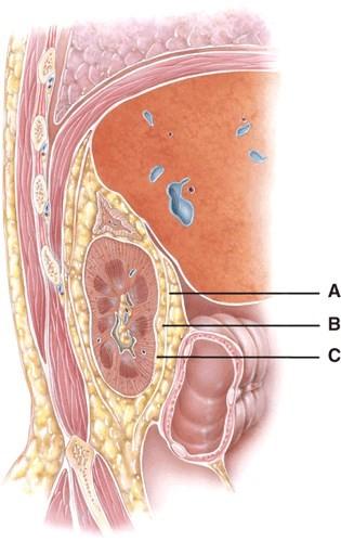
This is composed of dense irregular tissue that runs continuous with the ureter.

  1. A
  2. B
  3. C

back 28

C

front 29

This layers main function is to protect the kidney from trauma and hold it in place within the abdominal cavity.

  1. A
  2. B
  3. C

back 29

B

front 30

This layer runs deep to the peritoneum on the anterior surface of the kidneys.

  1. A
  2. B
  3. C

back 30

A

front 31

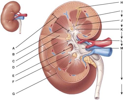
This can be divided into the cortical zone and the juxtamedullary zone.

  1. A
  2. D
  3. E
  4. F
  5. G

back 31

A

front 32

Where is the parenchyma?

  1. B
  2. M
  3. C
  4. D
  5. F

back 32

C

front 33

Each kidney can have anywhere from 8 to 18 of these.

  1. I
  2. J
  3. K
  4. L
  5. H

back 33

C

front 34

This structure is the apex of a renal pyramid.

  1. F
  2. D
  3. G
  4. K
  5. L

back 34

A

front 35

This is where the hilum extends into the kidney.

  1. E
  2. D
  3. G
  4. K
  5. L

back 35

A

front 36

Urine formed by the nephrons first drains into these.

  1. H
  2. I
  3. J
  4. K
  5. L

back 36

B

front 37

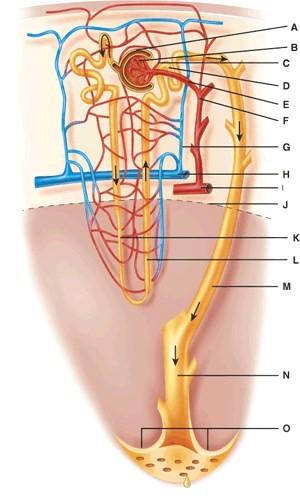
What two structures comprise the renal corpuscle?

  1. A and B
  2. C and D
  3. E and F
  4. K and L
  5. N and O

back 37

A

front 38

Where is the distal convoluted tubule?

  1. D
  2. E
  3. J
  4. K
  5. L

back 38

A

front 39

Where is the arcuate vein?

  1. E
  2. F
  3. G
  4. H
  5. I

back 39

D

front 40

Where is the ascending limb of the Loop of Henle?

  1. C
  2. D
  3. J
  4. K
  5. L

back 40

E

front 41

Where is the corticomedullary junction?

  1. G
  2. J
  3. M
  4. N
  5. O

back 41

B

front 42

Where is the papillary duct?

  1. C
  2. F
  3. H
  4. N
  5. O

back 42

D

front 43

What does line “M” point to?

  1. Arcuate artery
  2. Arcuate vein
  3. Collecting duct
  4. Descending loop
  5. Efferent arteriole

back 43

C

front 44

What is line “C” pointing to?

  1. Distal convoluted tubule
  2. Interlobular artery
  3. Efferent arteriole
  4. Arcuate artery
  5. Corticomedullary junction

back 44

C

front 45

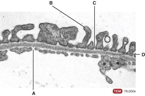
What is line “A” pointing to?

  1. Fenestrations
  2. Pedicels
  3. Filtration slit
  4. Basal lamina
  5. Lumen of the glomerulus

back 45

A

front 46

What is line “B” pointing to?

  1. Fenestrations
  2. Pedicels
  3. Filtration slit
  4. Basal lamina
  5. Lumen of the glomerulus

back 46

B

front 47

What is line “C” pointing to?

  1. Fenestrations
  2. Pedicels
  3. Filtration slit
  4. Basal lamina
  5. Lumen of the glomerulus

back 47

C

front 48

What is line “D” pointing to?

  1. Fenestrations
  2. Pedicels
  3. Filtration slit
  4. Basal lamina
  5. Lumen of the glomerulus

back 48

D

front 49

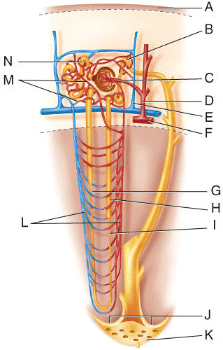
Fluid flowing from point N in the figure will go to which structure next?

  1. G
  2. H
  3. C
  4. B
  5. K

back 49

A

front 50

Fluid flowing from point G in the figure will go to which structure next?

  1. N
  2. H
  3. I
  4. M
  5. K

back 50

C

front 51

Fluid flowing from point H in the figure will go to which structure next?

  1. L
  2. B
  3. I
  4. G
  5. N

back 51

B

front 52

Fluid flowing from point B in the figure will go to which of the following structures?

  1. J
  2. M
  3. I
  4. G
  5. H

back 52

A

front 53

Which vessel in the diagram is the afferent arteriole?

  1. C
  2. D
  3. E
  4. F
  5. L

back 53

A

front 54

Which vessel in the diagram is the interlobular vein?

  1. C
  2. D
  3. E
  4. F
  5. L

back 54

C

front 55

Which vessel(s) in the diagram are the vasa recta?

  1. C
  2. D
  3. E
  4. M
  5. L

back 55

E

front 56

Which vessel in the diagram is the interlobular artery?

  1. D
  2. E
  3. F
  4. L

back 56

A

front 57

Which vessel in the diagram is the arcuate artery?

  1. C
  2. D
  3. E
  4. F
  5. L

back 57

D

front 58

Which vessel(s) in the diagram are the peritubular capillaries?

  1. C
  2. D
  3. E
  4. M
  5. L

back 58

D

front 59

An increase in permeability of the filtration membrane due to disease, injury, or irritation of kidney cells by substances such as bacterial toxins, ether, or heavy metals indicates which condition?

  1. albuminuria
  2. lucosuria
  3. robilinogenuria
  4. ketonuria
  5. bilirubinuria

back 59

A

front 60

Anorexia, starvation, or a diet too low in carbohydrates indicates which condition?

  1. albuminuria
  2. glucosuria
  3. urobilinogenuria
  4. ketonuria
  5. bilirubinuria

back 60

D

front 61

Stress, causing excessive amounts of epinephrine secretion which stimulates glycogen breakdown, indicates which condition? This condition can also indicate diabetes mellitus.

  1. albuminuria
  2. glucosuria
  3. urobilinogenuria
  4. hematuria
  5. bilirubinuria

back 61

B

front 62

Excessive urine concentration of a normal breakdown product of hemoglobin, caused by pernicious anemia, infectious hepatitis, jaundice or cirrhosis, indicates which condition?

  1. albuminuria
  2. glucosuria
  3. urobilinogenuria
  4. hematuria
  5. bilirubinuria

back 62

C

front 63

This substance gives bile its major pigmentation:

  1. ketone bodies
  2. erythrocytes
  3. casts
  4. bilirubin
  5. glucose

back 63

D

front 64

These are tiny masses of material, hardened in the lumen of the urinary tubule and are flushed out when filtrate builds up behind them:

  1. ketone bodies
  2. erythrocytes
  3. casts
  4. microbes
  5. urobilinogen

back 64

C

front 65

Candida albicans and E. coli are which type of abnormal constituent of urine:

  1. ketone bodies
  2. erythrocytes
  3. casts
  4. microbes
  5. urobilinogen

back 65

D

front 66

Which is the normal pH range of urine in humans?

  1. 8.1 – 10.6
  2. 4.6 – 8.0
  3. 1.0 – 3.0
  4. 3.1 – 4.0
  5. 10.0 – 12.0

back 66

B

front 67

What is the normal volume of urine produced in humans?

  1. 1L / hr
  2. 2L / day
  3. 2L / hr
  4. 3L / week
  5. 10L / 24 hours

back 67

B

front 68

What is the normal specific gravity range of urine in humans?

  1. 2.350 – 3.700
  2. 0.002 – 1.000
  3. 4.6 – 8.0
  4. 1.001 – 1.035
  5. 1.04 – 2.60

back 68

D

front 69

This hormone is released when the blood volume increases.

  1. Parathyroid Hormone
  2. Renin
  3. ADH
  4. Atrial Natriuretic Peptide
  5. Aldosterone

back 69

D

front 70

Consumption of salty food will cause an increase in this hormone.

  1. Aldosterone
  2. Renin
  3. ANH
  4. Angiotensin-II
  5. ADH

back 70

E

front 71

Absence of angiotensin converting enzyme will lead to

  1. decreased blood pressure
  2. increased blood pressure
  3. will not have any effect on blood pressure
  4. All of these choices
  5. None of these choices

back 71

A