Instructions for Side by Side Printing
- Print the notecards
- Fold each page in half along the solid vertical line
- Cut out the notecards by cutting along each horizontal dotted line
- Optional: Glue, tape or staple the ends of each notecard together
chapter 18: the heart
front 1 the first sound of the heart is valuable in diagnosis because it provides information about the function of the heat's pulmonary and aortic valves | back 1 true |
front 2 the aortic valves prevent back flow of blood from the aorta into right ventricle | back 2 False |
front 3 the visceral and parietal layers of pericardium are the layers of serous pericardium | back 3 True |
front 4 Superior and inferior vena cava returns venous blood from the upper and lower half of the body into the right atrium of the heart | back 4 True |
front 5 Increase of systemic blood pressure will increase the preload | back 5 False |
front 6 Preload is the stretching of right atrium, the greater the venous return more will be the preload | back 6 true |
front 7 Foramen ovale connects the two ventricles in fetal heart | back 7 False |
front 8 coarctatioin of aorta may increase the afterload and causing left ventricular hypertrophy | back 8 True |
front 9 pulmonary stenosis will lead to right ventricular hypertrophy | back 9 True |
front 10 cor-pulmonnale is the condition of lung congestion secondary to left ventricular failure | back 10 False |
front 11 the dicrotic notch refers to the brief rise in aortic pressure caused by back flow of blood rebounding off semilunar valves of aorta during ventricular diastole | back 11 True |
front 12 inotropic drugs can increase the force of cardiac muscle contraction | back 12 True |
front 13 abnormal heart sounds are called murmurs | back 13 True |
front 14 cardioacceleratory center is located in the medulla oblongata, which innervates SA and AV nodes, heart muscles, and coronary arteries through sympathetic nerves. | back 14 false |
front 15 the difference between resting and maximum state of cardiac output is called bainbridge reflex in heart | back 15 False |
front 16 anterior interventricular artery is a branch of? | back 16 left coronary artery |
front 17 circumflex coronary artery is a branch of? | back 17 left coronary artery |
front 18 right marginal artery is a branch of: | back 18 right coronary artery |
front 19 posterior interventricular artery is a branch of | back 19 right coronary artery |
front 20 endothelial layer of the inner myocardial surface is called | back 20 endocardium |
front 21 left ventricle pumps blood into the | back 21 aorta |
front 22 the semilunar valve lies between the right atrium and pulmonary trunk is called | back 22 pulmonary valve |
front 23 The first heart sound is due to the closure of AV valves | back 23 True |
front 24 the second peak in left atrial pressure is due to atrial systole | back 24 False |
front 25 hemorrhage with a large loss of blood causes | back 25 a lowering of blood pressure due to change in cardiac output |
front 26 the left ventricular wall of the heart is thicker than the right wall in order to | back 26 pump blood with greater pressure |
front 27 blood within the pulmonary veins returns to the | back 27 left atrium |
front 28 If cardiac muscle is deprived of its normal blood supply, primarily damage result from | back 28 a decreased delivery of oxygen |
front 29 the pericardial cavity | back 29 is the region of the thoracic cavity that contains the heart |
front 30 circulation of the blood through the heart | back 30 sup/inf vena cava -> right atrium -> through tricuspid valve to the right ventricle -> through pulmonary valve to the pulmonary trunk -> right and left pulmonary artery -> lungs -> right and left pulmonary veins -> left atrium -> through bicuspid valve to left ventricle -> aortic valves into aorta -> body |
front 31 List the layers of the sac around human heart | back 31 pericardium, fibrous pericardium, serous pericardium (Parietal and visceral layers) |
front 32 space between the pericardia layers is called what? and the content is called? | back 32 pericardial cavity serous fluid |
front 33 collection of excess amount of fluid int he pericardial space is called | back 33 cardiac temponade |
front 34 general term for the arteries that supply blood to the heart is | back 34 coronary arteries |
front 35 the major blood vessels entering the right atrium are | back 35 superior/interior vena cava, coronary sinus, anterior cardiac vein |
front 36 the right side of the hear is filled with | back 36 deoxygenated blood |
front 37 a valve between the right atrium and right ventricle is called | back 37 tricuspid valve |
front 38 a valve between the left atrium and left ventricle is called | back 38 mitral valve |
front 39 the property of self-stimulation of heart by speacialized tissure in the right atrium is called | back 39 autorhythmic |
front 40 what are those autorhythmic tissue in the walls of the right atrium called | back 40 SA node |
front 41 SA node generates electrical stimulation about | back 41 60-100 times per minute |
front 42 list the ECG waves in the order of occurence | back 42 P waves QRS complex T waves |
front 43 a wave obtained in the ECG tracing during atrial depolarization is | back 43 P wave |
front 44 a wave obtained in ECG tracing during ventricular depolarization is | back 44 QRS complex |
front 45 a wave obtained in ECG tracing during ventricular repolarization is | back 45 T wav |
front 46 define cardiac output whats the formula | back 46 cardiac output is the amount of blood pumped out of each ventricle in one minute CO = HR x SV |
front 47 define heart rate | back 47 number of beats per minute |
front 48 define stroke volume formula | back 48 volume of blood pumped out of each ventricle per beat SV = EDV - ESV |
front 49 define end diastolic volume | back 49 maximum amount of blood ventricles will contain in the cycle |
front 50 define end systolic volume | back 50 blood remaining in chambers when ventricles relaxes |
front 51 what is cardiac reserve | back 51 difference between resting and maximal CO |
front 52 the amount of blood pumped by each ventricle in one minute can be expressed as | back 52 stroke volume X heart rate |
front 53 atrial cotraction occurs | back 53 following P wave |
front 54 if the vagus nerves to the heart were cut, the result will be that | back 54 the heart rate would increase |
front 55 the AV valves are supported by______ so that they do not blow back up into the atria during ventricular contraction | back 55 chordae tendineae |
front 56 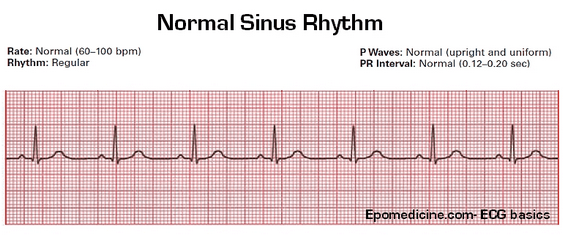 normal sinus rhythm | back 56 normal ECG trace |
front 57  Junctional rhythm | back 57 The SA node is nonfunctional. As a result: P waves are absent AV node paces heart 40-60 beats/minute |
front 58  second degree heart block | back 58 the AV node fails to conduct some SA node impulses. As a result: there are more P waves than QRS waves |
front 59  Ventricular fibrillation | back 59 electrical activity is disorganized.results in chaotic, grossly abnormal ECG deflections |
front 60 all events associated with blood flow through the heart during systole and diastole is called | back 60 cardiac cycle |
front 61 ventricular repolarization wave: | back 61 T wave |
front 62 the first sound of the heart s valuable in diagnosis because it provides information about the function of the heat's pulmonary and aortic valves | back 62 true |
front 63 the mediastinal cavity | back 63 is the region of the thoracic cavity that contains the heart |
front 64 the difference between resting and maximal cardiac output is called | back 64 cardiac reserve |
front 65 the apex of the heart is directed towards | back 65 the left |
front 66 tetralogy of fallot is a common finding in | back 66 infants with down syndrome |
front 67 when the second stimulation is provided to the hear immediately before the ventricular repolarization starts: | back 67 second depolarization will not take place |
front 68 cardiac center in the brain is located in | back 68 medulla oblongata |
front 69 the direction of the blood flow during ventricular contraction in an infant with ventricular septal defect will be | back 69 left ventricle to right ventricle |
front 70 the left coronary arteries arises from the | back 70 ascending aorta |
front 71 the coronary arteries arise from the | back 71 aorta |
front 72 left coronary artery is a branch of ascending aorta | back 72 true |
front 73 all of the structures below are components of conduction system of the heart. Except | back 73 fossa ovalis |
front 74 to ausculate the aortic valve, you would place your stethoscope in the | back 74 second intercostal space to the right of the sternum |
front 75 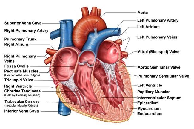 | back 75 no data |
front 76 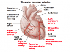 coronary arteries | back 76 no data |
front 77 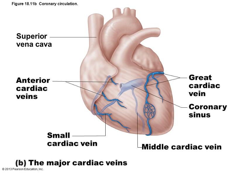 Veins of the heart | back 77 no data |
front 78 volume of blood pumped by each ventricle with each contraction is the | back 78 stroke volume |
front 79 the difference between resting and maximal cardiac output is | back 79 cardiac reserve |
front 80 increase in the heart rate initiated by the increased blood in atria which in turn causes the stimulation of the SA node is the | back 80 bainbridge reflex |
front 81 the degree of stretch of cardiac muscle cells before they contract is | back 81 Frank-starling's law |
front 82 relaxation of the ventricular muscle is called | back 82 diastole |
front 83 amount of blood collected in a ventricle during full relaxation | back 83 end diastolic volume |
front 84 pressure exerted by blood in the large arteries leaving the hear, against which heart must force the blood to eject out the ventricles is the | back 84 afterload |
front 85 which of the following occurs as AV valves close and signifies beginning of ventricular contraction | back 85 first heart beat |
front 86 all events associated with blood flow through the heart during relaxation and contraction | back 86 cardiac cycle |
front 87 which of the following occurs when semilunar valves of great vessels closes at the beginning of the ventricular diastole? | back 87 second heart sound |
front 88 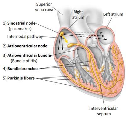 intrinsic conducting system of the heart | back 88 no data |