Instructions for Side by Side Printing
- Print the notecards
- Fold each page in half along the solid vertical line
- Cut out the notecards by cutting along each horizontal dotted line
- Optional: Glue, tape or staple the ends of each notecard together
Exercise 5-6 in the Lab Manual
front 1 A group of similar cells and associated structures(the matrix) that function together to carry out specific activities | back 1 Tissue |
front 2 What are the basic types of tissue in the human body? | back 2 Epithelial tissue, connective tissue, muscle tissue and nervous tissue |
front 3 What covers body surfaces and/or lines hollows organs, body cavities, and ducts. It can also form certain types of glands? | back 3 Epithelial Tissue |
front 4 What kind of tissue is often found connecting structures together. It protects and supports the body or specific parts of the body, secures organs to other structures, serves as an energy reserve(fat), provides immunity and transports substances through out the body(blood)? | back 4 Connective tissue |
front 5 What is two or more tissues that function together to perform specific activities? | back 5 Organ |
front 6 Epithelium consisting or one layer of thin flat, scale-like cells. | back 6 Simple squamous epithelium |
front 7 What location do you find simple squamous epithelium cells in the body? | back 7 kidneys, blood vessels |
front 8 What is the function of simple squamous epithelium cells? | back 8 Allows passage of materials by diffusion and filtration in sites where protection is not important; secrets lubrication substances in serosae. |
front 9 composed of one layer of cells with cells that are somewhat cube shaped and are tightly joined together forming tine tubes | back 9 simple cuboidal epithelium |
front 10 line the cavities and surfaces of structures throughout the body, and also form many glands | back 10 simple cuboidal epithelium |
front 11 Where is the lumen in a simple cuboidal epithelium | back 11 in the center of the cubed cell |
front 12 what are some locations of simple cuboidal epithelium tissue? | back 12 ducts and secretory portions of small glands, ovary surface, and kidney |
front 13 What is the function of simple cuboidal epithelium tissue | back 13 secretion and absorption |
front 14 Epithelium tissue that is longer than it is wide with the nucluei found in the base of the cell; often you will see goblet cells in this tissue | back 14 simple columnar epithelium |
front 15 Where in the body are simple columnar epithelium tissue found? | back 15 gallbladder, digestive track and some regions of the uterus |
front 16 What is the function of simple columnar epithelium tissue? | back 16 absorption and secretion of mucus, enzymes; ciliated types propels mucus by ciliated action |
front 17 Epithelium made up of cells that reach the basement membrane and appear to be stratified because their nuclei are at different levels. | back 17 pseudostratified ciliated columnar epithelium |
front 18 What is the function of pseudostratified ciliated columnar epithelium? | back 18 secretion, particularly of mucus, propulsion of mucus by cillary action |
front 19 Where in the body do you find pseudostratified ciliated columnar epithelium | back 19 trachea, most of the upper respiratory tract, and male sperm |
front 20 The nuclei of the cells can be seen at different levels and not all of the cells seems to reach the surface; However each cell attaches to the basement membrane, so there is only one layer of cells even though there appears to be more | back 20 pseudostratified ciliated columnar epithelium |
front 21 Epithelium that occurs in layers where the surface cells are squamous in shape | back 21 statified ciliated epithelium |
front 22 Where is stratified ciliated epithelium found in the body? | back 22 esophagus, mouth, vagina and epidermis of the skin |
front 23 What is the function of stratified ciliated epithelium tissue? | back 23 Protects underlying tissues in areas subjected to abrasions |
front 24 In the upper layers of the cell of a stratified squamous epithelium the shape of the nuclei are _________? | back 24 squamous or flat like |
front 25 In the deeper layers of the cell of a stratified squamous epithelium the shape of the nuclei are _________? | back 25 Round |
front 26 A type of loose connective tissue | back 26 Areolar connective tissue |
front 27 ________ tissue is composed of a loose arrangement of cells which lie in a matrix that is composed of ground substance and fibers. | back 27 Connective Tissue |
front 28 Collagen, elastic and reticular fibers are found in which type of tissue | back 28 Areolar connective tissue |
front 29 Areolar tissue is prevalently found where? | back 29 under epithella of body; mucus membranes; packages organs, and surrounds capillaries |
front 30 What is the function of Areolar connective tissue? | back 30 wraps and cushions organs; holds and conveys tissue fluids |
front 31 Composed of dense, regular connective tissue. | back 31 Tendons |
front 32 In addition to tendons what other structures are composed of dense connective tissue | back 32 most ligaments and aponeuroses |
front 33 What is the function of dense connective tissue? | back 33 Attaches muscle to bone or muscle to muscle |
front 34 You will find Collagen fibers, and fibroblast in what connective tissue? | back 34 dense connective tissue |
front 35 You will find collagen fibers, fat cells, elastic fibers, fibroblast and ground substance in which connective tissue? | back 35 Areolar connective tissue |
front 36 the gel-like material in which connective tissue cells and fibers are embedded. | back 36 ground substance |
front 37 A connective tissue found along with areolar connective tissue | back 37 Adipose connective tissue |
front 38 Fat cells are called what? | back 38 adipocytes |
front 39 Where in the body to you find adipose tissue? | back 39 under skin, around kidneys in the breast |
front 40 What are some functions of adipose tissue? | back 40 reserve food fuel, insulates against heat loss, supports and protects organs |
front 41 Has more rigidity than other types of connective tissue | back 41 Hyaline cartilage |
front 42 What is chondroitin sulfate? | back 42 jelly like material in the ground substance |
front 43 Where in the body do you find hyaline cartilage? | back 43 long bones, embrionic skeleton, costal cartilages of the ribs |
front 44 What is the function of hyaline cartilage? | back 44 supports and reinforces; has resilient cushioning properties |
front 45 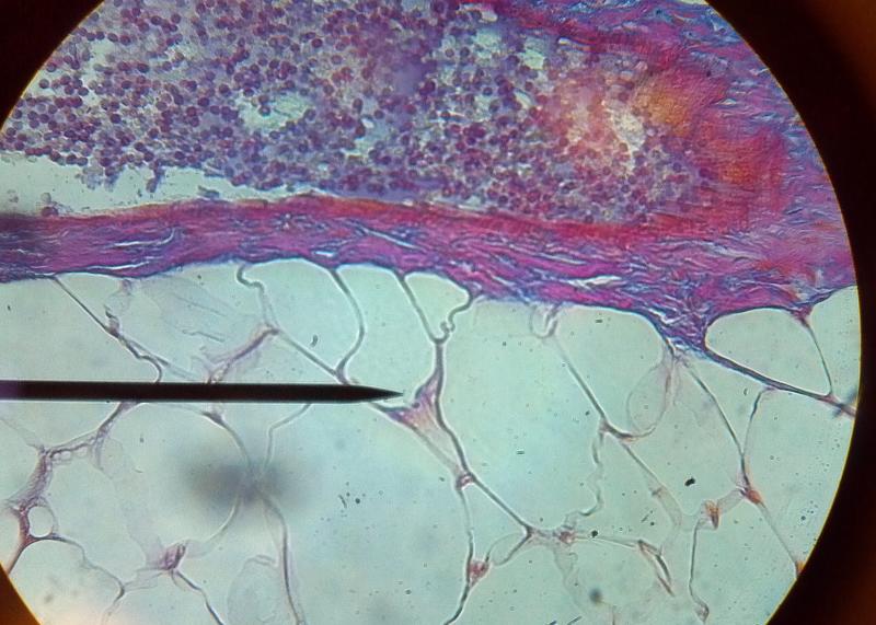 What kind of tissue is this? | back 45 Adipose tissue |
front 46 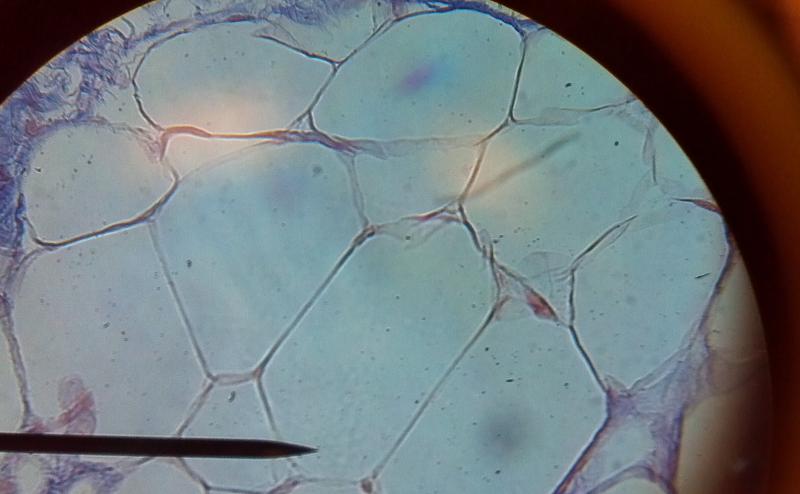 What kind of tissue is this? | back 46 Adipose Tissue |
front 47 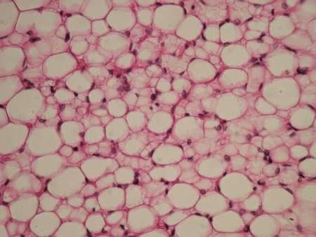 What kind of tissue is this? | back 47 Adipose Tissue |
front 48 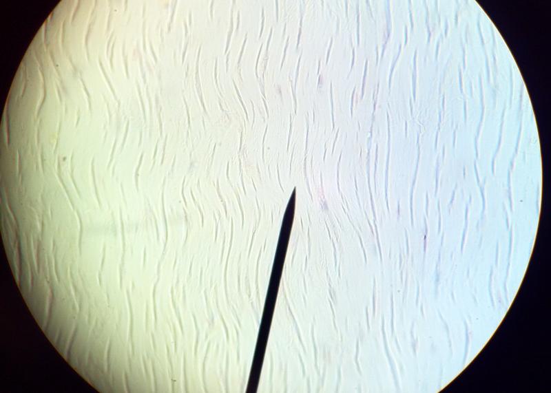 What kind of tissue is this? | back 48 Dense regular connective tissue |
front 49 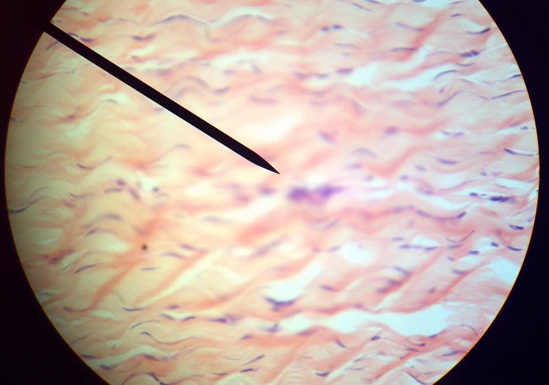 What kind of tissue is this? | back 49 Dense regular connective tissue |
front 50 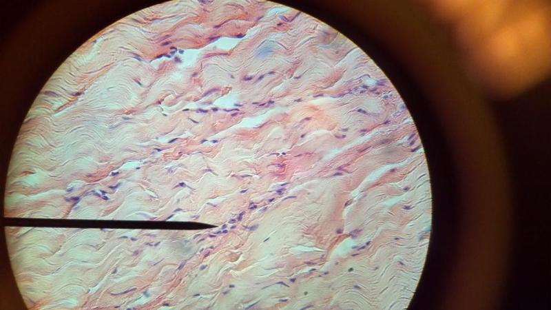 What kind of tissue is this? | back 50 Dense regular connective tissue |
front 51 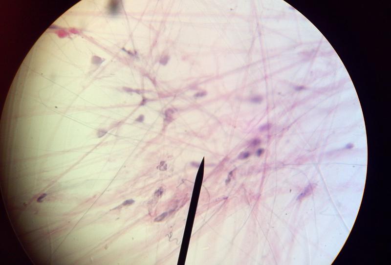 What kind of tissue is this? | back 51 Loose Areolar connective tissue |
front 52 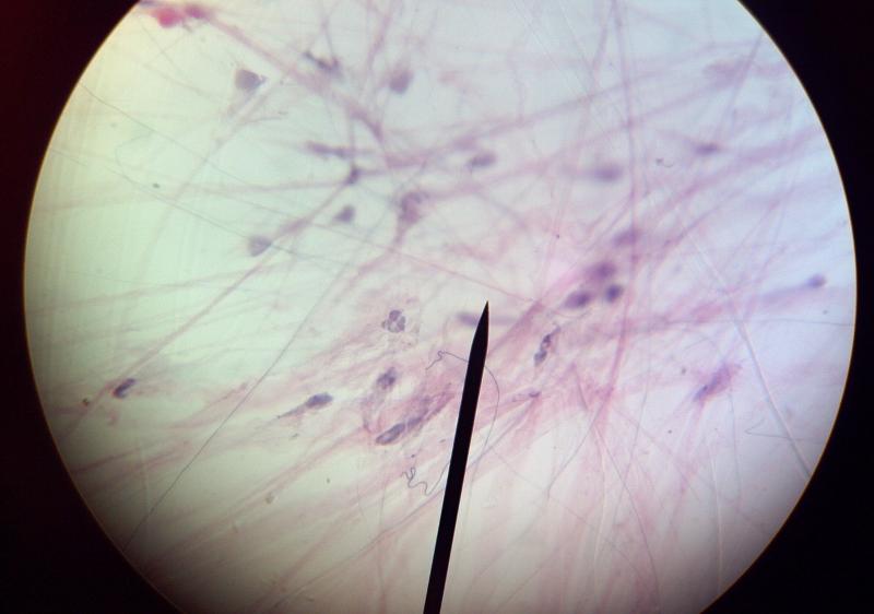 What kind of tissue is this? | back 52 Loose Areolar connective tissue |
front 53 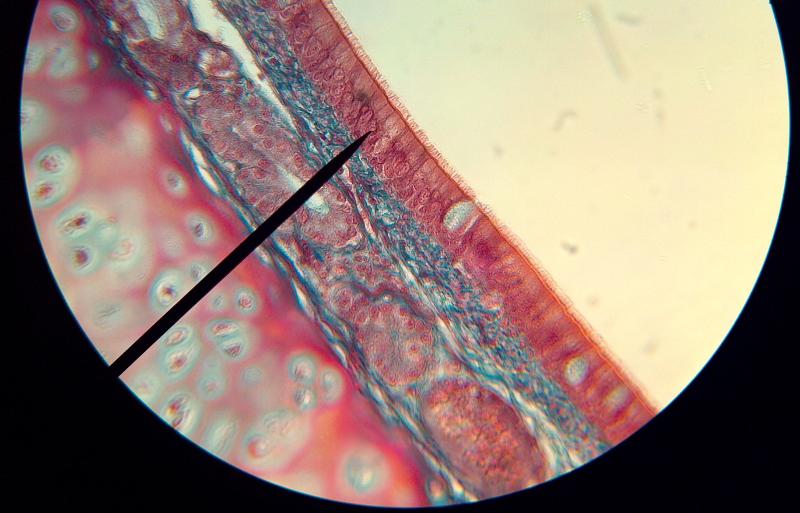 What kind of tissue is this? | back 53 Pseudostratified ciliated columnar |
front 54 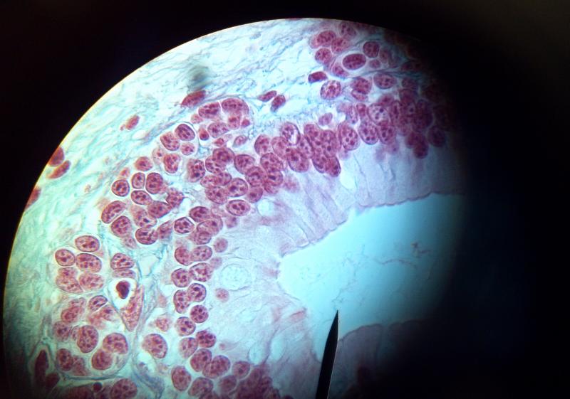 What kind of tissue is this? | back 54 Simple Columnar |
front 55 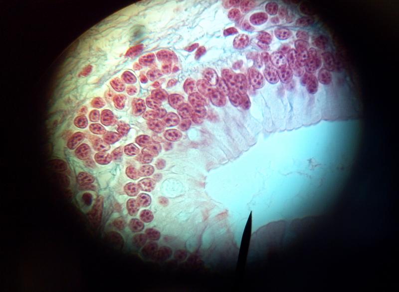 What kind of tissue is this? | back 55 Simple Columnar |
front 56 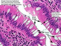 What kind of tissue is this? | back 56 Simple Columnar |
front 57  What kind of tissue is this? | back 57 Simple Columnar |
front 58 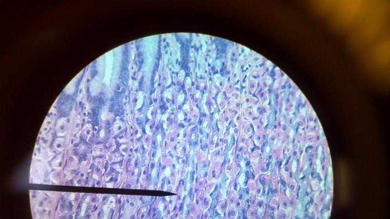 What kind of tissue is this? | back 58 Simple columnar |
front 59 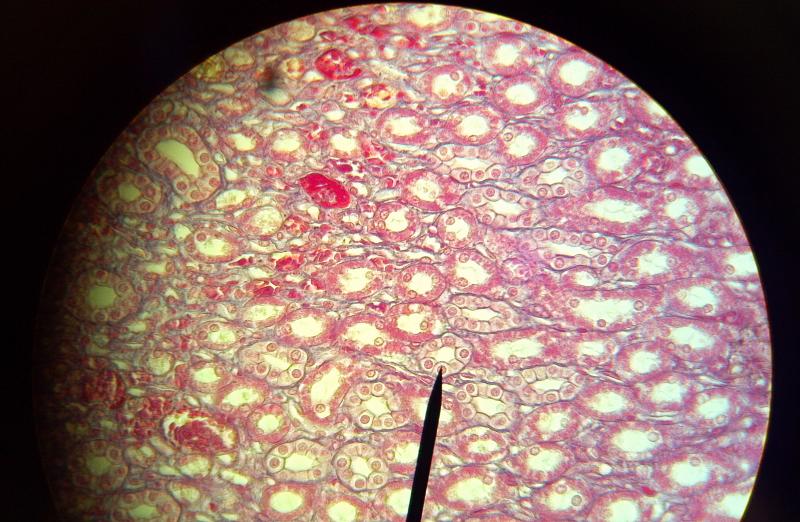 What kind of tissue is this? | back 59 Simple Cuboidal |
front 60 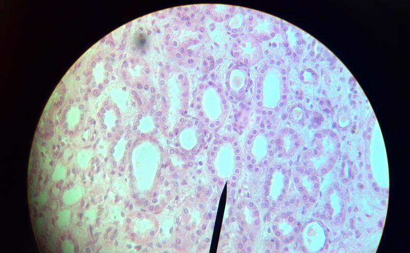 What kind of tissue is this? | back 60 Simple Cuboidal |
front 61 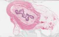 What kind of tissue is this? | back 61 Simple Squamous |
front 62 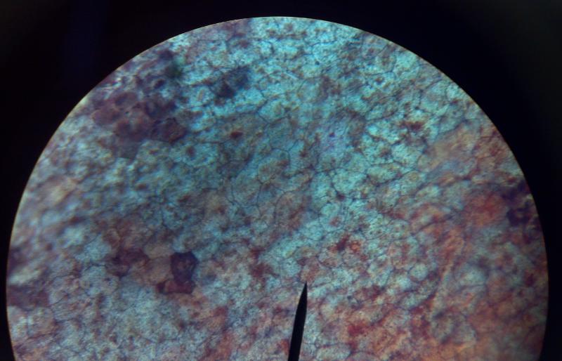 What kind of tissue is this? | back 62 Simple Squamous |
front 63 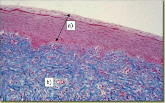 What kind of tissue is this? | back 63 Stratified Squamous |
front 64  What kind of tissue is this? | back 64 Hyaline Cartilage |
front 65 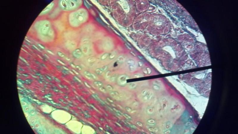 What kind of tissue is this? | back 65 Hyaline Cartilage |
front 66 Name the types of Connective Tissue? | back 66 Areolar, Adipose, hyaline, dense regular connective |
front 67 Name the one layer Epithelium tissue. | back 67 Simple Squamous, Simple Cuboidal, Simple Columnar, and Psuedostratified Ciliated Columnar |
front 68  What is A in this picture? | back 68 Epidermis |
front 69 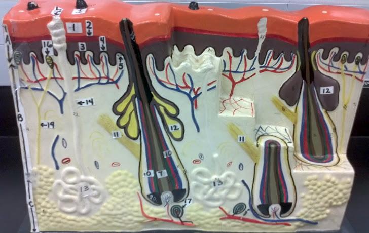 What is B in this picture? | back 69 Dermis |
front 70  What is C in this picture? | back 70 Hypodermis |
front 71  What is 1 in this picture? | back 71 Stratum Corneum |
front 72  What is 2 in this picture? | back 72 Stratum Granulosum |
front 73  What is 3 in this picture? | back 73 Stratum Spinosum |
front 74  What is 4 in this picture? | back 74 Stratum Basale |
front 75  What is 5 in this picture? | back 75 Dermal Papilla |
front 76  What is 6 in this picture? | back 76 Hair Shaft |
front 77  What is 7 in this picture? | back 77 Hair Root |
front 78  What is 8 in this picture? | back 78 Hair Bulb |
front 79  What is 9 in this picture? | back 79 Hair Papilla |
front 80 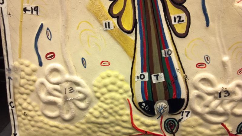 What is 10 in this picture? | back 80 Hair Follicle |
front 81  What is 11 in this picture? | back 81 Arrector Pili muscle |
front 82  What is 12 in this picture? | back 82 Sebaceous gland(sweat) |
front 83  What is 13 in this picture? | back 83 Sudoriferous gland(oil) |
front 84 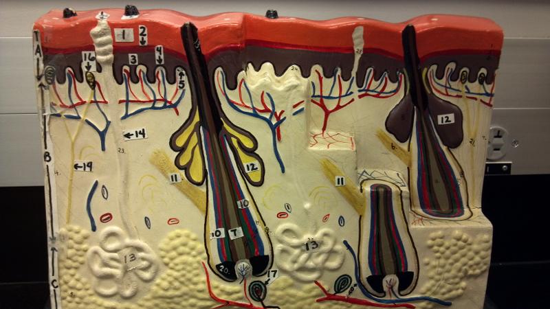 What is 14 in this picture? | back 84 Duct of the Sudoriferous gland |
front 85 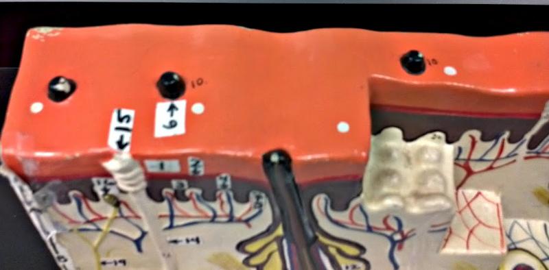 What is 15 in this picture? | back 85 Pore of the Sudoriferous |
front 86  What is 16 in this picture? | back 86 meissner's corpuscle(touch, light pressure) |
front 87  What is 17 in this picture? | back 87 pacinian corpuscle(heavy pressure) |
front 88 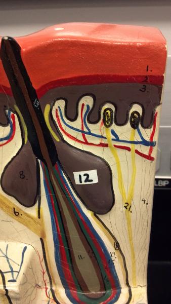 What is 18 in this picture? | back 88 Adipose Tissue |
front 89  What is 19 in this picture? | back 89 Nerve |
front 90  What is 20 in this picture? | back 90 blood vessels(red and blue) |
front 91 Is the largest and most versatile organ in the body | back 91 Skin |
front 92 What kind of tissue is skin composed of? | back 92 stratified squamous epithelium, dense connective tissue, areolar connective tissue, adipose connective tissue |
front 93 A group of organs that function for a common purpose | back 93 system |
front 94 What is the primary function of skin? | back 94 Protection |
front 95 A chemical that helps waterproof skin is called? | back 95 keratin |
front 96 What is the most superficial layer of the epidermis? | back 96 sratum corneum |
front 97 What is the deepest layer of the epidermis? | back 97 Stratum basale(germinativum) |
front 98 Where does an arrector pili muscle attach? | back 98 hair follicle |
front 99 What is the function of stratum corneum? | back 99 barrier function of the skin |
front 100 What is the function of stratum gerinativum (basale) | back 100 skin regeneration |
front 101 What is the function of keratin? | back 101 strengthens skin and helps waterproof |
front 102 What is the function of the papilla? | back 102 holds capillaries that provide nutrients to growing hair follicles |
front 103 What is the function of sudoriferous glands | back 103 body temp regulation |
front 104 What is the function of sebaceous glands? | back 104 oil secretion |
front 105 What is the function of Arrector pili? | back 105 raise hair when cold or frightened |
front 106 The dermis contains what layers? | back 106 hypodermis |
front 107 What kind of connective tissue is in the hypodermis? | back 107 areolar and adipose connective tissue |
front 108 The outermost cells of the epidermis is? | back 108 stratum corneum |
front 109 The bottom layer of epidermis that constantly makes new cells and pushes them toward the top surface is? | back 109 stratum basale(germinativum) |
front 110 The three major layers of the skin are? | back 110 epidermis, dermis and hypodermis |
front 111  What is 9 in this picture? | back 111 Hair papilla |
front 112  What is 10 of this picture? | back 112 Hair follicle |
front 113  What is 11 in this picture? | back 113 arrector pillae muscle |
front 114  What is 12 in this picture? | back 114 sebaceous gland |
front 115  What is 13 in this picture? | back 115 sudoriferous gland |
front 116 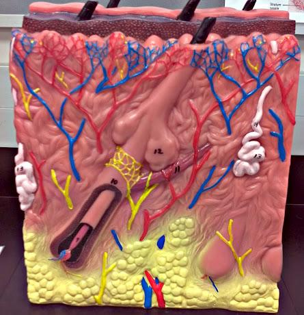 What is 14 in this picture? | back 116 duct of sudoriferous gland |
front 117  What is A in this picture? | back 117 Epidermis |
front 118  What is B in this picture? | back 118 Dermis |
front 119  What is C in this picture? | back 119 Hypodermis |
front 120  What is 1 in this picture? | back 120 Stratum corneum |
front 121  What is 2 in this picture? | back 121 Stratum granulosum |
front 122 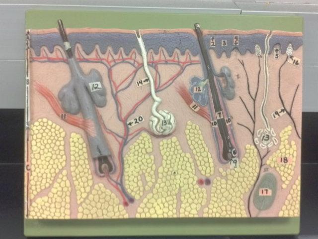 What is 3 in this picture? | back 122 Stratum spinosum |
front 123  What is 4 in this picture? | back 123 Stratum basale |
front 124  What is 5 in this picture? | back 124 Dermal Papilla |
front 125  What is 6 in this picture? | back 125 hair shaft |
front 126  What is 7 in this picture? | back 126 hair root |
front 127  What is 8 in this picture? | back 127 hair bulb |
front 128  What is 9 in this picture? | back 128 hair papilla |
front 129  What is 10 in this picture? | back 129 Hair follicle |
front 130  What is 11 in this picture? | back 130 Arrector Pilli muscle |
front 131  What is 12 in this picture? | back 131 Sebaceous gland(oil) |
front 132  What is 13 in this picture? | back 132 sudoriferous gland(sweat) |
front 133  What is 14 in this picture? | back 133 duct of sudoriferous |
front 134  What is 16 in this picture? | back 134 meissner's corpuscle |
front 135  What is 17 in this picture? | back 135 pacinian corpuscle |
front 136  What is 18 in this picture? | back 136 adipose tissue |
front 137  What is 19 in this picture? | back 137 Nerve |
front 138  What is 20 in this picture? | back 138 Blood vessel |
front 139 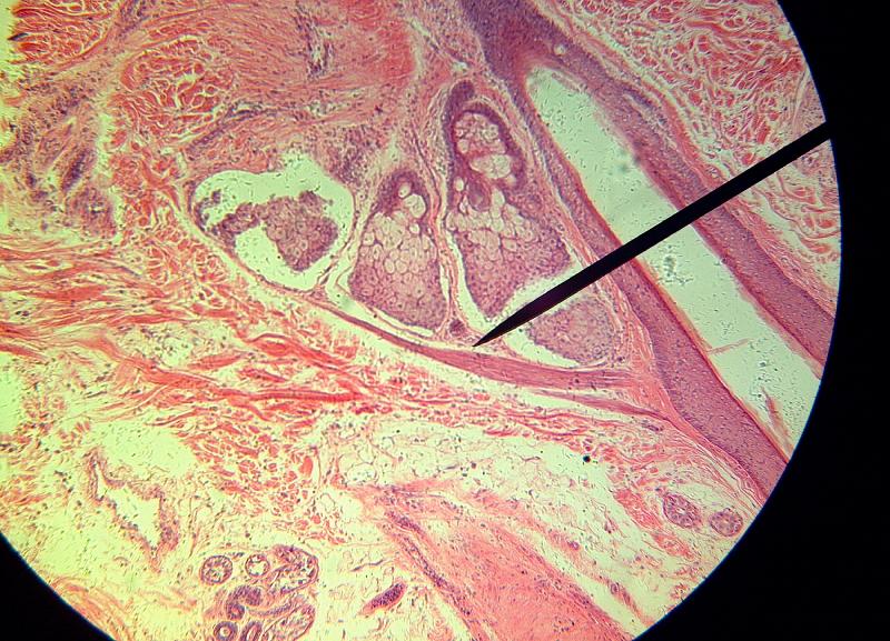 What is the pointer on in this picture? | back 139 Arrector Pillae |
front 140 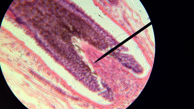 What is the pointer on in this picture? | back 140 Hair bulb/ Papilla |
front 141 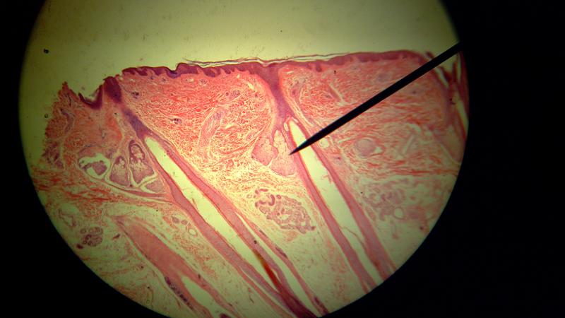 What is the pointer on in this picture? | back 141 Sebaceous gland |
front 142 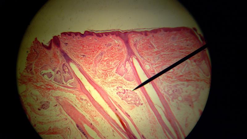 What is the pointer on in this picture? | back 142 Sudorifereous (sweat) gland |
front 143 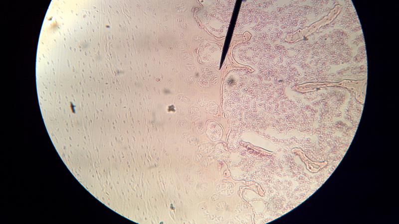 What is to the left of the pointer in this picture? | back 143 Hyaline cartilage |
front 144  What is to the right of this picture? | back 144 Spongy bone |
front 145 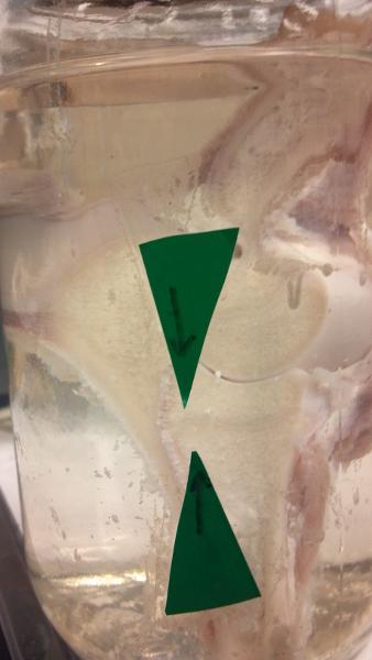 What are the arrows pointing to in this picture? | back 145 Epiphyseal plate |
front 146  What is the thick white stuff in this picture? | back 146 Articular(hyaline) cartilage |