The relationship between the height of a grid's lead strips and the distance between them is referred to as grid
A ratio
B radius
C frequency
D focusing distance
A ratio
-Grids are used in radiography to trap scattered radiation that otherwise would cause fog on the radiograph. Grid ratio is defined as the ratio of the height of the lead strips to the distance between them. Grid frequency refers to the number of lead strips per inch. Focusing distance and grid radius are terms denoting the distance range with which a focused grid may be used.
What pixel size has a 1024 × 1024 matrix with a 35-cm FOV?
A 30 mm
B 0.35 mm
C 0.15 mm
D 0.03 mm

In digital imaging, pixel size is determined by dividing the FOV by the matrix. In this case, the FOV is 35 cm; since the answer is expressed in millimeters, first change 35 cm to 350 mm. Then 350 divided by 1024 equals 0.35 mm.
The FOV and matrix size are independent of one another, that is, either can be changed and the other will remain unaffected. However, pixel size is affected by changes in either the FOV or matrix size. For example, if the matrix size is increased, pixel size decreases. If FOV increases, pixel size increases. Pixel size is inversely related to resolution. As pixel size increases, resolution decreases.

A part whose width is 6 inches will be imaged at 44 inches SID. The part to be imaged lies 9 inches from the IR. What will be the projected image width of the part?
A 8 inches
B 10 inches
C 12 inches
D 20 inches

A 8 inches
-As the object-to-image receptor distance (OID) increases, magnification of that object increases. Depending upon the information provided, we can determine the magnification factor, the percentage magnification, and image width. In the stated scenario, we are looking for image width. The formula used to determine image width is:
Substituting known factors the equation becomes:
35x = 264
x = 7.5 inches projected image width
A positive contrast agent
- absorbs x-ray photons
- results in a dark area on the radiograph
- is composed of elements having low atomic number
A 1 only
B 1 and 2 only
C 2 and 3 only
D 1, 2, and 3
A 1 only
-Radiopaque contrast agents appear white on the finished image because many x-ray photons are absorbed. These are referred to positive contrast agents—composed of dense (i.e., high atomic number) material through which x-rays will not pass easily. Radiolucent contrast agents appear black on the finished image because x-ray photons pass through easily. An example of a radiolucent contrast agent is air.
The functions of automatic beam limitation devices include
- reducing the production of scattered radiation
- increasing the absorption of scattered radiation
- changing the quality of the x-ray beam
A 1 only
B 2 only
C 1 and 2 only
D 1, 2, and 3
A 1 only
-Beam restrictors function to limit the size of the irradiated field. In so doing, they limit the volume of tissue irradiated (thereby decreasing the percentage of scattered radiation generated in the part) and help to reduce patient dose. Beam restrictors do not affect the quality (energy) of the x-ray beam—that is, the function of kilovoltage and filtration. Beam restrictors do not absorb scattered radiation—that is a function of grids
If 300 mA has been selected for a particular exposure, what exposure time should be selected to produce 18 mAs?
A 40 ms
B 60 ms
C 400 ms
D 600 ms
B 60 ms
-The exposure factor that regulates receptor exposure is milliampere-seconds (mAs). The equation used to determine mAs is mA × s = mAs. Substituting known factors:

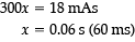
The radiograph seen below illustrates incorrect use of
A collimator
B grid
C AEC
D focal spot
B grid
-An upside-down focused grid presents its lead strips in the opposite direction to that of the x-ray beam. This results in severe grid cutoff everywhere except in the central portion of the radiographic image. Severe grid cutoff of chest anatomy can be seen outside the central exposed area. A misaligned collimator would not show such symmetrical loss of receptor exposure, nor would an incorrectly selected AEC photocell. Focal spot is unrelated to receptor exposure.
The best way to control voluntary motion is
A immobilization of the part.
B careful explanation of the procedure.
C short exposure time.
D physical restraint.
B careful explanation of the procedure.
-Patients who are able to cooperate are usually able to control voluntary motion if they are provided with an adequate explanation of the procedure. Once patients understand what is needed, most will cooperate to the best of their ability (by suspending respiration and holding still for the exposure). Certain body functions and responses, such as heart action, peristalsis, pain, and muscle spasm, cause involuntary motion that is uncontrollable by the patient. The best and only way to control involuntary motion is by always selecting the shortest possible exposure time. Involuntary motion may also be minimized by careful explanation, immobilization, and (as a last resort and only in certain cases) restraint.
If 92 kV and 12 mAs were used for a particular abdominal exposure with single-phase equipment, what mAs would be required to produce a similar radiograph with three-phase, six-pulse equipment?
A 36
B 24
C 8
D 3
C 8
-Single-phase radiographic equipment is much less efficient than three-phase equipment because it has a 100% voltage ripple. With three-phase equipment, voltage never drops to zero, and x-ray intensity is noticeably greater. To produce similar receptor exposure, only two thirds of the original mAs would be used for three-phase, six-pulse equipment (2/3 × 12 = 8 mAs). With 3-phase, 12-pulse equipment, the original mAs would be cut in half.
How is SID related to exposure rate and receptor exposure?
A As SID increases, exposure rate increases and radiographic receptor exposure increases.
B As SID increases, exposure rate increases and radiographic receptor exposure decreases.
C As SID increases, exposure rate decreases and radiographic receptor exposure increases.
D As SID increases, exposure rate decreases and radiographic receptor exposure decreases.
D As SID increases, exposure rate decreases and radiographic receptor exposure decreases.
-According to the inverse-square law of radiation, the intensity or exposure rate of radiation is inversely proportional to the square of the distance from its source. Thus, as distance from the source of radiation increases, exposure rate decreases. Because exposure rate and receptor exposure are directly proportional, if the exposure rate of a beam directed to an IR is decreased, the resulting receptor exposure would be decreased proportionately.
All the following are related to spatial resolution except
A milliamperage
B focal-spot size
C source-to-object distance
D OID
A milliamperage
-The focal-spot size selected will determine the amount of focal-spot, or geometric, blur produced in the image. OID is responsible for image magnification and hence spatial resolution. Source-to-object distance can vary with changes in SID and/or OID, and therefore impact magnification and resolution. The milliamperage is unrelated to spatial resolution; it affects the quantity of x-ray photons produced and thus receptor exposure and patient dose.
If 85 kVp, 400 mA, and ⅛ s were used for a particular exposure using single-phase equipment, which of the following milliamperage or time values would be required, all other factors being constant, to produce a similar receptor exposure using three-phase, 12-pulse equipment?
A 200 mA
B 600 mA
C 0.125 s
D 0.25 s
A 200 mA
-With three-phase equipment, the voltage never drops to zero, and x-ray intensity is significantly greater. When changing from single-phase to three-phase, six-pulse equipment, two-thirds of the original milliampere-seconds are required to produce a radiograph with similar receptor exposure. (When going from three-phase, six-pulse to single-phase, add one-third more milliampere-seconds.) When changing from single-phase to three-phase, 12-pulse equipment, only one-half of the original milliampere-seconds is required. (Going from three-phase, 12-pulse to single-phase requires twice the milliampere-seconds.) In this instance, we are changing from single-phase to three-phase, 12-pulse equipment; therefore, the new milliampere-seconds value should be half the original 50 mAs, or 25 mAs. The only selection that will provide 25 mAs is (A), 200 mA. (B) will produce 75 mAs (600 mA × ⅛ s = 75 mAs); (C) will produce 50 mAs (400 mA × 0.125 s = 50 mAs); (D) will produce 100 mAs (400 × 0.25 = 100 mAs).
Comparison of technical factors required
Single phase Three phase Three phase
X mAs 6-pulse 12-pulse
⅔ x mAs ½ x mAs
Misalignment of the tube–part–IR relationship results in
A shape distortion
B size distortion
C magnification
D blur
A shape distortion
-Shape distortion (e.g., foreshortening or elongation) is caused by improper alignment of the tube, part, and IR. Size distortion, or magnification, is caused by too great an OID or too short an SID. Focal-spot blur is caused by the use of a large focal spot.
As grid ratio is decreased,
A the scale of contrast becomes longer
B the scale of contrast becomes shorter
C receptor exposure decreases
D radiographic distortion decreases
A the scale of contrast becomes longer
-Because lead content decreases when grid ratio decreases, a smaller amount of scattered radiation is trapped before reaching the IR. More grays, therefore, are recorded, and a longer scale of contrast results. Receptor exposure would increase with a decrease in grid ratio. Grid ratio is unrelated to distortion.
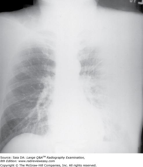
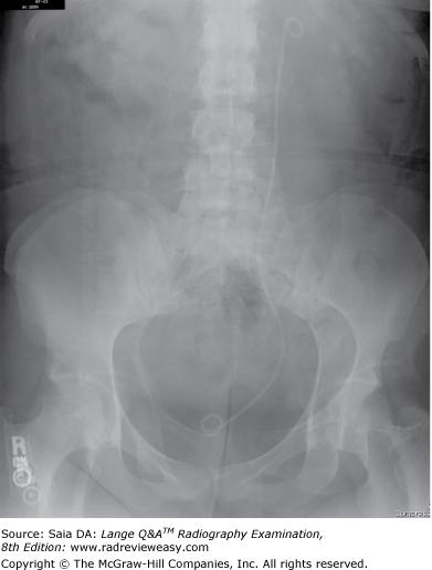
What is the correct critique of the CR image seen below?
A double exposure
B grid centering error
C incorrect AEC photocell
D inverted focused grid
B grid centering error
-This is an example of both off-focus and lateral decentering errors. Note the asymmetric cutoff from right to left. The individual grid errors, as well as the result of both errors together, is summarized below. Off-focus errors: Grid cutoff will occur if the SID is below the lower limits, or above the upper limits, of the specified focal range. This type of error is also referred to as focus–grid distance decentering. Off-focus errors are usually characterized by loss of receptor exposure at the periphery of the image. Off-center errors: If the x-ray beam is not centered to the grid (i.e., if it is shifted laterally) grid cutoff will occur. This type of error is referred to as lateral decentering and characterized by a uniform receptor exposure loss across the radiographic image.
If the x-ray beam is both off-center and off-focus below the focusing distance, the portion of the image below the focus will show increased receptor exposure; if the x-ray beam is off-center and off-focus above the focusing distance, the image below the focus will show decreased receptor exposure.
In which of the following examinations would a IR front with very low absorption properties be especially desirable?
A Extremity radiography
B Abdominal radiography
C Mammography
D Angiography
C Mammography
-Because mammographic techniques operate at very low kilovoltage levels, the IR front material becomes especially important. The use of soft, low-energy x-ray photons is the underlying principle of mammography; any attenuation of the beam would be most undesirable. Special plastics that resist impact and heat softening, such as polystyrene and polycarbonate, are used frequently as IR front material.
Both radiographic images shown in the figure below were made of the same subject using identical exposure factors. Which of the following statements correctly describe(s) these images?
- Image A demonstrates less receptor exposure because a shorter SID was used.
- Image A demonstrates more receptor exposure because the subject was turned PA.
- Image B demonstrates more receptor exposure because a shorter SID was used.
A 1 only
B 2 only
C 3 only
D 1 and 2 only
C 3 only
-In the figure, image B is darker and, therefore, has greater receptor exposure. Receptor exposure is largely determined by milliampere-seconds, SID, and exposure rate. In this case, there is a difference in SID between the two images. As SID decreases, exposure rate increases and receptor exposure increases. Image B is darker (demonstrates greater receptor exposure) than image A because image B was exposed at a shorter SID (and, therefore, a higher exposure rate).
Which of the following function(s) to reduce the amount of scattered radiation reaching the IR?
1.Grid devices
2.Restricted focal spot size
3.Beam restrictors
A 1 only
B 1 and 2 only
C 1 and 3 only
D 1, 2, and 3
C 1 and 3 only
-There are several ways to reduce the amount of scattered radiation reaching the IR. First, the use of optimum kVp is essential; excessive kVp will increase the production of scattered radiation. Second, conscientious use of the beam restrictor (collimator) will reduce scattered radiation; the smaller the volume of irradiated tissue, the less scattered radiation is produced. The use of grids helps clean up scattered radiation before it reaches the IR. The size of the tube focus has an impact on image geometry and spatial resolution, but it has no effect on scattered radiation.
Which of the following terms is used to express spatial resolution?
A Kiloelectronvolts (keV)
B Modulation transfer function (MTF)
C Relative speed
D Latitude
B Modulation transfer function (MTF)
-Resolution describes how closely fine details may be associated and still be recognized as separate details before seeming to blend into each other and appear as one. The degree of resolution transferred to the image receptor is a function of the resolving power of each of the system components and can be expressed in line pairs per millimeter (lp/mm), line-spread function (LSP), or modulation transfer function (MTF). Line pairs per millimeter can be measured using a resolution test pattern; a number of resolution test tools are available. LSP is measured using a 10-μm x-ray beam; MTF measures the amount of information lost between the object and the IR.
The reduction in x-ray photon intensity as the photon passes through material is termed
A absorption
B scattering
C attenuation
D divergence
C attenuation
-Absorption occurs when an x-ray photon interacts with matter and disappears, as in the photoelectric effect. Scattering occurs when there is partial transfer of energy to matter, as in the Compton effect. The reduction in the intensity of an x-ray beam as it passes through matter is called attenuation.
When the collimated field must extend past the edge of the body, allowing primary radiation to strike the tabletop, as in a lateral lumbar spine radiograph, what may be done to prevent excessive receptor exposure owing to undercutting?
A Reduce the milliampere-seconds.
B Reduce the kilovoltage.
C Use a shorter SID.
D Use lead rubber to absorb tabletop primary radiation
D Use lead rubber to absorb tabletop primary radiation.
-When the primary beam is restricted to an area near the periphery of the body, sometimes part of the illuminated area overhangs the edge of the body. If the exposure is then made, scattered radiation from the tabletop (where there is no absorber) will undercut the part, causing excessive receptor exposure. If, however, a lead rubber mat is placed on the overhanging illuminated area, most of this scatter will be absorbed. This is frequently helpful in lateral lumbar spine and AP shoulder radiographs.
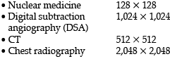
Typical examples of digital imaging include
- MRI
- CT
- CR
A 1 only
B 1 and 2 only
C 1 and 3 only
D 1, 2, and 3
D 1, 2, and 3
-CT (Computed Tomography), MRI (Magnetic Resonance Imaging), and CR (Computed Radiography) are three common examples of digital imaging. Special equipment is also available for direct digital radiography (DR)—images produced by either a fan-shaped x-ray beam received by linearly arrayed radiation detectors or a traditional fan-shaped x-ray beam received by a light-stimulated phosphor plate. Digital images can also be obtained in digital subtraction angiography (DSA), nuclear medicine, and diagnostic sonography. Analog images are conventional images; they can be converted to digital images with a device called a digitizer.
Exposure values arising from excessive kV, insufficient collimation, or thick anatomic structures are termed
A fog.
B matrix.
C artifact.
D resolution.
A fog.
-Scattered radiation produces fog, which can add unwanted exposure values to the x-ray image and impair its diagnostic value. Scattered radiation production is encouraged at high kV, insufficient beam restriction, and thick anatomic parts. Scattered radiation can be removed from the remnant beam with the use of grids.
According to the line-focus principle, an anode with a small angle provides
- improved spatial resolution.
- improved heat capacity.
- less heel effect.
A 1 and 2 only
B 1 and 3 only
C 2 and 3 only
D 1, 2, and 3
A 1 and 2 only
-The line-focus principle illustrates that as the target angle decreases, the effective focal spot decreases (providing improved spatial resolution), but the actual area of electron interaction remains much larger (allowing for greater heat capacity). It must be remembered, however, that a steep (small) target angle increases the heel effect, and part coverage may be compromised.
To produce a just perceptible increase in receptor exposure, the radiographer should increase the
A mAs by 30%
B mAs by 15%
C kV by 15%
D kV by 30%
A mAs by 30%
-If an x-ray image lacks sufficient receptor exposure, an increase in milliampere-seconds is required. The milliampere-seconds value regulates the number of x-ray photons produced at the target. An increase or decrease in milliampere-seconds of at least 30% is necessary to produce a perceptible effect. Increasing the kilovoltage by 15% will have about the same effect as doubling the milliampere-seconds.
Of the following groups of technical factors, which will produce the greatest receptor exposure?
A 10 mAs, 74 kV, 44-in. SID
B 10 mAs, 74 kV, 36-in. SID
C 5 mAs, 85 kV, 48-in. SID
D 5 mAs, 85 kV, 40-in. SID
B 10 mAs, 74 kV, 36-in. SID
-If (A) and (B) are reduced to 5 mAs for consistency, the kilovoltage will increase to 85 kV in both cases, thereby balancing receptor exposures. Thus, the greatest receptor exposure is determined by the shortest SID (greatest exposure rate).
A patient is being positioned for a particular radiographic examination. The x-ray tube, image recorder, and grid are properly aligned, but the body part is angled. Which of the following will result?
A Grid cutoff at the periphery of the image
B Grid cutoff along the center of the image
C Increased receptor exposure at the periphery
D Image distortion
D Image distortion
-Proper alignment of the x-ray tube, body part, and image recorder is required to avoid image distortion in the form of foreshortening or elongation. Foreshortening will usually result when the part is out of alignment. Elongation is often a result of angulation of the x-ray tube. Grid lines or grid cutoff will occur when the grid itself is off-center or not in alignment with the x-ray tube. Grid lines/grid cut off indicates absorption of the useful beam by the misaligned grid.
If 300 mA has been selected for a particular exposure, what exposure time would be required to produce 60 mAs?
A 1/60 second
B 1/30 second
C 1/10 second
D 1/5 second
D 1/5 second
-The mAs is the technical factor that regulates receptor exposure. The equation used to determine mAs is mA × s = mAs. Substituting the known factors,
300x = 60
x = 0.2 (1/5) second
Foreshortening can be caused by
A the radiographic object being placed at an angle to the IR
B excessive distance between the object and the IR
C insufficient distance between the focus and the IR
D excessive distance between the focus and the IR
A the radiographic object being placed at an angle to the IR
-Aligning the x-ray tube, anatomic part, and IR so that they are parallel reduces shape distortion.Angulation of the long axis of the part with respect to the IR results in foreshortening of the object. Tube angulation causes elongation of the part. Size distortion (magnification) is inversely proportional to SID and directly proportional to OID. Decreasing the SID and increasing the OID serve to increase size distortion.
An increase in the kilovoltage applied to the x-ray tube increases the
- x-ray wavelength
- exposure rate
- patient absorption
A 1 only
B 2 only
C 2 and 3 only
D 1, 2, and 3
B 2 only
-As the kilovoltage is increased, a greater number of electrons are driven across to the anode with greater force. Therefore, as energy conversion takes place at the anode, more high-energy (short-wavelength) photons are produced. However, because they are higher-energy photons, there will be less patient absorption.
If a particular grid has lead strips 0.40 mm thick, 4.0 mm high, and 0.25 mm apart, what is its grid ratio?
A 8:1
B 10:1
C 12:1
D 16:1
D 16:1
-Grid ratio is defined as the ratio between the height of the lead strips and the width of the distance between them (i.e., their height divided by the distance between them). If the height of the lead strips is 4.0 mm and the lead strips are 0.25 mm apart, the grid ratio must be 16:1 (4.0 divided by 0.25). The thickness of the lead strip is unrelated to grid ratio.
In digital imaging, as the size of the image matrix increases,
- FOV increases
- pixel size decreases
- spatial resolution increases
A 1 only
B 1 and 2 only
C 2 and 3 only
D 1, 2, and 3
C 2 and 3 only
-The FOV and matrix size are independent of one another; that is, either can be changed, and the other will remain unaffected. However, pixel size is affected by changes in either the FOV or matrix size. For example, if the matrix size is increased, pixel size decreases. If FOV increases, pixel size increases. Pixel size is inversely related to resolution. As pixel size decreases, resolution increases. FOV and matrix size are related to pixel size according to the equation Pixel size = FOV / Matrix.
The quantity of scattered radiation reaching the IR can be reduced through the use of
- a fast imaging system
- an air gap
- a stationary grid
A 1 and 2 only
B 1 and 3 only
C 2 and 3 only
D 1, 2, and 3
C 2 and 3 only
-Scattered radiation adds unwanted degrading densities to the x-ray image. The single most important way to reduce the production of scattered radiation is to collimate. Although collimation, optimalkilovoltage, and compression can be used, a large amount of scattered radiation is still generated within the part being imaged, and because it adds unwanted non–information-carrying densities, it can have a severely degrading effect on image quality. A grid is a device interposed between the patient and IR that functions to absorb a large percentage of scattered radiation before it reaches the IR. Imaging system speed is unrelated to scattered radiation. A grid is constructed of alternating strips of lead foil andradiolucent filler material. X-ray photons traveling in the same direction as the primary beam pass between the lead strips. X-ray photons, having undergone interactions within the body and deviated in various directions, are absorbed by the lead strips; this is referred to as cleanup of scattered radiation. An air gap introduced between the object and IR can have an effect similar to that of a grid. As energetic scattered radiation emerges from the body, it continues to travel in its divergent fashion and much of the time will bypass the IR. It is usually necessary to increase the SID to reduce magnification caused by increased OID.
A decrease from 90 to 77 kVp will result in an increase in
- wavelength
- gray scale
- scattered radiation
A 1 only
B 1 and 2 only
C 2 and 3 only
D 1, 2, and 3
A 1 only
-As kilovoltage is decreased, fewer electrons are driven to the anode at a slower speed and with less energy. This results in production of fewer and lower energy, longer wavelength x-ray photons. Thus, kV affects both quantity and quality of the x-ray beam. However, although kilovoltage and receptor exposure are directly related, they are not directly proportional; that is, twice the receptor exposure does not result from doubling the kilovoltage. With respect to the effect of kilovoltage on ireceptor exposure, there is a convenient rule (15% rule) that can be followed. If it is desired to double the receptor exposure yet impossible to adjust the mAs, a similar effect can be achieved by increasing the kV by 15%. Conversely, the receptor exposure may be cut in half by decreasing the kV by 15%.
Which of the following statements is (are) true regarding the images below?
- Image A was made using a higher kilovoltage than image B.
- Image A was made with a higher-ratio grid than image B.
- Image A demonstrates shorter-scale contrast than image B.
A 1 only
B 1 and 2 only
C 2 and 3 only
D 1, 2, and 3
C 2 and 3 only
-Image A was made using 80 kV at 75 mAs; image B was made using 100 kV at 18 mAs; all other exposure factors remained the same. As kilovoltage is increased, the percentage of scattered radiation relative to primary radiation increases—hence, the grayer appearance of image B. Use of optimal kilovoltage for each anatomic part is helpful in keeping scatter to a minimum. The production of scattered radiation also will be limited if the field size is as small as possible. A grid is the most effective way to remove scattered photons from those exiting the patient. Grids are designed to selectively absorb scattered radiation while absorbing as little of the useful beam as possible. Images produced with higher-ratio grids are likely to evidence the effect of less scattered radiation than those made with lower-ratio grids.
Which of the following has the greatest effect on receptor exposure?
A Aluminum filtration
B Kilovoltage
C SID
D Scattered radiation
C SID
-Receptor exposure is greatly affected by changes in the SID, as expressed by the inverse-square law of radiation. As distance from the radiation source increases, exposure rate decreases, and receptor exposure decreases. Exposure rate is inversely proportional to the square of the SID. Aluminum filtration, kilovoltage, and scattered radiation all have a significant effect on receptor exposure, but they are not the primary controlling factors.
Exposure rate will decrease with an increase in
- SID
- kilovoltage
- focal-spot size
A 1 only
B 1 and 2 only
C 2 and 3 only
D 1, 2, and 3
A 1 only
-Exposure rate decreases with an increase in SID according to the inverse-square law of radiation. The quantity of x-ray photons produced at the focal spot is the function of milliampere-seconds. The quality(i.e., wavelength, penetration, and energy) of x-ray photons produced at the target is the function of kilovoltage. The kilovoltage also has an effect on exposure rate because an increase in kilovoltage will increase the number of high-energy x-ray photons produced at the anode.
In amorphous selenium flat-panel detectors, the term amorphous refers to a
A crystalline material having typical crystalline structure.
B crystalline material lacking typical crystalline structure.
C toxic crystalline material.
D homogeneous crystalline material.
B crystalline material lacking typical crystalline structure.
-Flat-panel detectors used in DR are often made of an amorphous selenium (a-Se)–coated thin-film transistor (TFT) array. They function to convert the x-ray energy (emerging from the radiographed part) into an electrical signal. The TFT capacitors send the electrical signal to the analog-to-digital converter (ADC) to be changed to a digital signal. Amorphous selenium refers to a crystalline material (selenium) that lacks its crystalline structure. Amorphous selenium or silicon is used to produce the direct-conversion flat-panel detectors used in DR.
The primary function of filtration is to reduce
A patient skin dose.
B operator dose.
C image noise.
D scattered radiation.
A patient skin dose.
-It is our ethical responsibility to minimize radiation dose to patients. X-rays produced at the target make up a heterogeneous primary beam. There are many “soft” (low-energy) photons that, if not removed, would contribute only to greater patient dose. They are too weak to penetrate the patient and expose the IR. These soft x-rays penetrate only a small thickness of tissue before being absorbed.
Cassette-front material can be made of which of the following?
- Carbon fiber
- Magnesium
- Lead
A 1 only
B 1 and 2 only
C 1 and 3 only
D 1, 2, and 3
B 1 and 2 only
-The cassette–IR front material must not attenuate the remnant beam yet must be sturdy enough to withstand daily use. Bakelite has long been used as the material for tabletops and IR fronts, but now it has been replaced largely by magnesium and carbon fiber. Lead would not be a suitable material because it would absorb the remnant beam, and no image would be formed.
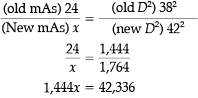
A satisfactory radiograph was made without a grid, using a 72-inch SID and 8 mAs. If the distance is changed to 40 inches and an 8:1 ratio grid is added, what should be the new mAs?
A 10 mAs
B 18 mAs
C 20 mAs
D 32 mAs

A 10 mAs
-According to the inverse square law of radiation, as the distance between the radiation source and the IR decreases, the exposure rate increases. Therefore, a decrease in technical factors is first indicated to compensate for the distance change. The following formula (exposure maintenance formula) is used to determine new mAs values, when changing distance:
Substituting known values,
To then compensate for adding an 8:1 grid, you must multiply the 2.4 mAs by a factor of 4. Thus, 9.6 mAs is required to produce a receptor exposure similar to the original radiograph. The following are the factors used for mAs conversion from nongrid to grid:
(Bushong, 8th ed., pp. 69, 252)
No grid= 1 × original mAs
5:1 grid = 2 × original mAs
6:1 grid = 3 × original mAs
8:1 grid = 4 × original mAs
12:1 grid = 5 × original mAs
16:1 grid = 6 × original mAs
The exposure factors of 400 mA, 70 ms, and 78 kV were used to produce a particular receptor exposure. A similar radiograph can be produced using 500 mA, 90 kV, and
A 14 ms
B 28 ms
C 56 ms
D 70 ms
B 28 ms
-First, evaluate the change(s): The kilovoltage was increased by 15% (78 + 15% = 90). A 15% increase in kilovoltage will double the receptor exposure; therefore, it is necessary to use half the original milliampere-seconds value to maintain the original receptor exposure. The original milliampere-seconds value was 28 mAs (400 mA × 0.07 second [70 ms] 28 mAs), so we now need 14 mAs, using 500 mA. Because mA × s mAs:
All the following affect the exposure rate of the primary beam except
A milliamperage
B kilovoltage
C distance
D field size
D field size
-Exposure rate is regulated by milliamperage. Distance significantly affects the exposure rate according to the inverse-square law of radiation. Kilovoltage also has an effect on exposure rate because an increase in kilovoltage will increase the number of high-energy photons produced at the target. The size of the x-ray field determines the volume of tissue irradiated, and hence the amount of scattered radiation generated, but is unrelated to the exposure rate.
A grid usually is employed in which of the following circumstances?
- When radiographing a large or dense body part
- When using high kilovoltage
- When a lower patient dose is required
A 1 only
B 3 only
C 1 and 2 only
D 1, 2, and 3
C 1 and 2 only
-Significant scattered radiation is generated within the part when imaging large or dense body parts and when using high kilovoltage. A radiographic grid is made of alternating lead strips and interspace material; it is placed between the patient and the IR to absorb energetic scatter emerging from the patient. Although a grid prevents much of the scattered radiation from reaching the radiograph, its use does necessitate a significant increase in patient exposure.
If a radiograph were made of an average-size knee using automatic exposure control (AEC) and all three photocells were selected, the resulting radiograph would demonstrate
A underexposed image.
B overexposed image.
C poor spatial resolution.
D adequate exposure.
A underexposed image.
-Proper functioning of the AEC depends on accurate positioning by the radiographer. The correct photocell(s) must be selected, and the anatomic part of interest must completely cover the photocell(s) to achieve the appropriate exposure. If a photocell is left uncovered, scattered radiation from the part being examined will cause premature termination of exposure and an underexposed radiograph.
Which of the following groups of exposure factors would be most appropriate for a sthenic adult IVU?
A 300 mA, 0.02 s, 72 kVp
B 300 mA, 0.01 s, 82 kVp
C 150 mA, 0.01 s, 94 kVp
D 100 mA, 0.03 s, 82 kVp
A 300 mA, 0.02 s, 72 kVp
-IVU requires the use of iodinated contrast media. Low kilovoltage (about 70 kVp) is usually used to enhance the photoelectric effect and, in turn, to better visualize the renal collecting system. High kilovoltage will produce excessive scattered radiation and obviate the effect of the contrast agent. A higher milliamperage with a short exposure time generally is preferable.
Which of the following affect(s) both the quantity and the quality of the primary beam?
- Half-value layer (HVL)
- Kilovoltage (kV)
- Milliamperage (mA)
A 1 only
B 2 only
C 1 and 2 only
D 1, 2, and 3
C 1 and 2 only
-Kilovoltage and the HVL affect both the quantity and the quality of the primary beam. The principal qualitative factor for the primary beam is kilovoltage, but an increase in kilovoltage will also create an increase in the number of photons produced at the target. HVL is defined as the amount of material necessary to decrease the intensity of the beam to one-half its original value, thereby effecting a change in both beam quality and quantity. The milliampere-seconds value is adjusted to regulate the number of x-ray photons produced at the target. X-ray-beam quality is unaffected by changes in milliampere-seconds.
Of the following groups of exposure factors, which will produce the greatest receptor exposure?
A 400 mA, 30 ms, 72-in. SID
B 200 mA, 30 ms, 36-in. SID
C 200 mA, 60 ms, 36-in. SID
D 400 mA, 60 ms , 72-in. SID
C 200 mA, 60 ms, 36-in. SID
-The formula mA × s = mAs is used to determine each milliampere-second setting (remember to first change milliseconds to seconds). The greatest receptor exposure will be produced by the combination of highest milliampere-seconds value and shortest SID. The groups in choices (B) and (D) should produce identical receptor exposure, according to the inverse-square law, because group (D) includes twice the distance and 4 times the milliampere-seconds value of group (B). The group in (A) has twice the distance of the group in (B) but only twice the milliampere-seconds; therefore, it has the least receptor exposure. The group in (C) has the same distance as the group in (B) and twice the milliampere-seconds, making group in (C) the group of technical factors that will produce the greatest receptor exposure.
Decreasing field size from 14 × 17 in. to 8 × 10 in., with no other changes, will
A decrease receptor exposure and decrease the amount of scattered radiation generated within the part
B decrease receptor exposure and increase the amount of scattered radiation generated within the part
C increase receptor exposure and increase the amount of scattered radiation generated within the part
D increase receptor exposure and decrease the amount of scattered radiation generated within the part
A decrease receptor exposure and decrease the amount of scattered radiation generated within the part
-Limiting the size of the radiographic field (irradiated area) serves to limit the amount of scattered radiation produced within the anatomic part. As the amount of scattered radiation production decreases, so does the resultant receptor exposure. Therefore, as field size decreases, scattered radiation production decreases, and overall receptor exposure decreases. Limiting the size of the radiographic field is a very effective means of reducing the quantity of non–information-carrying scattered radiation (fog) produced. Limiting the size of the radiographic field is also the most effective means of patient radiation protection.
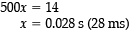
A satisfactory radiograph of the abdomen was made at a 38-in. SID using 400 mA, 60-ms exposure, and 80 kV. If the distance is changed to 42 in., what new exposure time would be required?
A 25 ms
B 50 ms
C 73 ms
D 93 ms
C 73 ms
-According to the inverse square law, as the distance between the radiation source and the IR decreases, the exposure rate increases. Therefore, a decrease in technical factors is indicated, and the exposure-maintenance formula is used to determine new milliampere-seconds values when changing distance:
Thus, x = 29.31 mAs at 42-in. SID. Then, to determine the new exposure time (mA × s = mAs),
Thus, x = 0.073 second (73 ms) at 400 mA.
With all other factors constant, as digital image matrix size increases,
1.pixel size decreases.
2.resolution increases.
3.pixel size increases.
A 1 only
B 2 only
C 1 and 2 only
D 2 and 3 only
C 1 and 2 only
-A digital image is formed by a matrix of pixels (picture elements) in rows and columns. A matrix that has 512 pixels in each row and column is a 512 × 512 matrix. The term field of view is used to describe how much of the patient (eg, 150-mm diameter) is included in the matrix. The matrix and the field of view can be changed independently, without one affecting the other, but changes in either will change pixel size. As in traditional radiography, spatial resolution is measured in line pairs per mm (lp/mm). As matrix size is increased, there are more and smaller pixels in the matrix, and therefore improved resolution. Fewer and larger pixels result in a poor resolution, "pixelly" image, that is, one in which you can actually see the individual pixel boxes.
If a radiograph exposed using a 12:1 ratio grid exhibits a loss of receptor exposure at its lateral edges, it is probably because the
A SID was too great
B grid failed to move during the exposure
C x-ray tube was angled in the direction of the lead strips
D central ray was off-center
A SID was too great
-If the SID is above or below the recommended focusing distance, the primary beam will not coincide with the angled lead strips at the lateral edges. Consequently, there will be absorption of the useful beam, termed grid cutoff. If the grid failed to move during the exposure, there would be grid lines throughout. Central ray angulation in the direction of the lead strips is appropriate and will not cause grid cutoff. If the central ray were off-center, there would be uniform loss of receptor exposure.
Which of the following statements is (are) true with respect to the radiograph shown in Figure 4–29?
- The image exhibits long-scale contrast.
- The image exhibits soft tissue fold.
- The image demonstrates motion blur.
A 1 and 2 only
B 1 and 3 only
C 2 and 3 only
D 1, 2, and 3
D 1, 2, and 3
-The abdomen radiograph shown in the figure demonstrates motion blur. This can be seen particularly in the upper abdomen and in the bowel gas patterns. Motion obliterates spatial resolution. Patients who are in pain often are unable to cooperate as fully as patients who are not in pain. Careful positioning and patient instruction are helpful, but it remains useful to use the shortest exposure time possible. The radiograph also demonstrates good long-scale contrast that enables visualization of many tissue densities. The dark horizontal line across the abdomen is a soft tissue fold accentuated by a taut elastic underwear waist-band.
Radiographic contrast is the result of
A transmitted electrons
B differential absorption
C absorbed photons
D milliampere-seconds selection
B differential absorption
-Differential absorption refers to the x-ray absorption characteristics of neighboring anatomic structures—determined by the atomic number of the tissue being examined. The radiographic representation of these various tissue density structures is referred to as radiographic contrast; it may be enhanced with high-contrast technical factors, especially using low kilovoltage levels in analog imaging. At low kilovoltage levels, the photoelectric effect predominates. If photons are absorbed, there will be no contrast. The technical factor milliampere-seconds is used to regulate receptor exposure.
The direction of electron travel in the x-ray tube is
A filament to cathode
B cathode to anode
C anode to focus
D anode to cathode
B cathode to anode
-The x-ray tube is a diode tube; that is, it has two electrodes—a negative and a positive. The cathode assembly is the negative terminal of the x-ray tube, and the anode is the positive terminal. Electrons are released by the cathode filament (thermionic emission) as it is heated to incandescence. When kilovoltage is applied, the electrons are driven across to the anode's focal spot. Upon sudden deceleration of electrons at the anode surface, x-rays are produced. Hence, electrons travel from cathode to anode within the x-ray tube.
Characteristics of high-ratio focused grids, compared with lower-ratio grids, include which of the following?
- They allow more positioning latitude.
- They are more efficient in collecting SR.
- They absorb more of the useful beam.
A 1 only
B 1 and 2 only
C 2 and 3 only
D 1, 2, and 3
C 2 and 3 only
-Two of a grid's physical characteristics that determine its degree of efficiency in the removal of scattered radiation are grid ratio (the height of the lead strips compared with the distance between them) and the number of lead strips per inch. As the lead strips are made taller or the distance between them decreases, scattered radiation is more likely to be trapped before reaching the IR. A 12:1 ratio grid will absorb more scattered radiation than an 8:1 ratio grid. An undesirable but unavoidable characteristic of grids is that they do absorb some primary/useful photons as well as scattered photons. The higher the ratio grid, the more scatter radiation the grid will clean up, but more useful photons will be absorbed as well. The higher the primary to scattered photon transmission ratio, the more desirable is the grid. Higher-ratio grids restrict positioning latitude more severely—grid centering must be more accurate (than with lower-ratio grids) to avoid grid cutoff.
Greater latitude is available to the radiographer in which of the following circumstances?
- Using high-kV technical factors
- Using a low-ratio grid
- Using low-kV technical factors
A 1 only
B 1 and 2 only
C 2 and 3 only
D 3 only
B 1 and 2 only
-In the low-kilovoltage ranges, a difference of just a few kilovolts makes a very noticeable radiographic difference, therefore offering little margin for error/latitude. High-kilovolt technical factors offer much greater margin for error; in the high-kV ranges, an error of a few kV makes little/no difference in the resulting image. Lower-ratio grids offer more tube-centering latitude than high-ratio grids.
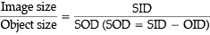
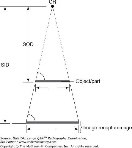
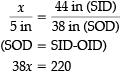
In an AP abdomen radiograph taken at 105-cm SID during an IVU series, one renal shadow measures 9 cm in width. If the OID is 18 cm, what is the actual width of the kidney?
A 5 cm
B 7.5 cm
C 11 cm
D 18 cm
B 7.5 cm
-As OID increases, magnification increases. Viscera and structures within the body will be varying distances from the IR depending on their location within the body and the position used for the exposure. The size of a particular structure or image can be calculated using the following formula:
Substituting known quantities:
Thus, x = 7.45 cm (approximate actual size). The relationship between SID, SOD, and OID is illustrated in Figure 7–23. (Bushong, 10th ed., p. 174)
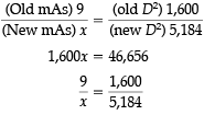
A particular radiograph was produced using 6 mAs and 110 kVp with an 8:1 ratio grid. The radiograph is to be repeated using a 16:1 ratio grid. What should be the new mAs?
A 3
B 6
C 9
D 12

C 9
-To change nongrid exposures to grid exposures, or to adjust exposure when changing from one grid ratio to another, you must remember the factor for each grid ratio:
No grid = 1 × the original mAs
5:1 grid = 2 × the original mAs
6:1 grid = 3 × the original mAs
8:1 grid = 4 × the original mAs
12:1 grid = 5 × the original mAs
16:1 grid = 6 × the original mAs
To adjust exposure factors, you simply compare the old with the new:
x = 9 mAs using 16:1 grid.
Which of the following can affect radiographic contrast?
1.LUT
2.Pathology
3.OID
A 1 only
B 1 and 2 only
C 1 and 3 only
D 1, 2, and 3
D 1, 2, and 3
-All three factors can affect radiographic contrast. The look up table (LUT) can alter the contrast. Since pathology can alter the degree of attenuation of the x-ray beam, it can affect contrast. The type of pathology will determine how contrast is affected. An additive pathology such as Paget's disease will increase contrast, while a destructive disease such as osteoporosis will decrease contrast. OID can affect contrast when it is used as an air gap. If a 6-inch air gap (OID) is introduced between the part and the IR, much of the scattered radiation emitted from the body will not reach the IR; the air gap thus acts as a grid and increases image contrast.
An exposure was made using 600 mA and 18 ms. If the mA is changed to 400, which of the following exposure times would most closely approximate the original receptor exposure?
A 16 ms
B 0.16 second
C 27 ms
D 0.27 second
C 27 ms
-Since 18 ms is equal to 0.018 s, and since mA × time = mAs, the original mAs was 10.8. Now it is only necessary to determine what exposure time must be used with 400 mA to provide the same 10.8 mAs (and thus the same receptor exposure) because mA × time = mAs,
400x = 10.8
x = 0.027 second (27 milliseconds)
Which of the following factors impact(s) spatial resolution?
- Focal spot size
- Subject motion
- SOD
A 1 only
B 1 and 2 only
C 2 and 3 only
D 1, 2, and 3
D 1, 2, and 3
-Focal-spot size affects spatial resolution by its effect on focal-spot blur: The larger the focal-spot size, the greater is the blur produced. Spatial resolution is affected significantly by distance changes because of their effect on magnification. As SID increases and as OID decreases, magnification decreases and spatial resolution increases. SOD is determined by subtracting OID from SID.
The attenuation of x-ray photons is not influenced by
- pathology
- effective atomic number
- photon quantity
A 1 only
B 3 only
C 2 and 3 only
D 1, 2, and 3
B 3 only
-Attenuation (decreased intensity through scattering or absorption) of the x-ray beam is a result of its original energy and its interactions with different types and thicknesses of tissue. The greater the original energy/quality (the higher the kilovoltage) of the incident beam, the less is the attenuation. The greater the effective atomic number of the tissues (tissue type and pathology determine absorbing properties), the greater is the beam attenuation. The greater the volume of tissue (subject density and thickness), the greater is the beam attenuation.
Which of the following are methods of limiting the production of scattered radiation?
- Using moderate ratio grids
- Using the prone position for abdominal examinations
- Restricting the field size to the smallest practical size
A 1 and 2 only
B 1 and 3 only
C 2 and 3 only
D 1, 2, and 3
C 2 and 3 only
-If a fairly large patient is turned prone, the abdominal measurement will be significantly different from the AP measurement as a result of the effect of compression. Thus, the part is essentially “thinner,” and less scattered radiation will be produced. If the patient remains supine and a compression band is applied, a similar effect will be produced. Beam restriction is probably the single most effective means of reducing the production of scattered radiation. Grid ratio affects the cleanup of scattered radiation; it has no effect on the production of scattered radiation.
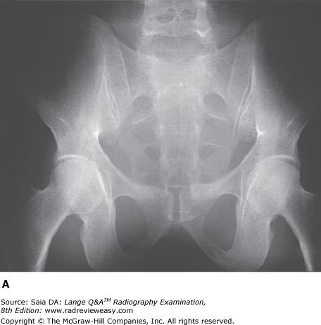
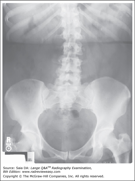
What is the correct critique of the CR image shown in Figure 4–3?
A double exposure
B inverted IP
C incomplete erasure
D image fading
A double exposure
-The image shown is a double exposure. Note the ilia and lower pelvic structures. Two pelves are clearly identifiable. Particularly noteworthy is how CR will “correct” the exposure values. The image does not appear overexposed, but the superimposed abdominal images are unmistakably evident. An inverted IP would have imaged the rear panel of the IP—a large grid-like appearance. An incomplete erasure or image fading would show only a portion of the image—here we have the entire superimposed abdomen.
Exposure factors of 90 kVp and 4 mAs are used for a particular nongrid exposure. What should be the new mAs if an 8:1 grid is added?
A 8
B 12
C 16
D 20
C 16
-To change nongrid to grid exposure or to adjust exposure when changing from one grid ratio to another, it is necessary to recall the factor for each grid ratio:
No grid = 1 × the original mAs
5:1 grid = 2 × the original mAs
6:1 grid = 3 × the original mAs
8:1 grid = 4 × the original mAs
12:1 (or 10:1) grid = 5 × the original mAs
16:1 grid = 6 × the original mAs
Therefore, to change from nongrid to an 8:1 grid, multiply the original mAs by a factor of 4. A new mAs of 16 is required.
If 84 kV and 8 mAs were used for a particular abdominal exposure with single-phase equipment, what milliampere-seconds value would be required to produce a similar radiograph with three-phase, 12-pulse equipment?
A 24 mAs
B 16 mAs
C 8 mAs
D 4 mAs
D 4 mAs
-Single-phase radiographic equipment is much less efficient than three-phase equipment because it has a 100% voltage ripple. With three-phase equipment, voltage never drops to zero, and x-ray intensity is significantly greater. To produce similar receptor exposure, only two-thirds of the original milliampere-seconds would be used for three-phase, six-pulse equipment ( 2 / 3 × 8 = 5.3 mAs). With three-phase, 12-pulse equipment, the original milliampere-seconds would be cut in half ( 1 / 2 × 8 = 4 mAs).
An increase in kilovoltage in analog imaging is most likely to
A produce a longer scale of contrast
B produce a shorter scale of contrast
C decrease the receptor exposure
D decrease the production of scattered radiation
A produce a longer scale of contrast
-An increase in kilovoltage increases the overall average energy of the x-ray photons produced at the target, thus giving them greater penetrability. (This can increase the incidence of Compton interaction and, therefore, the production of scattered radiation.) Greater penetration of all tissues serves to lengthen the scale of contrast. However, excessive scattered radiation reaching the IR will cause a fog and carries no useful information.
If exposure factors of 85 kVp, 400 mA, and 12 ms yield an output exposure of 150 mR, what is the milliroentgens per milliampere-seconds (mR/mAs)?
A 0.32
B 3.1
C 17.6
D 31
D 31
-Determining milliroentgens per milliampere-seconds output is often done to determine linearity among x-ray machines. However, all the equipment being compared must be of the same type (e.g., all single-phase or all three-phase, six-pulse). If there is linearity among these machines, then identical technique charts can be used. In the example given, 400 mA and 12 ms were used, equaling 4.8 mAs. If the output for 4.8 mAs was 150 mR, then 1 mAs is equal to 31.25 mR (150 mR ÷ 4.8 mAs = 31.25 mR/mAs).
A technique chart should be prepared for each AEC x-ray unit and should contain which of the following information for each type of examination?
- Photocell(s) used
- Optimum kilovoltage
- Backup time
A 1 only
B 1 and 2 only
C 2 and 3 only
D 1, 2, and 3
D 1, 2, and 3
-The AEC automatically adjusts the exposure required for adjacent body tissues/parts that have different thicknesses and tissue densities. Proper functioning of the AEC (phototimer or ionization chamber) depends on accurate positioning by the radiographer. The correct photocell (s) must be selected, and the anatomic part of interest must completely cover the photocell to achieve the desired receptor exposure. If collimation is inadequate and a field size larger than the part is used, excessive scattered radiation from the body or tabletop can cause the AEC to terminate the exposure prematurely, resulting in an underexposed image. Backup time always should be selected on the manual timer to prevent patient overexposure and to protect the x-ray tube from excessive heat production should the AEC malfunction. Selection of the optimal kilovoltage for the part being radiographed is essential—no practical amount of milliampere-seconds can make up for inadequate penetration (kilovoltage), and excessive kilovoltage can cause the AEC to terminate the exposure prematurely. A technique chart, therefore, is strongly recommended for use with AEC; it should indicate the optimal kilovoltage for the part, the photocells that should be selected, and the backup time that should be set.
The primary function of filtration is to reduce
A patient skin dose
B operator dose
C image noise
D scattered radiation
A patient skin dose
-It is our ethical responsibility to minimize the radiation dose to our patients. X-rays produced at the tungsten target make up a heterogeneous primary beam. There are many “soft” (low-energy) photons that, if not removed by filters, would only contribute to greater patient skin dose. They are too weak to penetrate the patient and contribute to the image-forming radiation; they penetrate a small thickness of tissue and are absorbed.
An increase in the kilovoltage applied to the x-ray tube increases the
- percentage of high-energy photons produced.
- beam intensity.
- patient absorption.
A 1 only
B 1 and 2 only
C 2 and 3 only
D 1, 2, and 3
B 1 and 2 only
-As the kilovoltage is increased, a greater number of electrons are driven across to the anode with greater force. Therefore, as energy conversion takes place at the anode, more high-energy photons are produced. However, because they are higher-energy photons, there will be less patient absorption.
An exposure was made at 40-in. SID using 5 mAs and 105 kVp with an 8:1 grid. In an effort to improve image contrast, the image is repeated using a 12:1 grid and 90 kVp. Which of the following exposure times will be most appropriate, using 400 mA, to maintain the original receptor exposure?
A 0.01 s
B 0.03 s
C 0.1 s
D 0.3 s

B 0.03 s
-The use of high kilovoltage with a fairly low-ratio grid will be ineffective in ridding the remnant beam of scattered radiation. To improve contrast in this example, it has been decided to decrease the kilovoltage by 15%, thus making it necessary to increase the milliampere-seconds from 5 mAs to 10 mAs. Because an increase in the grid ratio to 12:1 is also desired, another change in milliampere-seconds will be required (remember, 10 mAs is now the old mAs):
Thus, x = 12.5 mAs at 90 kVp. Now determine the exposure time required with 400 mA to produce 12.5 mAs:
A radiograph exposed using a 12:1 ratio grid may exhibit a loss of receptor exposure at its lateral edges because the
A SID was too great.
B grid failed to move during the exposure.
C x-ray tube was angled in the direction of the lead strips.
D CR was off-center.
A SID was too great.
-If the SID is above or below the recommended focusing distance, the primary beam at the lateral edges will not coincide with the angled lead strips. Consequently, there will be absorption of the useful beam, termed grid cutoff. If the grid failed to move during the exposure, there would be grid lines throughout. CR angulation in the direction of the lead strips is appropriate and will not cause grid cutoff. If the CR were off-center, there would be uniform loss of receptor exposure.
Phosphors classified as rare earth include
- lanthanum oxybromide.
- gadolinium oxysulfide.
- cesium iodide.
A 1 only
B 1 and 2 only
C 2 and 3 only
D 1, 2, and 3
B 1 and 2 only
-Rare earth phosphors have a greater conversion efficiency than do other phosphors. Lanthanum oxybromide is a blue-emitting phosphor, and gadolinium oxysulfide is a green-emitting phosphor. Cesium iodide is the phosphor used on the input screen of image intensifiers; it is not a rare earth phosphor.
Which of the following matrix sizes is most likely to produce the best image resolution?
A 128 × 128
B 512 × 512
C 1,024 × 1,024
D 2,048 × 2,048
D 2,048 × 2,048
-The matrix is the number of pixels in the xy direction. The larger the matrix size, the better is the image resolution. Typical image matrix sizes used in radiography are
A digital image is formed by a matrix of pixels in rows and columns. A matrix having 512 pixels in each row and column is a 512 × 512 matrix. The term field of view is used to describe how much of the patient (e.g., 150-mm diameter) is included in the matrix. The matrix or field of view can be changed without affecting the other, but changes in either will change pixel size. As in traditional radiography, spatial resolution is measured in line pairs per millimeter (lp/mm). As matrix size is increased, there are more and smaller pixels in the matrix and, therefore, improved spatial resolution. Fewer and larger pixels result in a poor-resolution “pixelly” image, that is, one in which you can actually see the individual pixel boxes.
Of the following groups of analog exposure factors, which is likely to produce the shortest scale of image contrast?
A 500 mA, 0.040 second, 70 kV
B 100 mA, 0.100 second, 80 kV
C 200 mA, 0.025 second, 92 kV
D 700 mA, 0.014 second, 80 kV
A 500 mA, 0.040 second, 70 kV
-The most important factor regulating radiographic contrast in analog imaging is kilovoltage. The lower the kilovoltage, the shorter is the scale of contrast. All the milliampere-seconds values in this problem have been adjusted for kilovoltage changes to maintain receptor exposure, but just a glance at each of the kilovoltage is often a good indicator of which will produce the longest scale or shortest scale contrast.
A lateral radiograph of the cervical spine was made at 40 in. using 300 mA and 0.03 second exposure. If it is desired to increase the distance to 72 in., what should be the new milliampere (mA) setting, all other factors remaining constant?
A 400 mA
B 800 mA
C 1000 mA
D 1200 mA
C 1000 mA
-When exposure rate decreases (as a result of increased SID), an appropriate increase in milliampere-seconds is required to maintain the original receptor exposure. The formula used to determine the new milliampere-seconds value (exposure-maintenance formula) is substituting known values:
Substituting known values:
Thus, x = 29.16 mAs at 72 in. SID. To determine the required milliamperes (mA × s = mAs),
0.03 x = 29.16
x = 972 mA
The radiographic accessory used to measure the thickness of body parts in order to determine optimal selection of exposure factors is the
A fulcrum
B caliper
C densitometer
D ruler
B caliper
-Radiographic technique charts are highly recommended for use with every x-ray unit. A technique chart identifies the standardized factors that should be used with that particular x-ray unit for various examinations/positions of anatomic parts of different sizes. To be used effectively, these technique charts require that the anatomic part in question be measured correctly with a caliper. A fulcrum is of importance in tomography; a densitometer is used in sensitometry and QA.
Geometric unsharpness is influenced by which of the following?
- Distance from object to image
- Distance from source to object
- Distance from source to image
A 1 only
B 1 and 2 only
C 1 and 3 only
D 1, 2, and 3
D 1, 2, and 3
-Geometric unsharpness is affected by all three factors listed. As OID increases, so does magnification. OID is directly related to magnification; i.e. as OID increases, so does magnification. Focal-object distance and SID are inversely related to magnification. As focal-object distance and SID decrease, magnification increases.
A 5-in. object to be radiographed at a 44-in. SID lies 6 in. from the IR. What will be the image width?
A 5.1 in.
B 5.7 in.
C 6.1 in.
D 6.7 in.
B 5.7 in
-Magnification is part of every radiographic image. Anatomic parts within the body are at various distances from the IR and, therefore, have various degrees of magnification. The formula used to determine the amount of image magnification is
Substituting known values:
The exposure factors of 400 mA, 17 ms, and 82 kV produce a milliampere-seconds value of
A 2.35
B 6.8
C 23.5
D 68
B 6.8
-To calculate milliampere-seconds, multiply milliamperage times exposure time. In this case, 400 mA × 0.017 second (17 ms) = 6.8 mAs. Careful attention to proper decimal placement will help to avoid basic math errors.
Which of the following is most likely to result from the introduction of a grid to a particular radiographic examination?
A Increased patient dose and increased scattered radiation fog
B Decreased patient dose and decreased scattered radiation fog
C Increased patient dose and decreased scattered radiation fog
D Decreased patient dose and increased scattered radiation fog
C Increased patient dose and decreased scattered radiation fog
-A grid is a device interposed between the patient and image receptor that absorbs a large percentage of scattered radiation before it reaches the image receptor. It is constructed of alternating strips of lead foil and radiolucent filler material. X-ray photons traveling in the same direction as the primary beam pass between the lead strips. X-ray photons, having undergone interactions within the body and deviated in various directions, are absorbed by the lead strips; this is referred to as "clean-up" of scattered radiation. When a grid is introduced, there is a very significant decrease in receptor exposure. To maintain a diagnostic image, the addition of a grid must be accompanied by an appropriately substantial increase in mAs, hence, increased patient dose.
A focal-spot size of 0.3 mm or smaller is essential for
A small-bone radiography
B magnification radiography
C long SID techniques
D fluoroscopy
B magnification radiography
-A fractional focal spot of 0.3 mm or smaller is essential for reproducing fine spatial resolution without focal-spot blurring in magnification radiography. As the object image is magnified, so will be any associated blur unless a fractional focal spot is used. Use of a fractional focal spot on a routine basis is unnecessary; it is not advised because it causes unnecessary wear on the x-ray tube and offers little radiographic advantage.

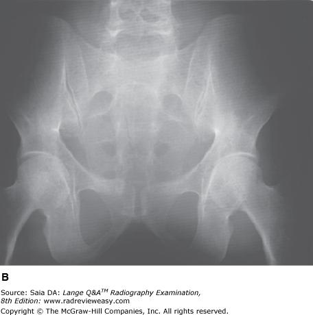
The radiograph of the pelvis shown in the figure below is unacceptable because of
A motion.
B inadequate penetration.
C scattered radiation fog.
D double exposure.
C scattered radiation fog.
-Radiographic contrast, especially in analog images, can be greatly affected by changes in kilovoltage (see figures below). As kilovoltage increases, a greater number of high-energy photons are produced at the target. These photons are more penetrating, but they also produce more scattered radiation, contributing to lower radiographic contrast as a result of scattered radiation fog. Radiograph B was made using 100 kVp and 18 mAs. Radiograph A was made of the same part using 80 kVp and 75 mAs, all other factors constant. The image details in radiograph A are far more perceptible as a result of the production of less scattered radiation
What are the effects of scattered radiation on a radiographic image?
- It produces fog.
- It increases contrast.
- It increases grid cutoff.
A 1 only
B 2 only
C 1 and 2 only
D 1, 2, and 3
A 1 only
-Scattered radiation is produced as x-ray photons travel through matter, interact with atoms, and are scattered (change direction). If these scattered rays are energetic enough to exit the body, they will strike the IR from all different angles. They, therefore, do not carry useful information and merely produce a flat, gray (low-contrast) fog over the image. Grid cutoff increases contrast and is caused by an improper relationship between the x-ray tube and the grid, resulting in absorption of some of the useful/primary beam.
Which of the following will contribute to the production of longer-scale radiographic contrast?
1.An increase in kV
2.An increase in grid ratio
3.An increase in photon energy
A 1 only
B 1 and 2 only
C 1 and 3 only
D 1, 2, and 3
C 1 and 3 only
-Increased photon energy is caused by an increase in kVp, resulting in more penetration of the part and a longer scale of contrast. Increasing the grid ratio will result in a larger percentage of scattered radiation being absorbed and hence a shorter scale of contrast.
If the x-ray image exhibits insufficient receptor exposure, this might be attributed to
- Insufficient kilovoltage
- Insufficient SID
- grid cutoff
A 1 only
B 1 and 2 only
C 1 and 3 only
D 1, 2, and 3
C 1 and 3 only
-As kilovoltage is reduced, the number of high-energy photons produced at the target is reduced; therefore, a decrease in receptor exposure occurs. If a grid has been used improperly (off-centered or out of focal range), the lead strips will absorb excessive amounts of primary radiation, resulting in grid cutoff and loss of receptor exposure. If the SID is inadequate (too short), an increase in receptor exposure will occur.
If a 4-inch collimated field is changed to a 14-inch collimated field, with no other changes, the image receptor will experience
A decreased receptor exposure.
B increased receptor exposure.
C more spatial resolution.
D less spatial resolution.
B increased receptor exposure.
-More scattered radiation is generated within a part as the kilovoltage is increased, as the size of the field is increased, and as the thickness and density of tissue increases. As the quantity of scattered radiation increases from any of these sources, receptor exposure increases. Beam restriction does not impact spatial resolution.
Which of the following is likely to contribute to the radiographic contrast present on an analog x-ray image?
- Atomic number of tissues radiographed
- Any pathologic processes
- Degree of muscle development
A 1 and 2 only
B 1 and 3 only
C 2 and 3 only
D 1, 2, and 3
D 1, 2, and 3
-The radiographic subject, the patient, is composed of many different tissue types that have varying tissue densities, resulting in varying degrees of photon attenuation and absorption. The atomic number of the tissues under investigation is directly related to their attenuation coefficient. This differential absorption contributes to the various shades of gray (scale of radiographic contrast) on the radiographic image. Normal tissue density may be altered significantly in the presence of pathologic processes. For example, destructive bone disease can cause a dramatic decrease in tissue density (and subsequent increase in receptor exposure). Abnormal accumulation of fluid (as in ascites) will cause a significant increase in tissue density. Muscle atrophy or highly developed muscles similarly will decrease or increase tissue density.
If 92 kV and 12 mAs were used for a particular abdominal exposure with single-phase equipment, what mAs would be required to produce a similar radiograph with three-phase, six-pulse equipment?
A 36
B 24
C 8
D 6
C 8
-Single-phase radiographic equipment is much less efficient than three-phase equipment because it has a 100% voltage ripple. With three-phase equipment, voltage never drops to zero, and x-ray intensity is significantly greater. To produce similar receptor exposure, only two thirds of the original mAs would be used for three-phase, six-pulse equipment (2/3 × 12 = 8 mAs). With 3-phase, 12-pulse equipment, the original mAs would be cut in half.
The exposure factors of 300 mA, 0.017 second, and 72 kVp produce an mAs value of
A 5.
B 50.
C 500.
D 5000.
A 5.
-To calculate mAs, multiply milliamperage times exposure time. In this case, 300 mA × 0.017 s = 5.10 mAs. Careful attention to proper decimal placement will help avoid basic math errors.
Which of the following statements is (are) most likely true regarding the figure below?
1.Image A was made using a higher kVp than image B.
2.Image A was made with a higher ratio grid than image B.
3.Image A demonstrates shorter scale contrast than image B.
A 1 only
B 1 and 2 only
C 2 and 3 only
D 1, 2, and 3

C 2 and 3 only
-Image A was made using 80 kVp at 75 mAs; Image B was made using 100 kVp at 18 mAs; all other exposure factors remained the same. As kVp is increased, the percentage of scattered radiation relative to primary radiation increases, hence the grayer appearance of image B. Use of optimal kilovoltage for each anatomic part is helpful in keeping scatter to a minimum. The production of scattered radiation will also be limited if the field size is as small as possible. A grid is the most effective way to remove scattered photons from those exiting the patient. Grids are designed to selectively absorb scattered radiation while absorbing as little of the primary radiation as possible. Images produced with higher ratio grids will possess fewer grays than those made with lower ratio grids.
Which of the following combinations is most likely to be associated with quantum mottle?
A Decreased milliampere-seconds, decreased SID
B Increased milliampere-seconds, decreased kilovoltage
C Decreased milliampere-seconds, increased kilovoltage
D Increased milliampere-seconds, increased SID
C Decreased milliampere-seconds, increased kilovoltage
-Quantum mottle is a grainy appearance on a finished image that is seen especially in fast-imaging systems. It is similar to the "pixelated" appearance of an enlarged digital image; it has a spotted or freckled appearance. Fast imaging systems using low-milliampere-seconds and high-kilovoltage factors are most likely to be the cause of quantum mottle.
Which of the following combinations will result in the most scattered radiation reaching the image receptor?
A Using more mAs and compressing the part
B Using more mAs and a higher ratio grid
C Using less mAs and more kVp
D Using less mAs and compressing the part
C Using less mAs and more kVp
-As x-ray photons travel through a part, they either pass all the way through to expose the image receptor, or they undergo interaction(s) that may result in their being absorbed by the part or deviated in direction. It is those that change direction (scattered radiation) that undermine the image. With respect to the radiographic image, it is responsible for the scattered radiation that reaches the image receptor. Scattered radiation adds unwanted, degrading exposure to the radiographic image.
The single most important way to reduce the production of scattered radiation is to collimate. Although collimation, use of lower kVp (with appropriately higher mAs), and compression can be used, a large amount of scattered radiation can still be generated within the part being radiographed. Because scattered radiation adds unwanted non information-carrying photons, it can have a degrading effect on image quality...thus the need for grids.
Which of the following conditions will require an increase in x-ray photon energy/penetration?
A Fibrosarcoma
B Osteomalacia
C Paralytic ileus
D Ascites
D Ascites
-The ability of x-ray photons to penetrate a body part has a great deal to do with the composition of that part (e.g., bone vs. soft tissue vs. air) and the presence of any pathologic condition. Pathologic conditions can alter the normal nature of the anatomic part. Some conditions, such as osteomalacia, fibrosarcoma, and paralytic ileus (obstruction), result in a decrease in body tissue density. When body tissue density decreases, x-rays will penetrate the tissues more readily; that is, there is more x-ray penetrability. In conditions such as ascites, where body tissue density increases as a result of the accumulation of fluid, x-rays will not readily penetrate the body tissues; that is, there is less x-ray penetrability.
A lateral (analog) radiograph of the lumbar spine was made using 200 mA, 1/2 second exposure, and 90 kV. If the exposure factors were changed to 200 mA, 0.25 second, and 104 kV, there would be an obvious change in which of the following?
- Receptor exposure
- Scale of grays/contrast
- Distortion
A 1 only
B 2 only
C 2 and 3 only
D 1, 2, and 3
B 2 only
-The original milliampere-seconds value (regulating receptor exposure) was 100. The original kilovoltage (impacting contrast) was 90. The milliampere-seconds value was cut in half, to 50. The kilovoltage was increased (by 15%) to compensate for the receptor exposure loss and thereby increase the scale of grays.
A graphic diagram of signal values representing various absorption properties within the part being imaged is called
a A processing algorithm
B DICOM
C histogram
D window
C histogram
-A histogram is a graph usually having several peaks and valleys representing the pixel values/absorbing properties of the various tissues, and so on that make up the imaged part. These various attenuators include such things as bone, muscle, air, contrast agents, foreign bodies, and pathology. The various pixel values, then, represent image contrast. If the histogram has a rather flat “tail,” this represents underexposed areas at the periphery of the image, which can skew the overall histogram analysis. The radiographer selects the particular processing algorithm on the computer/control panel that corresponds to the anatomic part and projection being performed. DICOM (Digital Imaging and Communications in Medicine) refers to the standard for communication between PACS and HIS/RIS systems. Windowing refers to the radiographer's post processing adjustment of contrast and brightness (at the workstation).
Which of the following is most likely to occur as a result of using a 30-in. SID with a 14 × 17 in. IR to radiograph a fairly homogeneous structure?
A Production of quantum mottle
B Receptor exposure variation between opposite ends of the IR
C Production of scatter radiation fog
D Excessively short-scale contrast
B Receptor exposure variation between opposite ends of the IR
-Since x-ray photons are produced at the tungsten target, they more readily diverge toward the cathode end of the x-ray tube. As they try to diverge toward the anode, they interact with and are absorbed by the anode “heel.” Consequently, there is a greater intensity (quantity) of x-ray photons at the cathode end of the x-ray beam. This phenomenon is known as the anode heel effect. Because shorter SIDs and larger IR sizes require greater divergence of the x-ray beam to provide coverage, the anode heel effect will be accentuated.
Which of the following terms is used to describe unsharp edges of tiny radiographic details?
A Diffusion
B Mottle
C Blur
D Umbra
C Blur
-Spatial resolution is evaluated by how sharply tiny anatomic details are imaged on the x-ray image. The area of blurriness that may be associated with tiny image details is termed geometric blur. The blurriness can be produced by using a large focal spot, increased OID, or decreased SID. The image proper (i.e., without blur) is often termed the umbra. Mottle is a grainy appearance associated with insufficient receptor exposure.
Which of the following can contribute to the image contrast?
- Tissue density
- Pathology
- Muscle development
A 1 and 2 only
B 1 and 3 only
C 2 and 3 only
D 1, 2, and 3
D 1, 2, and 3
-The radiographic subject (the patient) is composed of many different tissue types of varying tissue densities, resulting in varying degrees of photon attenuation and absorption. This differential absorption contributes to the various shades of gray. Normal tissue density may be significantly altered in the presence of pathology. For example, destructive bone disease can cause a dramatic decrease in tissue density. Abnormal accumulation of fluid (as in ascites) will cause a significant increase in tissue density. Muscle atrophy or highly developed muscles similarly will decrease or increase tissue density.
A radiograph made using 300 mA, 0.1 second, and 75 kVp exhibits motion unsharpness, but otherwise satisfactory technical quality. The radiograph will be repeated using a shorter exposure time. Using 86 kV and 500 mA, what should be the new exposure time?
A 0.12 second
B 0.06 second
C 0.03 second
D 0.01 second
C 0.03 second
-The mAs formula is milliamperage × time = mAs. With two of the factors known, the third can be determined. To find the mAs that was originally used, substitute the known values:
300 × 0.1 = 30
We have increased the kilovoltage to 86, an increase of 15%, which has an effect similar to that of doubling the mAs. Therefore, only 15 mAs is now required as a result of the kV increase:
mA × s = mAs
500 x = 15
x = 0.03-second exposure
How is source-to-image distance (SID) related to exposure rate and receptor exposure?
A As SID increases, exposure rate increases and receptor exposure increases.
B As SID increases, exposure rate increases and receptor exposure decreases.
C As SID increases, exposure rate decreases and receptor exposure increases.
D As SID increases, exposure rate decreases and receptor exposure decreases.
D As SID increases, exposure rate decreases and receptor exposure decreases.
-According to the inverse-square law of radiation, the intensity or exposure rate of radiation from its source is inversely proportional to the square of the distance. Thus, as distance from the source of radiation increases, exposure rate decreases. Because exposure rate and receptor exposure are directly proportional, if the exposure rate of a beam directed to the IR is decreased, the resulting receptor exposure would be decreased proportionally.
Changes in milliampere-seconds can affect all the following except
A quantity of x-ray photons produced
B exposure rate
C receptor exposure
D spatial resolution
D spatial resolution
-Milliampere-seconds (mAs) are the product of milliamperes (mA) and exposure time (seconds). Any combinations of milliamperes and time that will produce a given milliampere-seconds value will produce identical receptor exposure. This is known as the reciprocity law. The milliampere-seconds value is a quantitative factor. The mAs is directly proportional to x-ray-beam intensity, exposure rate,quantity, or number of x-ray photons produced and patient dose. If the mAs value is doubled, twice the receptor exposure and twice the patients dose. If the milliampere-seconds value is cut in half, the receptor exposure and patient dose are cut in half. The milliampere-seconds value has no effect on spatial resolution.
How are mAs and receptor exposure related in the process of image formation?
A mAs and receptor exposure are inversely proportional
B mAs and receptor exposure are directly proportional
C mAs and receptor exposure are related to image unsharpness
D mAs and receptor exposure are unrelated
B mAs and receptor exposure are directly proportional
-The milliampere-seconds value regulates the number of x-ray photons produced at the target and thus regulates receptor exposure. If it is desired to double the receptor exposure, one simply doubles the milliampere-seconds; therefore, milliampere-seconds and receptor exposure are directly proportional.
Which of the following is (are) associated with subject contrast?
- Patient thickness
- Tissue density
- Kilovoltage
A 1 only
B 1 and 2 only
C 1 and 3 only
D 1, 2, and 3
D 1, 2, and 3
-Several factors influence subject contrast, each as a result of beam-attenuation differences in the irradiated tissues. As patient thickness and tissue density increase, attenuation increases, and subject contrast is increased. As kilovoltage increases, higher-energy photons are produced, beam attenuation is decreased, and subject contrast decreases.
Which of the following absorbers has the highest attenuation coefficient?
A Bone
B Muscle
C Fat
D Air
A Bone
-The radiographic subject, the patient, is composed of many different tissue types that have varying tissue densities, resulting in varying degrees of photon attenuation and absorption. The atomic number (Z) of the tissues under investigation is directly related to its attenuation coefficient. This differential absorption contributes to the various shades of gray (scale of radiographic contrast) on the finished x-ray image. Air has an effective Z number of 7.78, fat is about 6.46, water is 7.51, muscle is 7.64, and boneis 12.31.
Using a short (25–30 in.) SID with a large (14 × 17 in.) IR is likely to
A increase the scale of contrast
B increase the anode heel effect
C cause malfunction of the AEC
D cause premature termination of the exposure
B increase the anode heel effect
-Use of a short SID with a large-size IR (and also with anode angles of 10 degrees or less) causes the anode heel effect to be much more apparent. The x-ray beam needs to diverge more to cover a large-size IR, and it needs to diverge even more for coverage as the SID decreases. The x-ray beam has no problem diverging toward the cathode end of the beam, but as it tries to diverge toward the anode end of the beam, it is eventually stopped by the anode (x-ray photons are absorbed by the anode). This causes a decrease in beam intensity at the anode end of the beam and is characteristic of the anode heel effect.
Geometric unsharpness will be least obvious
- at long SIDs.
- with small focal spots.
- at the anode end of the image.
A 1 only
B 1 and 2 only
C 2 and 3 only
D 1, 2, and 3
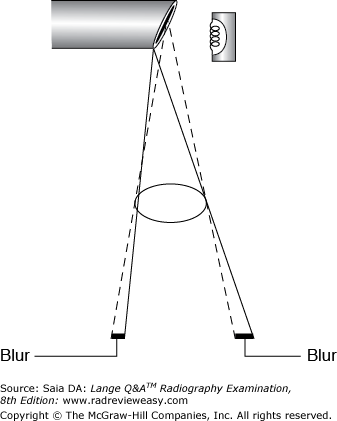
D 1, 2, and 3
-The x-ray tube anode is designed according to the line-focus principle, that is, with the focal track beveled (Figure 6–24). This allows a larger actual focal spot to project a smaller effective focal spot, resulting in improved spatial resolution with less blur. However, because of the target angle, penumbral blur varies along the longitudinal tube axis, being greater at the cathode end of the image and less at the anode end of the image. Therefore, better spatial resolution will be appreciated using small focal spots at the anode end of the x-ray beam and at longer SIDs.
Which of the following pathologic conditions would require an increase in exposure factors?
A Pneumoperitoneum
B Obstructed bowel
C Renal colic
D Ascites
D Ascites
-Because pneumoperitoneum is an abnormal accumulation of air or gas in the peritoneal cavity, it would require a decrease in exposure factors. Obstructed bowel usually involves distended, air- or gas-filled bowel loops, again requiring a decrease in exposure factors. With ascites, there is an abnormal accumulation of fluid in the abdominal cavity, necessitating an increase in exposure factors. Renal colic is the pain associated with the passage of renal calculi; no change from the normal exposure factors is usually required
A satisfactory radiograph was made using a 40-inch SID, 10 mAs, and a 12:1 grid. If the examination will be repeated at a distance of 48 inches and using an 8:1 grid, what should be the new mAs to maintain the original receptor exposure?
A 5.6
B 8.8
C 11.5
D 14.4

C 11.5
-According to the exposure maintenance formula, if the SID is changed to 48 inches, 14.4 mAs is required to maintain the original receptor exposure.
Then, to compensate for changing from a 12:1 grid to an 8:1 grid, the mAs becomes 11.5:
Thus, 11.5 mAs is required to produce a receptor exposure similar to that of the original image.
The following are the factors used for mAs conversion from nongrid to grid:
No grid = 1 × the original mAs
5:1 grid = 2 × the original mAs
6:1 grid = 3 × the original mAs
8:1 grid = 4 × the original mAs
12:1 grid = 5 × the original mAs
16:1 grid = 6 × the original mAs
Combinations of milliamperage and exposure time that produce a particular milliampere-seconds value will produce identical receptor exposure. This statement is an expression of the
A inverse-square law
B line-focus principle
C reciprocity law
D D log E curve
C reciprocity law
-A number of milliamperage and exposure time settings can produce the same milliampere-seconds value. Each of the following milliamperage and time combinations produces 10 mAs: 100 mA and 0.1 s, 200 mA and 0.05 s, 300 mA and, and 400 mA and 0.025 s. These milliamperage and exposure-time combinations should produce identical receptor exposures. This is known as the reciprocity law. The radiographer can make good use of the reciprocity law when manipulating exposure factors to decrease exposure time and decrease motion unsharpness.
Better resolution is obtained with
A high SNR.
B low SNR.
C windowing.
D smaller matrix.
A high SNR.
-Spatial resolution increases as SNR (signal-to-noise ratio) increases. A high SNR (e.g., 1000:1) indicates that there is far more signal than noise. A lower SNR (e.g., 200:1) indicates a "noisy" image. Windowing is unrelated to resolution; it permits post-processing image manipulation. Image matrix has a great deal to do with resolution. A larger image matrix (1800 × 1800) offers better resolution than a smaller image matrix (700 × 700). Smaller image matrices look "pixelly."
The advantage(s) of high-kilovoltage chest radiography is (are) that
- exposure latitude is increased
- it produces long-scale contrast
- it reduces patient dose
A 1 only
B 1 and 2 only
C 2 and 3 only
D 1, 2, and 3
D 1, 2, and 3
-The chest is composed of tissues with widely differing densities (bone and air). In an effort to “even out” these tissue densities and better visualize pulmonary vascular markings, high kilovoltage generally is used. This produces more uniform penetration and results in a longer scale of contrast with visualization of the pulmonary vascular markings as well as bone (which is better penetrated) and air densities. The increased kilovoltage also affords the advantage of greater exposure latitude (an error of a few kilovolts will make little, if any, difference). The fact that the kilovoltage is increased means that the milliampere-seconds value is reduced accordingly, and thus patient dose is reduced as well. A grid usually is used whenever high kilovoltage is required.
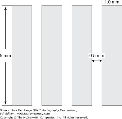
What grid ratio is represented in Figure 4–8?
A 3:1
B 5:1
C 10:1
D 16:1
C 10:1
-Grid ratio is defined as the height of the lead strips compared with (divided by) the width of the interspace material. The width of the lead strips has no bearing on the grid ratio. The height of the lead strips is 5 mm; the width of the interspace material (same as the distance between the lead strips) is 0.5 mm. Therefore, the grid ratio is 5/0.5, or a 10:1 grid ratio.
Which of the following adult radiographic examinations usually require(s) use of a grid?
- Ribs
- Vertebrae
- Shoulder
A 1 only
B 1 and 2 only
C 1 and 3 only
D 1, 2, and 3
D 1, 2, and 3
-Generally speaking, anatomic parts measuring in excess of 10 cm require a grid. (The major exception to this rule can be the chest). The larger the part, the more scattered radiation is generated. To avoid degradation of the image as a result of scattered radiation fog, a grid is used to absorb scatter. Parts generally requiring the use of a grid include the skull, spine, ribs, pelvis, shoulder, and femur.
Which of the following is (are) classified as rare earth phosphors?
- Lanthanum oxybromide
- Gadolinium oxysulfide
- Cesium iodide
A 1 only
B 1 and 2 only
C 2 and 3 only
D 1, 2, and 3
B 1 and 2 only
-Rare earth phosphors have a greater conversion efficiency than do other phosphors. Lanthanum oxybromide is a blue-emitting rare earth phosphor, and gadolinium oxysulfide is a green-emitting rare earth phosphor. Cesium iodide is the phosphor used on the input screen of image intensifiers; it is not a rare earth phosphor.
If 300 mA has been selected for a particular exposure, what exposure time would be required to produce 6 mAs?
A 5 ms
B 10 ms
C 15 ms
D 20 ms
D 20 ms
-Milliampere-seconds (mAs) is the exposure factor that regulates receptor exposure. The equation used to determine mAs is mA × s = mAs. Substituting the known factors:
In which of the following examinations should 70 kV not be exceeded?
A Upper GI (UGI)
B Barium enema (BE)
C Intravenous urogram (IVU)
D Chest
C Intravenous urogram (IVU)
-The iodine-based contrast material used in IVU gives optimal opacification at 60 to 70 kV. Use of higher kilovoltage will negate the effect of the contrast medium; a lower contrast will be produced, and poor visualization of the renal collecting system will result. GI and BE examinations employ high-kilovoltage exposure factors (about 120 kV) to penetrate through the barium. In chest radiography, high-kilovoltage technical factors are preferred for maximum visualization of pulmonary vascular markings made visible with long-scale contrast.
Using a 48-in. SID, how much OID must be introduced to magnify an object two times?
A 8-in. OID
B 12-in. OID
C 16-in. OID
D 24-in. OID
D 24-in. OID
-Magnification radiography may be used to delineate a suspected hairline fracture or to enlarge tiny, contrast-filled blood vessels. It also has application in mammography. To magnify an object to twice its actual size, the part must be placed midway between the focal spot and the IR.
What is the best way to reduce magnification distortion?
A Use a small focal spot.
B Increase the SID.
C Decrease the OID.
D Avoid tube angle techniques.
C Decrease the OID
-There are two types of distortion: size and shape. Shape distortion relates to the alignment of the x-ray tube, the part to be radiographed, and the image recorder. There are two kinds of shape distortion:elongation and foreshortening. Size distortion is magnification, and it is related to the OID and the SID. Magnification can be reduced by either increasing the SID or decreasing the OID. However, an increase in SID must be accompanied by an increase in mAs to maintain receptor exposure. It is therefore preferable, in the interest of exposure, to reduce OID whenever possible.
If 0.05 second was selected for a particular exposure, what mA would be necessary to produce 15 mAs?
A 900
B 600
C 500
D 300
D 300
-The formula for mAs is mA × s = mAs. Substituting known values,
0.05x = 15
x = 300 mA
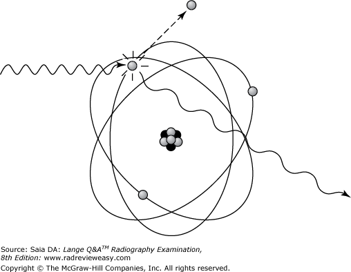
Which of the following is most closely related to the figure below?
A low kV
B high kV
C low mA
D high mA
B high kV
-n Compton scatter, a high-energy (high kilovoltage) x-ray photon ejects an outer-shell electron in tissue or other absorber. The ejected electron is called a recoil electron. Although the x-ray photon is deflected with somewhat reduced energy (modified scatter), it retains most of its original energy and exits the body as an energetic scattered photon. Because the scattered photon exits the body, it does not pose a radiation hazard to the patient. It can, however, contribute to image fog and pose a radiation hazard to personnel (as in fluoroscopic procedures). In the photoelectric effect, a relatively low-energy (low kilovoltage) x-ray photon uses all its energy (true/total absorption) to eject an inner-shell electron, leaving an orbital vacancy. An electron from the shell above drops down to fill the vacancy and in so doing gives up energy in the form of a characteristic ray. The photoelectric effect is more likely to occur in absorbers having high atomic number (e.g., bone or positive contrast media) and contributes significantly to patient dose because all the photon energy is absorbed by the patient (and, therefore, is responsible for the production of short-scale contrast). Brems and characteristic x-rays are produced at the focal spot as high-speed electrons are rapidly decelerated.
The exposure factors of 300 mA, 0.07 second, and 95 kVp were used to deliver a particular analog receptor exposure and contrast. A similar analog x-ray image can be produced using 500 mA, 80 kVp, and
A 0.01 second.
B 0.04 second.
C 0.08 second.
D 0.16 second.
C 0.08 second.
-First, evaluate the change(s): The kVp was decreased by about 15% [95–15% = 80.7]. A 15% decrease in kVp will cut the receptor exposure in half; therefore, it is necessary to use twice the original mAs to maintain the original receptor exposure. The original mAs was 21, and so we now need 42 mAs, using the 500-mA station. Because mA × s = mAs,
500x = 42
x = 0.084 second
Which of the following can have an effect on radiographic contrast?
1.Beam restriction
2.Grids
3.Focal spot size
A 1 only
B 1 and 2 only
C 2 and 3 only
D 1, 2, and 3
B 1 and 2 only
-Radiographic contrast the result of x-ray absorption difference between tissue densities resulting in scale of grays. Since the function of grids is to collect scattered radiation, they serve to shorten the scale of contrast. Beam restrictors function to limit the x-ray field size, thereby reducing the production of scattered radiation and shortening the scale of contrast. Focal spot size is one of the geometric factors affecting spatial resolution; it has no effect on the scale of contrast or receptor exposure. It is the function of radiographic contrast to make details visible.
If the radiographer is unable to achieve a short OID because of the structure of the body part or patient condition, which of the following adjustments can be made to minimize magnification distortion?
A A smaller focal-spot size should be used.
B A longer SID should be used.
C A smaller FOV should be used.
D A lower-ratio grid should be used.
B A longer SID should be used.
-An increase in SID will help to decrease the effect of excessive OID. For example, in the lateral projection of the cervical spine, there is normally a significant OID that would result in obvious magnification at a 40-in. SID. This effect is decreased by the use of a 72-in. SID. However, especially with larger body parts, increased SID usually requires a significant increase in exposure factors. Focal-spot size, FOV size, and grid ratio are unrelated to magnification.
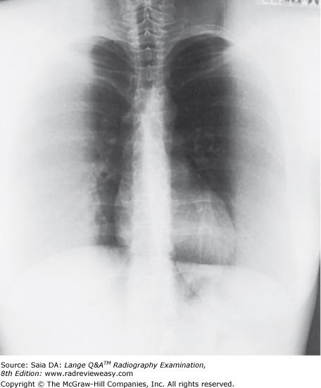
The radiograph shown in the image below exhibits a loss of receptor exposure as a result of
A x-ray tube angulation across grid lines.
B exceeding the focusing distance.
C incorrect grid placement.
D insufficient SID.
C incorrect grid placement.
-If the x-ray tube is angled significantly across the lead strips of a focused grid, there is uniform loss of receptor exposure (grid cutoff). Insufficient or excessive distance with focused grids causes loss of receptor exposure (grid cutoff) along the periphery of the image. Figure 6–10 demonstrates grid cutoff everywhere except along a central vertical strip of the image. This receptor exposure loss is due to the focused grid's being placed upside down. Thus, the middle vertical lead strips allow x-rays to pass, but because the lead strips cant laterally, they are directly opposite to the direction of the x-ray photons (rather than parallel to them), and severe grid cutoff results.
Which of the following groups of exposure factors would be most appropriate to control involuntary motion?
A 400 mA, 0.03 second
B 200 mA, 0.06 second
C 600 mA, 0.02 second
D 100 mA, 0.12 second
C 600 mA, 0.02 second
-Control of motion, both voluntary and involuntary, is an important part of radiography. Patients are unable to control certain types of motion, such as heart action, peristalsis, and muscle spasm. In these circumstances, it is essential to use the shortest possible exposure time in order to have a “stop action” effect
If a 6-in. OID is introduced during a particular radiographic examination, what change in SID will be necessary to overcome objectionable magnification?
A The SID must be increased by 6 in..
B The SID must be increased by 18 in..
C The SID must be decreased by 6 in..
D The SID must be increased by 42 in..
D The SID must be increased by 42 in..
-As OID is increased, spatial resolution is diminished as a result of magnification distortion. If the OID cannot be minimized, an increase in SID is required to reduce the effect of magnification distortion. However, the relationship between OID and SID is not an equal relationship. In fact, to compensate for every 1 in. of OID, an increase of 7 in. of SID is required. Therefore, an OID of 6 in. requires an SID increase of 42 in.. This is why a chest radiograph with a 6-in. air gap usually is performed at a 10-ft SID.
Which of the following will improve the spatial resolution of image-intensified images?
- A very thin coating of cesium iodide on the input phosphor
- A smaller-diameter input screen/phosphor
- Increased total brightness gain
A 1 only
B 1 and 2 only
C 1 and 3 only
D 1, 2, and 3
B 1 and 2 only
-An image's spatial resolution refers to the sharpness of its image details. As the input screen/phosphor layer is made thinner, spatial resolution increases. Also, the smaller the input phosphor diameter, the greater is the spatial resolution. A brighter image is easier to see but does not affect resolution.
An increase in kilovoltage with appropriate compensation of milliampere-seconds will result in
- increased part penetration.
- higher contrast.
- increased receptor exposure.
A 1 only
B 1 and 2 only
C 2 and 3 only
D 1 and 3 only
A 1 only
-As the kilovoltage is increased, photon energy increases and more part penetration will occur. As the milliampere-seconds value is decreased to compensate for the increased kilovoltage, receptor exposure should remain the same.
Compression of the breast during mammographic imaging improves the technical quality of the image because
- geometric blurring is decreased
- less scattered radiation is produced
- patient motion is reduced
A 1 only
B 3 only
C 2 and 3 only
D 1, 2, and 3
D 1, 2, and 3
-Compression of the breast tissue during mammographic imaging improves the technical quality of the image for several reasons. Compression brings breast structures into closer contact with the IR, thus reducing geometric blur and improving resolution. As the breast tissue is compressed and essentially becomes thinner, less scattered radiation is produced. Compression serves as excellent immobilization as well.
A compensating filter is used to
A absorb the harmful photons that contribute only to patient dose
B even out widely differing tissue densities
C eliminate much of the scattered radiation
D improve fluoroscopy
B even out widely differing tissue densities
-A compensating filter is used to make up for widely differing tissue densities. For example, it is difficult to obtain a satisfactory image of the mediastinum and lungs simultaneously without the use of a compensating filter to “even out” the densities. With this device, the chest is radiographed using mediastinal factors, and a trough-shaped filter (thicker laterally) is used to absorb excess photons that would overexpose the lungs. The middle portion of the filter lets the photons pass to the mediastinum almost unimpeded. Filters that absorb the photons contributing to skin dose are inherent and added filters. Compensating filtration is unrelated to elimination of scattered radiation or fluoroscopy.
Underexposure of a radiograph can be caused by all the following except insufficient
A milliamperage (mA)
B exposure time
C Kilovoltage
D SID
D SID
-Insufficient milliamperage and/or exposure time will result in decreased receptor exposure. Insufficient kilovoltage will result in under penetration. Insufficient SID, however, will result in increased exposure rate and overexposure of the IR.
Foreshortening of an anatomic structure means that
A it is projected on the IR smaller than its actual size
B its image is more lengthened than its actual size
C it is accompanied by geometric blur
D it is significantly magnified
A it is projected on the IR smaller than its actual size
-If a structure of a given length is not positioned parallel to the recording medium (PSP or film), it will be projected smaller than its actual size (foreshortened). An example of this can be a lateral projection of the third digit. If the finger is positioned so as to be parallel to the IR, no distortion will occur. If, however, the finger is positioned so that its distal portion rests on the cassette while its proximal portion remains a distance from the IR, foreshortening will occur.
For which of the following examinations might the use of a grid not be necessary in an adult patient?
A Hip
B Knee
C Abdomen
D Lumbar spine
B Knee
-The abdomen is a thick structure that contains many structures of similar tissue density, and thus it requires increased exposure and a grid to absorb scattered radiation. The lumbar spine and hip are also dense structures requiring increased exposure and use of a grid. The knee, however, is frequently small enough to be imaged without a grid. The general rule is that structures measuring more than 10 cm should be imaged with a grid.
A particular radiograph was produced using 12 mAs and 85 kV with a 16:1 ratio grid. The radiograph is to be repeated using an 8:1 ratio grid. What should be the new milliampere-seconds value?
A 3
B 6
C 8
D 10

C 8
-To change nongrid exposures to grid exposures, or to adjust exposure when changing from one grid ratio to another, you must remember the factor for each grid ratio:
To adjust exposure factors, you simply compare the old with the new:
If a duration of 0.05 second was selected for a particular exposure, what milliamperage would be necessary to produce 30 mAs?
A 900
B 600
C 500
D 300

B 600
-The formula for mAs is mA × s = mAs. Substituting known values:
Exposure factors of 100 kVp and 6 mAs are used with a 6:1 grid for a particular exposure. What should be the new milliampere-seconds value if a 12:1 grid is substituted?
A 7.5 mAs
B 10 mAs
C 13 mAs
D 18 mAs

B 10 mAs
-To change nongrid to grid exposure, or to adjust exposure when changing from one grid ratio to another, recall the factor for each grid ratio:
The grid conversion formula is
Substituting known quantities:
Thus, x = 10 mAs with a 12:1 grid.
Distortion can be caused by
- tube angle
- the position of the organ or structure within the body
- the radiographic positioning of the part
A 1 only
B 1 and 2 only
C 2 and 3 only
D 1, 2, and 3
D 1, 2, and 3
-Distortion is caused by improper alignment of the tube, body part, and IR. Anatomic structures within the body are rarely parallel to the IR in a simple recumbent position. In an attempt to overcome this distortion, we position the part to be parallel with the IR or angle the central ray to “open up” the part. Examples of this technique are obliquing the pelvis to place the ilium parallel to the IR or angling the central ray cephalad to “open up” the sigmoid colon.
Exposure factors of 90 kV and 3 mAs are used for a particular nongrid exposure. What should be the new milliampere-seconds (mAs) value if a 12:1 grid is added?
A 86
B 9
C 12
D 15
D 15
-To change nongrid to grid exposure or to adjust exposure when changing from one grid ratio to another, it is necessary to recall the factor for each grid ratio:
Therefore, to change from nongrid to a 12:1 grid, multiply the original milliampere-seconds value by a factor of 5. A new milliampere-seconds value of 15 is required.
The exposure factors used for a particular nongrid x-ray image were 300 mA, 4 ms, and 90 kV. Another image, using an 8:1 grid, is requested. Which of the following groups of factors is most appropriate?
A 400 mA, 3 ms, 110 kV
B 400 mA, 12 ms, 90 kV
C 300 mA, 8 ms, 100 kV
D 200 mA, 240 ms, 90 kV
B 400 mA, 12 ms, 90 kV
-The addition of a grid will help to clean up the scattered radiation produced by higher kilovoltage, but the grid requires an adjustment of milliampere-seconds. According to the grid conversion factors listed here, the addition of an 8:1 grid requires that the original milliampere-seconds be multiplied by a factor of 4:
The original milliampere-seconds value is 1.2. The ideal adjustment, therefore, requires a 4.8 mAs at 90 kV. Although 2.4 mAs with 100 kV (choice C), or 1.2 mAs with 110 kV (choice A), also might seem workable, an increase in kilovoltage would further compromise contrast, nullifying the effect of the grid. Additionally, kilovoltage exceeding 100 should not be used with an 8:1 grid.
Compared with a low-ratio grid, a high-ratio grid will
- allow more centering latitude
- absorb more scattered radiation
- absorb more useful radiation
A 1 only
B 1 and 2 only
C 2 and 3 only
D 1, 2, and 3
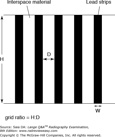
C 2 and 3 only
-Grid ratio is defined as the height of the lead strips to the width of the interspace material (Figure 4–32). The higher the lead strips (or the smaller the distance between the strips), the higher the grid ratio, and the greater the percentage of scattered radiation absorbed. However, a grid does absorb some of the useful radiation as well. The higher the lead strips, the more critical is the need for accurate centering because the lead strips will more readily trap photons whose direction does not parallel them.
The factors that impact spatial resolution include
1.Focal spot size
2.Type of rectification
3.SID
A 1 and 2 only
B 1 and 3 only
C 2 and 3 only
D 1, 2, and 3
B 1 and 3 only
-Focal spot size affects spatial resolution by its effect on focal spot blur: The larger the focal spot size, the greater the blur produced. Spatial resolution is significantly affected by distance changes because of their effect on magnification. As SID increases, magnification decreases and spatial resolution increases. The method of rectification has no controlling effect on spatial resolution. Single-phase rectified units produce intermittent radiation at fluctuating voltage, whereas three-phase units produce almost constant potential. Single phase equipment exposures could require longer exposures, possibly resulting in motion unsharpness, though that equipment is seldom used today.
A 15% increase in kVp accompanied by a 50% decrease in mAs will result in
A decreased patient dose
B increase in contrast.
C increased brightness.
D spatial resolution.
A decreased patient dose
-Since mAs is directly proportional to receptor exposure and patient dose, the receptor exposure is cut in half and patient dose is cut in half. Spatial resolution is unaffected by changes in kVp. Brightness is a function of the computer software.
Which of the following may be used to reduce the effect of scattered radiation on the radiographic image?
- Grids
- Collimators
- Compression bands
A 1 only
B 1 and 3 only
C 2 and 3 only
D 1, 2, and 3
D 1, 2, and 3
-Collimators restrict the size of the irradiated field, thereby limiting the volume of irradiated tissue, and hence less scattered radiation is produced. Once radiation has scattered and emerged from the body, it can be trapped by the grid's lead strips. Grids effectively remove much of the scattered radiation in the remnant beam before it reaches the IR. Compression can be applied to reduce the effect of excessive fatty tissue (e.g., in the abdomen), in effect reducing the thickness of the part to be radiographed.
Which type of error results in grid cutoff at the periphery of the radiographic image?
A Off-focus
B Off-center
C Off-level
D Off-angle
A Off-focus
-The lead strips in a focused grid are made to parallel the x-ray beam. Therefore, scattered radiation, which radiates in directions other than that of the primary beam, will be absorbed by the grid. When the x-ray beam does not parallel the lead strips, some type of grid cutoff occurs. If the x-ray beam is not centered to the grid, or if the x-ray tube and grid surface are not parallel (level), there will be a fairly uniform decrease in receptor exposure across the entire image. However, if the grid is not used within its recommended SID (focus) range (i.e., if the SID is too great or too little), there will be a decrease in receptor exposure at the periphery of the image.
In radiography of a large abdomen, which of the following is (are) effective way(s) to minimize the amount of scattered radiation reaching the image receptor (IR)?
- Use of optimal collimation
- Use of low mAs
- Use of a low-ratio rather than high-ratio grid
A 1 only
B 1 and 2 only
C 1 and 3 only
D 1, 2, and 3
A 1 only
-One way to minimize scattered radiation reaching the IR is to use optimal kilovoltage; excessive kilovoltage increases the production of scattered radiation. Maximum collimation is exceedingly important because the smaller the volume of irradiated material, the less scattered radiation will be produced. The mAs selection has no impact on scattered radiation production or cleanup. Low-ratio grids allow a greater percentage of scattered radiation to reach the IR. Use of a high-ratio grid will clean up a greater amount of scattered radiation before it reaches the IR. Use of a compression band, or the prone position, in a large abdomen has the effect of making the abdomen “thinner”; it will, therefore, generate less scattered radiation.
Decreasing field size from 14 × 17 into 8 × 10 inches will
A decrease receptor exposure and increase the amount of scattered radiation generated within the part.
B increase receptor exposure and increase the amount of scattered radiation generated within the part.
C increase receptor exposure and decrease the amount of scattered radiation generated within the part.
D decrease receptor exposure and decrease the amount of scattered radiation generated within the part.
D decrease receptor exposure and decrease the amount of scattered radiation generated within the part.
-Limiting the size of the radiographic field serves to limit the amount of scattered radiation produced within the anatomic part. As the amount of scattered radiation generated within the part decreases, so does the resultant signal or amount of radiation received by the image receptor. Hence, beam restriction is a very effective means of reducing the quantity of non-information-carrying scattered radiation (fog) produced.
A 3-inch object to be radiographed at a 36-inch SID lies 4 inches from the image recorder. What will be the image width?
A 2.6 inches
B 3.3 inches
C 26 inches
D 33 inches
B 3.3 inches
-Magnification is part of every radiographic image. Anatomic parts within the body are at various distances from the image recorder and therefore have various degrees of magnification. The formula used to determine the amount of image magnification is:
Substituting known values:
x = 3.37 inches image width
Which of the following will produce the greatest distortion?
A AP projection of the skull
B PA projection of the skull
C 37° AP axial of the skull
D 20° PA axial of the skull
C 37° AP axial of the skull
-Distortion is the result of misalignment of the x-ray tube, the anatomic part, and the IR. If these three parts are not parallel with one another, shape distortion occurs. The greater the misalignment, the greater the distortion. In the example cited, the image made with the greatest tube angle will produce the greatest distortion. Distortion is often introduced intentionally to visualize some structure to better advantage. The 37° (caudad) AP axial projection of the skull, for example, projects the facial bones inferiorly so that the occipital bone can be visualized to better advantage.
Disadvantages of moving grids over stationary grids include which of the following?
- They can prohibit the use of very short exposure times.
- They increase patient radiation dose.
- They can cause phantom images when anatomic parts parallel their motion.
A 1 only
B 1 and 2 only
C 2 and 3 only
D 1, 2, and 3
B 1 and 2 only
-One generally thinks in terms of moving grids being totally superior to stationary grids because moving grids function to blur the images of the lead strips on the radiographic image. Moving grids do, however, have several disadvantages. First, their complex mechanism is expensive and subject to malfunction. Second, today's sophisticated x-ray equipment makes possible the use of extremely short exposures, a valuable feature whenever motion may be a problem (as in pediatric radiography). However, grid mechanisms frequently are not able to oscillate rapidly enough for the short exposure times, and as a result, the grid motion is “stopped,” and the lead strips are imaged. Third, patient dose is increased with moving grids. Since the central ray is not always centered to the grid because it is in motion, lateral decentering occurs (resulting in diminished density), and consequently, an increase in exposure is needed to compensate (either manually or via AEC).
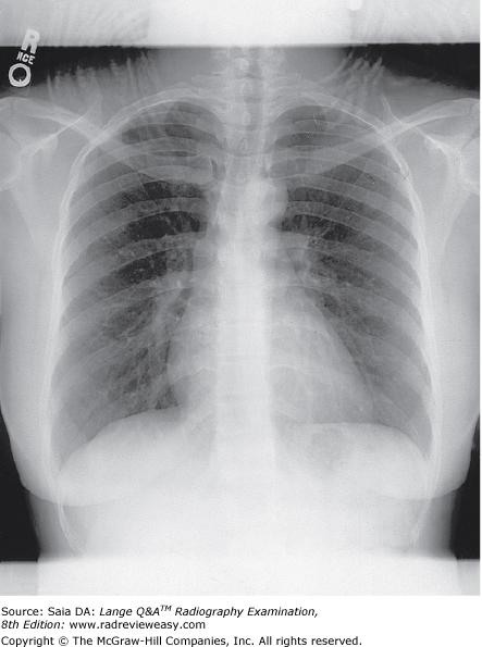
The radiograph shown in Figure 4–12 demonstrates an example of
A overexposure
B motion
C laser jitter
D exposure artifact
D exposure artifact
-The radiograph shown is that of an adult PA erect chest. The image is well positioned and exposed, but observe the braids of hair that extend past the neck and superimpose on the pulmonary apices. Braided hair should be pinned up or otherwise removed from superimposition on thoracic structures. The braided hair was imaged during the exposure of the PA chest and is, therefore, referred to as an exposure artifact. Laser jitter is an example of a processing artifacts occurring in the PSP scanner/leader.
Which of the following groups of analog exposure factors is most likely to produce the longest scale of contrast?
A 200 mA, 0.25 second, 70 kVp, 12:1 grid
B 500 mA, 0.10 second, 90 kVp, 8:1 grid
C 400 mA, 0.125 second, 80 kVp, 12:1 grid
D 300 mA, 0.16 second, 70 kVp, 8:1 grid
B 500 mA, 0.10 second, 90 kVp, 8:1 grid
-Of the given factors, kilovoltage and grid ratio will have a significant effect on the scale of radiographic contrast. The mAs values are almost identical. Because an increased kilovoltage and low-ratio grid combination would allow the greatest amount of scattered radiation to reach the IR, thereby producing more gray tones, B is the best answer. Group D also uses a low-ratio grid, but the kilovoltage is too low to produce as many gray tones as B.
Which of the following groups of exposure factors will produce the greatest receptor exposure?
A 100 mA, 50 ms
B 200 mA, 40 ms
C 400 mA, 70 ms
D 600 mA, 30 ms
C 400 mA, 70 ms
-Milliampere-seconds (mAs) is the exposure factor that determines receptor exposure. Using the equation milliamperage × time = mAs, determine each mAs: (A) = 5 mAs, (B) = 8 mAs, (C) = 28 mAs, (D) = 18 mAs. Group C will produce the greatest receptor exposure.
A satisfactory radiograph was made without a grid using a 72-in. SID and 8 mAs. If the distance is changed to 40 in. and an 12:1 ratio grid is added, what should be the new milliampere-seconds value?
A 9.5 mAs
B 12 mAs
C 21 mAs
D 26 mAs
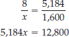
B 12 mAs
-According to the inverse-square law of radiation, as the distance between the radiation source and the IR decreases, the exposure rate increases. Therefore, a decrease in technical factors is first indicated to compensate for the distance change. The following formula (exposure-maintenance formula) is used to determine new milliampere-seconds values when changing distance:
Substituting known values:
Thus, x = 2.47 mAs at 40-in. SID. To then compensate for adding a 12:1 grid, you must multiply the 2.47 mAs by a factor of 5. Thus, 12 mAs is required to produce a receptor exposure similar to the original image. The following are the factors used for milliampere-seconds conversion from nongrid to grid:
Spatial resolution is inversely related to
- SID
- OID
- grid ratio
A 1 only
B 2 only
C 1 and 2 only
D 1, 2, and 3
B 2 only
-SID is directly related to spatial resolution because as SID increases, so does resolution (because magnification is decreased). OID is inversely related to spatial resolution because as OID increases, spatial resolution decreases. Grid ratio is not associated with spatial resolution. Therefore, of the given choices, OID is inversely related to spatial resolution. SID is directly related to spatial resolution.
Focal-spot blur is greatest
A directly along the course of the central ray
B toward the cathode end of the x-ray beam
C toward the anode end of the x-ray beam
D as the SID is increased
B toward the cathode end of the x-ray beam
-Focal-spot blur, or geometric blur, is caused by photons emerging from a large focal spot. The actual focal spot is always larger than the effective (or projected) focal spot, as illustrated by the line-focus principle. In addition, the effective focal-spot size varies along the longitudinal tube axis, being greatest in size at the cathode end of the beam and smallest at the anode end of the beam. Because the projected focal spot is greatest at the cathode end of the x-ray tube, geometric blur is also greatest at the corresponding part (cathode end) of the radiograph.
Exposure factors of 110 kVp and 12 mAs are used with an 8:1 grid for a particular exposure. What should be the new mAs if a 12:1 grid is substituted?
A 3
B 9
C 15
D 18

C 15
-To change nongrid to grid exposure, or to adjust exposure when changing from one grid ratio to another, recall the factor for each grid ratio:
The grid conversion formula is
Substituting known quantities:
(Saia, p 328)
No grid = 1 × the original mAs5:1 grid = 2 × the original mAs6:1 grid = 3 × the original mAs8:1 grid = 4 × the original mAs12:1 grid = 5 × the original mAs16:1 grid = 6 × the original mAs
The effect that differential absorption has on radiographic contrast of a high-subject-contrast part can be minimized by
- using a compensating filter.
- using high-kilovoltage exposure factors.
- increased collimation.
A 1 only
B 1 and 2 only
C 2 and 3 only
D 1, 2, and 3
B 1 and 2 only
-Differential absorption refers to the different attenuation, or absorption, properties of adjacent body tissues. Two parts with widely differing absorption characteristics will produce a high radiographic contrast. Frequently, exposure factors that would properly expose one part will severely overexpose or underexpose the neighboring part (as with lungs vs. the thoracic spine). This effect can be minimized by the use of a compensating filter or by the use of high kilovoltage (for more uniform penetration). Increased collimation is important in the control of patient dose and scattered radiation, not differential absorption.
Methods that help to reduce the production of scattered radiation include using
- compression
- beam restriction
- a grid
A 1 and 2 only
B 1 and 3 only
C 2 and 3 only
D 1, 2, and 3
A 1 and 2 only
-Limiting the size of the irradiated field is a most effective method of decreasing the production of scattered radiation. The smaller the volume of tissue irradiated, the smaller is the amount of scattered radiation generated; this can be accomplished using compression (prone position instead of supine or a compression band). Use of a grid does not affect the production of scattered radiation but rather removes it once it has been produced.
Which of the following fluoroscopic modes delivers the smallest patient dose?
A 30 cm field
B 25 cm field
C 12 cm field
D 9 cm field
A 30 cm field
-In magnification fluoroscopic imaging, the charge on the electrostatic focusing lenses is increased in order to confine electrons to a smaller portion of the input phosphor. This magnifies the image, but at the expense of less brightness. In order to increase brightness to a diagnostic level, the mA is increased. Smaller input phosphor field sizes (D) produce magnified images of the anatomical areas being evaluated, but with an increase in patient dose. Larger input phosphor field sizes (A) produce little or no magnification of the anatomical areas being evaluated, and with decreased patient dose.
Because of the anode heel effect, the intensity of the x-ray beam is greatest along the
A path of the central ray
B anode end of the beam
C cathode end of the beam
D transverse axis of the IR
C cathode end of the beam
-Because the anode's focal track is beveled (angled, facing the cathode), x-ray photons can freely diverge toward the cathode end of the x-ray tube. However, the “heel” of the focal track prevents x-ray photons from diverging toward the anode end of the tube. This results in varying intensity from anode to cathode, with fewer photons at the anode end and more photons at the cathode end. The anode heel effect is most noticeable when using large IRs, short SIDs, and steep target angles.
What should be done to correct for magnification when using air-gap technique?
A Decrease OID
B Increase OID
C Decrease SID
D Increase SID
D Increase SID
-OID is used to effect an increase in contrast in the absence of a grid, usually in chest radiography. If a 6-in. air gap (OID) is introduced between the part and the IR, much of the scattered radiation emitted from the body will not reach the IR; thus, the OID acts as a low-ratio grid and increases image contrast. However, the 6-in. OID air gap will make a very noticeable increase in magnification. To correct for this, the SID must be increased. Generally speaking, the SID needs to be increased 7 in. for every 1 in. of OID. With a 6-in. OID, the SID usually is increased from 6 to 10 ft (120 in.).
A radiograph made using 300 mA, 0.1 second, and 75 kV exhibits motion unsharpness but otherwise satisfactory technical quality. The radiograph will be repeated using a shorter exposure time. Using 86 kV and 400 mA, what should be the new exposure time?
A 25 ms
B 37 ms
C 50 ms
D 75 ms

B 37 ms
-The milliampere-seconds (mAs) formula is milliamperage × time = mAs. With two of the factors known, the third can be determined. To find the milliampere-seconds value that was used originally, substitute the known values:
We have increased the kilovoltage to 86 kV, an increase of 15%, which has an effect similar to that of doubling the milliampere-seconds. Therefore, only 15 mAs is now required as a result of the kilovoltage increase:
Thus, x = 0.0375-s exposure = 37.5 ms.
Which of the following is most likely to produce a radiograph with a long scale of contrast?
A Increased photon energy
B Increased OID
C Increased mAs
D Increased SID
A Increased photon energy
-An increase in photon energy accompanies an increase in kilovoltage. Kilovoltage regulates the penetrability of x-ray photons; it regulates their wavelength—the amount of energy with which they are associated. The higher the related energy of an x-ray beam, the greater its penetrability (kilovoltage and photon energy are directly related; kilovoltage and wavelength are inversely related). Adjustments in kilovoltage can have a big impact on radiographic contrast in analog imaging: As kilovoltage (photon energy) is increased, the number of grays increases, thereby producing a longer scale of contrast. An increase in OID would, if anything (air-gap), result in an increase in contrast. An increase in mAs is frequently accompanied by an appropriate decrease in kilovoltage, which would also shorten the contrast scale. SID and image contrast are unrelated.
Compared to a low ratio grid, a high ratio grid will
1.absorb more primary radiation.
2.absorb more scattered radiation.
3.allow more centering latitude.
A 1 only
B 1 and 2 only
C 2 and 3 only
D 1, 2, and 3
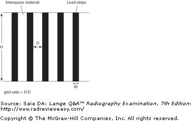
B 1 and 2 only
-Grid ratio is defined as the height of the lead strips to the width of the interspace material (see the figure below). The higher the lead strips (or the smaller the distance between the strips), the greater the grid ratio and the greater the percentage of scattered radiation absorbed. However, a grid does absorb some primary radiation as well. The higher the lead strips, the more critical the need for accurate centering, as the lead strips will more readily trap photons whose direction do not parallel them.
Which of the following groups of technical factors would be most appropriate for the radiographic examination shown in Figure 4–30?
A 400 mA, 1/30 s, 72 kV
B 300 mA, 1/50 s, 82 kV
C 300 mA, 1/120 s, 94 kV
D 50 mA, ¼ s, 72 kV
A 400 mA, 1/30 s, 72 kV
-A 15-minute oblique image of an IVU is pictured. IVU requires the use of iodinated contrast medium.Low kilovoltage (about 70 kV) usually is employed to enhance the photoelectric effect and, in turn, better visualize the renal collecting system. High kilovoltage will produce excessive scattered radiation and obviate the effect of the contrast agent. A higher milliamperage with a shorter exposure time is preferred to decrease the possibility of motion.
In a PA projection of the chest being used for cardiac evaluation, the heart measures 15.2 cm between its widest points. If the magnification factor is known to be 1.3, what is the actual diameter of the heart?
A 9.7 cm
B 11.7 cm
C 19.7 cm
D 20.3 cm

B 11.7 cm
-The formula for magnification factor is MF = image size/object size. In the stated problem, the anatomic measurement is 15.2 cm, and the magnification factor is known to be 1.3. Substituting the known factors in the appropriate equation,
x = 11.69 cm (actual anatomic size)
Shape distortion is influenced by the relationship between the
- x-ray tube and the part to be imaged
- part to be imaged and the IR
- IR and the x-ray tube
A 1 only
B 1 and 2 only
C 1 and 3 only
D 1, 2, and 3
D 1, 2, and 3
-Shape distortion is caused by misalignment of the x-ray tube, the part to be radiographed, and the IR/film.An object can be falsely imaged (foreshortened or elongated) by incorrect placement of the tube, the body part, or the IR. Only one of the three need be misaligned for distortion to occur.
Which of the following is most likely to produce a high-quality image?
A Small image matrix
B High signal-to-noise ratio (SNR)
C Large pixel size
D Low resolution
B High signal-to-noise ratio (SNR)
-SNR can refer to home television images, magnetic resonance images (MRIs), ultrasound images, x-ray images, and so on. Noise interferes with visualization of anatomic image details, for example, scattered radiation fog, graininess from quantum mottle, and so on. The actual signal can be from x-rays, sound waves, and so on. The signal is desirable, the noise is not, therefore, a higher SNR produces a higher-quality image. Low SNR severely impairs contrast resolution.
An x-ray image of the ankle was made at 40-SID, 200 mA, 50 ms, 70 kV, 0.6 mm focal spot, and minimal OID. Which of the following modifications would result in the greatest increase in magnification?
A 1.2 mm focal spot
B 36-in. SID
C 44-in. SID
D 4-in. OID
D 4-in. OID
-All the factor changes affect spatial resolution, but focal spot size does not affect magnification. An increase in SID would decrease magnification. Although a decrease in SID will increase magnification, it does not have as significant an effect as an increase in OID. In general, it requires an increase of 7 in. SID to compensate for every inch of OID.
A part whose width is 6 inches will be imaged at 44 inches SID. The part to be imaged lies 9 inches from the IR. What will be the magnification factor?
A 1.25
B 1.86
C 4.9
D 7.3
A 1.25
-As the object-to-image receptor distance (OID) increases, magnification of that object increases. Depending upon the information provided, we can determine the magnification factor, the percentage magnification, and image width. In the stated scenario, we are looking for image width. The formula used to determine magnification factor is:
MF = SID/SOD
Substituting known factors the equation becomes:
MF = 44/35
MF = 1.257
The "1" in the answer represents the actual object, while the ".257" represents the degree of magnification. The percent magnification can be determined by moving the decimal two places to the right. Thus, the percent magnification is 25.7%.
Which one of the following is (are) used to control the production of scattered radiation?
- Collimators
- Optimal kV
- Use of grids
A 1 only
B 1 and 2 only
C 2 and 3 only
D 1, 2, and 3
B 1 and 2 only
-As kilovoltage is increased, x-ray photons begin to interact with atoms of tissue via the Compton scattered interaction. Scattered x-ray photons result, which serve only to add unwanted, undiagnostic densities (scattered radiation fog) to the radiologic image. (While Compton scatter reduces patient dose compared with photoelectric interactions, it can pose a significant radiation hazard to personnel during fluoroscopic procedures.) Therefore, the use of optimal kilovoltage is recommended to reduce the production of scattered radiation. Scattered radiation is also a function of the size and content of the irradiated field. The greater the volume and atomic number of the tissue, the greater is the production of scattered radiation. Although there is little that can be done about the atomic number of the structure to be radiographed, every effort can be made to keep the field size restricted to the essential area of interest in an effort to decrease production of scattered radiation. Grids have no effect on the production of scattered radiation, but they are very effective in removing scattered radiation from the beam before it strikes the IR.
Why is a very short exposure time essential in chest radiography?
A To avoid excessive focal-spot blur
B To maintain short-scale contrast
C To minimize involuntary motion
D To minimize patient discomfort
C To minimize involuntary motion
-Radiographers usually are able to stop voluntary motion using suspended respiration, careful instruction, and immobilization. However, involuntary motion also must be considered. To have a “stop action” effect on the heart when radiographing the chest, it is essential to use a short exposure time.
If 92 kV and 15 mAs were used for a particular abdominal exposure with single-phase equipment, what milliampere-seconds value would be required to produce a similar radiograph with three-phase, 12-pulse equipment?
A 36
B 24
C 10
D 7.5
D 7.5
-Single-phase radiographic equipment is less efficient than three-phase equipment because it has a 100% voltage ripple. With three-phase equipment, voltage never drops to zero, and x-ray intensity is significantly greater. To produce similar receptor exposure, only two-thirds of the original milliampere-seconds would be used for three-phase, six-pulse equipment. With three-phase, 12-pulse equipment, the original milliampere-seconds would be cut in half (one-half of 15 mAs = 7.5).
Which of the following pathologic conditions are considered additiveconditions with respect to selection of exposure factors?
- Osteoma
- Bronchiectasis
- Pneumonia
A 1 and 2 only
B 1 and 3 only
C 2 and 3 only
D 1, 2, and 3
D 1, 2, and 3
-All these conditions are considered technically additive because they all involve an increase in tissue density. Osteoma, or exostosis, is a (usually benign) bony tumor that can develop on bone. Bronchiectasis is a chronic dilatation of the bronchi with accumulation of fluid. Pneumonia is inflammation of the lung(s) with accumulation of fluid. Additional bony tissues and the pathologic presence of fluid are additive pathologic conditions and require an increase in exposure factors. Destructive conditions such as osteoporosis require a decrease in exposure factors.
The interaction between x-ray photons and matter illustrated in Figure 4–22 is most likely to be associated with
- high kilovoltage
- high contrast
- high-atomic-number absorber
A 1 only
B 1 and 2 only
C 2 and 3 only
D 1, 2, and 3
A 1 only
-Diagnostic x-ray photons interact with tissue in a number of ways, but mostly they are involved in the production of Compton scatter or the photoelectric effect. Compton scatter is pictured; it occurs when a relatively high-energy (kV) photon uses some of its energy to eject an outer-shell electron. In so doing, the photon is deviated in direction and becomes a scattered photon. Compton scatter causes objectionable scattered radiation fog in large structures such as the abdomen and poses a radiation hazard to personnel during procedures such as fluoroscopy. In the photoelectric effect, a relatively low-energy x-ray photon uses all its energy to eject an inner-shell electron, leaving a hole in the K shell. An L-shell electron then drops down to fill the K vacancy and in so doing emits a characteristic ray whose energy is equal to the difference between the binding energies of the K and L shells. The photoelectric effect occurs with high-atomic-number absorbers such as bone and positive contrast media and is responsible for the production of radiographic contrast. It is helpful for the production of the radiographic image, but it contributes to the dose received by the patient (because it involves complete absorption of the incident photon).
If a radiograph exhibits insufficient receptor exposure, this might be attributed to
1.inadequate kVp.
2.inadequate SID.
3.grid cutoff.
A 1 only
B 1 and 2 only
C 1 and 3 only
D 1, 2, and 3
C 1 and 3 only
-As kVp is reduced, the number of high-energy photons produced at the target is reduced; therefore, a decrease in receptor exposure occurs. If a grid has been used improperly (off-centered or out of focal range), the lead strips will absorb excessive amounts of the useful beam, resulting in grid cutoff and loss of receptor exposure. If the SID is inadequate (too short), an increased receptor exposure will result.
Exposure rate increases with an increase in
1.mA.
2.kVp.
3.SID.
A 1 only
B 1 and 2 only
C 2 and 3 only
D 1, 2, and 3
B 1 and 2 only
-The quantity of x-ray photons produced at the target is the function of mAs. The quality (wavelength, penetration, energy) of x-ray photons produced at the target is the function of kVp. The kVp also has an effect on exposure rate, because an increase in kVp will increase the number of high-energy x-ray photons produced at the target. Exposure rate decreases with an increase in SID.
Which of the following terms/units is used to express the resolution of a diagnostic image?
A Line pairs per millimeter (lp/mm)
B Speed
C Latitude
D Kiloelectronvolts (keV)
A Line pairs per millimeter (lp/mm)
-Resolution describes how closely fine details may be associated and still be recognized as separate details before seeming to blend into each other and appear “as one.” The degree of resolution transferred to the IR is a function of the resolving power of each of the system components and can be expressed in line pairs per millimeter (lp/mm). It can be measured using a resolution test pattern; a variety of resolution test tools are available. The star pattern generally is used for focal-spot-size evaluation, whereas the parallel-line type is used for evaluating image receptors. Resolution can also be expressed in terms of line-spread function (LSP) or modulation transfer function (MTF). LSP is measured using a 10-×m x-ray beam; MTF measures the amount of information lost between the object and the IR.
Acceptable method(s) of minimizing motion unsharpness is (are)
- suspended respiration.
- short exposure time.
- patient instruction.
A 1 only
B 1 and 2 only
C 1 and 3 only
D 1, 2, and 3
D 1, 2, and 3
-Motion causes unsharpness that destroys spatial resolution. Careful and accurate patient instruction is essential for minimizing voluntary motion. Suspended respiration eliminates respiratory motion. Using the shortest possible exposure time is essential to decrease involuntary motion. Immobilization can also be very useful in eliminating motion unsharpness.
What pixel size has a 2048 × 2048 matrix with an 80-cm FOV?
A 0.04 mm
B 0.08 mm
C 0.2 mm
D 0.4 mm

D 0.4 mm
-In digital imaging, pixel size is determined by dividing the field of view (FOV) by the matrix. In this case the FOV is 80 cm; since the answer is expressed in mm, first change 80 cm to 800 mm. Then 800 divided by 2048 equals 0.4 mm.
80 cm = 800 mm
The FOV and matrix size are independent of one another, that is, either can be changed and the other will remain unaffected. However, pixel size is affected by changes in either the FOV or matrix size. For example, if the matrix size is increased, pixel size decreases. If FOV increases, pixel size increases. Pixel size is inversely related to resolution. As pixel size increases, resolution decreases.
SID affects spatial resolution in which of the following way
A Spatial resolution is directly related to SID.
B Spatial resolution is inversely related to SID.
C As SID increases, spatial resolution decreases.
D SID is not a spatial resolution factor.
A Spatial resolution is directly related to SID.
-As the distance from focal spot to IR (SID) increases, so does spatial resolution. Because the part is being exposed by more perpendicular (less divergent) rays, less magnification and blur are produced. Although the best spatial resolution is obtained using a long SID, the necessary increase in exposure factors and resulting increased patient exposure become a problem. An optimal 40-in. SID is used for most radiography, with the major exception being chest examinations.
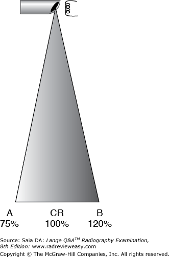
Figure 4–19 is representative of
A the anode heel effect
B the line-focus principle
C the inverse-square law
D the reciprocity law
A the anode heel effect
-The figure represents the anode heel effect. Because the anode's focal track is beveled, x-ray photons can freely diverge toward the cathode end of the x-ray tube. However, the “heel” of the focal track prevents x-ray photons from diverging toward the anode end of the tube. This results in varying intensity with fewer photons at the anode end (A) and more photons at the cathode end (B).
The line-focus principle relates to the anode's focal spot, and x-ray tube targets are constructed according to the line-focus principle—the focal spot is angled to the vertical. As the actual focal spot is projected downward, it is foreshortened; thus, the effective focal spot is always smaller than the actual focal spot.
The inverse-square law deals with the changing x-ray intensity with changes in distance. This principle is important in image formation and in radiation protection.
The effect described as differential absorption is
- responsible for radiographic contrast
- a result of attenuating characteristics of tissue
- minimized by the use of a high peak kilovoltage
A 1 only
B 1 and 2 only
C 1 and 3 only
D 1, 2, and 3
D 1, 2, and 3
-Differential absorption refers to the x-ray absorption characteristics of neighboring anatomic structures. The radiographic representation of these structures is referred to as radiographic contrast; it may be enhanced with high-contrast technical factors, especially using low kilovoltage levels. At low-kilovoltage levels, the photoelectric effect predominates.
A focal-spot size of 0.3 mm or smaller is essential for which of the following procedures?
A Bone radiography
B Magnification radiography
C Tomography
D Fluoroscopy
B Magnification radiography
-A fractional focal spot of 0.3 mm or smaller is essential for rendering fine detail without focal-spot blurring in magnification radiography. As the object image is magnified, so will be the associated blur unless the fractional focal spot is used. Fluoroscopic procedures probably would cause great wear on a fractional focal spot. Use of the fractional focal spot is not essential in bone radiography, although magnification of bony structures often is helpful in locating hairline fractures.
Which of the following focal-spot sizes should be employed for magnification radiography?
A 0.2 mm
B 0.6 mm
C 1.2 mm
D 2.0 mm
A 0.2 mm
-Proper use of focal spot size is of paramount importance in magnification radiography. A magnified image that is diagnostic can be obtained only by using a fractional focal spot of 0.3 mm or smaller. The amount of blur or geometric unsharpness produced by focal spots that are larger in size render the radiograph undiagnostic
The line-focus principle expresses the relationship between
A the actual and the effective focal spot
B exposure given the IR and resulting spatial resolution
C SID used and resulting receptor exposure
D grid ratio and lines per inch
A the actual and the effective focal spot
-The line-focus principle is a geometric principle illustrating that the actual focal spot is larger than the effective (projected) focal spot. The actual focal spot (target) is larger, to accommodate heat over a larger area, and is angled so as to project a smaller focal spot, thus maintaining spatial resolution by reducing blur. The relationship between the SID and resulting receptor exposure is expressed by the inverse-square law. Grid ratio and lines per inch are unrelated to the line-focus principle.
Which of the following technical changes would best serve to remedy the effect of very dissimilar tissue densities?
A Use of short exposure time
B Use of a high-ratio grid
C High-kilovoltage exposure factors
D High milliampere-seconds exposure factors
C High-kilovoltage exposure factors
-When tissue densities within a part are very dissimilar (e.g., the chest), the radiographic result (especially analog) can be unacceptably high contrast. To “even out” these exposure values and produce a more appropriate scale of grays, exposure factors using high kilovoltage should be employed. The higher the grid ratio, the higher is the resulting contrast. Use of short exposure time is always encouraged to reduce the possibility of motion unsharpness but has no impact on varying tissue densities. Exposure factors using high milliampere-seconds generally result in excessive receptor exposure, frequently obliterating much of the gray scale.
Which of the following is (are) characteristic(s) of a 16:1 grid?
1.It absorbs a high percentage of scattered radiation.
2.It has little positioning latitude.
3.It is used with high-kVp exposures.
A 1 only
B 1 and 3 only
C 2 and 3 only
D 1, 2, and 3
D 1, 2, and 3
-High-kilovoltage exposures produce large amounts of scattered radiation, and therefore high-ratio grids are used in an effort to trap more of this scattered radiation. However, accurate centering and positioning become more critical to avoid grid cutoff.
The term differential absorption is closely related to
- beam intensity
- subject contrast
- pathology
A 1 only
B 1 and 2 only
C 2 and 3 only
D 1, 2, and 3
C 2 and 3 only
-The radiographic subject, the patient, is composed of many different tissue types of varying densities (i.e., subject contrast), resulting in varying degrees of photon attenuation and absorption. This differential absorption contributes to the various shades of gray in the x-ray image, as does the energy/penetration characteristic of the beam as regulated by the kV. Beam intensity/quantity/mAs is not related to differential absorption. Normal tissue density may be significantly altered in the presence of pathology. For example, destructive bone disease can cause a dramatic decrease in tissue density. Abnormal accumulation of fluid (as in ascites) will cause a significant increase in tissue density. Muscle atrophy or highly developed muscles similarly will decrease or increase tissue density.
Which of the following groups of exposure factors will produce the shortest scale of contrast?
A 200 mA, 0.25 s, 70 kVp, 12:1 grid
B 500 mA, 0.10 s, 90 kVp, 8:1 grid
C 400 mA, 0.125 s, 80 kVp, 12:1 grid
D 300 mA, 0.16 s, 70 kVp, 8:1 grid
A 200 mA, 0.25 s, 70 kVp, 12:1 grid
-Of the given factors, kilovoltage and grid ratio will have a significant effect on the scale of radiographic contrast. The milliampere-seconds values are almost identical. Because decreased kilovoltage and high-ratio grid combination would allow the least amount of scattered radiation to reach the IR, thereby producing fewer gray tones, (A) is the best answer. Group (D) also uses low kilovoltage, but the grid ratio is lower, thereby allowing more scatter to reach the IR and producing more gray tones.
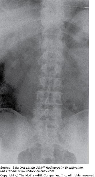
The image quality seen in the figure below is most likely the result of
A an off-level grid
B pronounced anode heel effect
C low-milliampere-seconds and high-kilovoltage factors
D low-kilovoltage and high-milliampere–seconds factors
C low-milliampere-seconds and high-kilovoltage factors
-Quantum noise, or mottle, is a grainy appearance having a spotted or freckled appearance. Low-milliampere-seconds and high-kilovoltage factors are most likely to be the cause of quantum noise/mottle. Grid cutoff is absorption of the useful x-ray beam by the grid and usually results in loss of receptor exposure and visibility of grid lines. The anode heel effect is most pronounced using short SIDs, large IRs, small anode angles, and imaging parts having uneven tissue densities; it is represented by a noticeable receptor exposure difference between the anode and cathode ends of the image.
The CR should be directed to the center of the part of greatest interest to avoid
A rotation distortion
B magnification
C foreshortening
D elongation
A rotation distortion
-Anatomic details placed away from the path of the CR will be exposed by more divergent rays, resulting in rotation distortion. This is why the CR must be directed to the midpoint of the part of greatest interest. For example, if bilateral hands are requested, they should be examined individually; if imaged simultaneously, the CR will be directed to no anatomic part (between the two hands) and rotation distortion will occur.
Magnification occurs when an OID is introduced, or with a decrease in SID. Foreshortening and elongation are the two types of shape distortion—caused by nonalignment of the x-ray tube, part/subject, and IR.
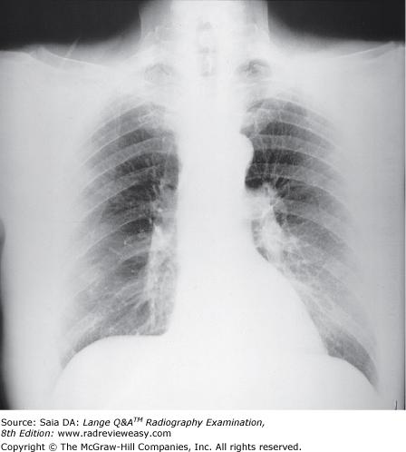
Which of the following statements is (are) true regarding Figure 7–10?
- Excessive kilovoltage was used.
- High contrast is demonstrated.
- Insufficient penetration is evident.
A 1 only
B 1 and 2 only
C 2 and 3 only
D 1, 2, and 3
C 2 and 3 only
-The radiograph shows evidence of very few grays; this is short scale, or high, contrast. There is inadequate penetration of the denser structures—the heart and the lung bases and apices. Penetration and contrast are a function of kilovoltage. Inadequate penetration and high contrast are a result of insufficient peak kilovoltage.
The fact that x-ray intensity across the primary beam can vary as much as 45% describes the
A line-focus principle.
B transformer law.
C anode heel effect.
D inverse-square law.
C anode heel effect.
-A beveled focal track extends around the periphery of the anode disk; when a small angle is used, the beveled edge allows for a smaller effective focal spot and better detail. The disadvantage, however, is that photons are noticeably absorbed by the “heel” of the anode, resulting in a smaller percentage of x-ray photons at the anode end of the x-ray beam and a concentration of x-ray photons at the cathode end of the beam. This is known as the anode heel effect and can cause a primary beam variation of up to 45%. The anode heel effect becomes more pronounced as the SID decreases, as IR size increases, and as target angle decreases.
A radiograph made with a parallel grid demonstrates decreased receptor exposure on its lateral edges. This is most likely due to
A static electrical discharge
B the grid being off-centered
C improper tube angle
D decreased SID
D decreased SID
-The lead strips in a parallel grid are parallel to one another and, therefore, are not parallel to the x-ray beam. The more divergent the x-ray beam, the more likely there is to be cutoff/decreased receptor exposure at the lateral edges of the image. This problem becomes more pronounced at short SIDs. If there were a centering or tube angle problem, there would be more likely to be a noticeable receptor exposure loss on one side or the other.
A 15% decrease in kilovoltage accompanied by a 50% increase in milliampere-seconds will result in a(n)
A increase in patient dose
B increase in exposure latitude
C decrease in receptor exposure
D decrease in spatial resolution
A increase in patient dose
-A 15% decrease in kilovoltage with a 50% increase in milliampere-seconds produces an image similar to the original but with significant differences. The receptor exposure and patient dose are doubled because of the increase in milliampere-seconds. E xposure latitude is wide in CR and DR, controlled by computer software. Spatial resolution is unaffected by changes in kilovoltage.
Focusing distance is associated with which of the following?
A Computed tomography
B Chest radiography
C Magnification radiography
D Grids
D Grids
-Focusing distance is the term used to specify the optimal SID used with a particular focused grid. It is usually expressed as focal range, indicating the minimum and maximum SID workable with that grid. Lesser or greater distances can result in grid cutoff. Although proper distance is important in computed tomography and chest and magnification radiography, focusing distance is unrelated to them.
In electronic imaging, as digital image matrix size increases
- pixel size decreases
- resolution decreases
- pixel depth decreases
A 1 only
B 2 only
C 1 and 2 only
D 2 and 3 only
A 1 only
-Pixel depth is directly related to shades of gray—called dynamic range—and is measured in bits. The greater the number of bits, the more shades of gray. For example, a 1-bit (2 1 ) pixel will demonstrate 2 shades of gray, whereas a 6-bit (2 6 ) pixel can display 64 shades and a 7-bit (2 7 ) pixel 128 shades. However, pixel depth is unrelated to resolution.
A digital image is formed by a matrix of pixels (picture elements) in rows and columns. A matrix that has 512 pixels in each row and column is a 512 × 512 matrix. The term field of view is used to describe how much of the patient (e.g., 150-mm diameter) is included in the matrix. The matrix and the field of view can be changed independently without one affecting the other, but changes in either will change pixel size. As in traditional radiography, spatial resolution is measured in line pairs per millimeter (lp/mm). As matrix size is increased (e.g., from 512 × 512 to 1,024 × 1,024) there are more and smaller pixels in the matrix and, therefore, improved resolution. Fewer and larger pixels result in poor resolution, a “pixelly” image, that is, one in which you can actually see the individual pixel boxes.
A particular milliampere-seconds value, regardless of the combination of milliamperes and time, will reproduce the same receptor exposure. This is a statement of the
A line-focus principle
B inverse-square law
C reciprocity law
D law of conservation of energy
C reciprocity law
-The reciprocity law states that a particular milliampere-seconds value, regardless of the milliamperage and exposure time used, will provide identical receptor exposure. Milliampere-seconds is directly proportional to beam intensity and receptor exposure
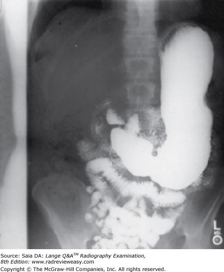
Which of the following errors is illustrated in the figure below?
A Patient not centered to IR
B X-ray tube not centered to grid
C Inaccurate collimation
D Unilateral grid cutoff
B X-ray tube not centered to grid
-The radiograph shown demonstrates a 1.5-in. unexposed strip along the length of the film. This occurred because, although the patient was centered correctly to the collimator light and x-ray field, the x-ray tube was not centered to the grid. If the patient was off-center, the entire image would be exposed, and the patient's spine would be off-center. Grid cutoff would not appear as such a sharply delineated line but rather as a gradually decreasing receptor exposure.
Geometric unsharpness is directly influenced by
- OID
- SOD
- SID
A 1 only
B 1 and 2 only
C 1 and 3 only
D 1, 2, and 3
A 1 only
-Geometric unsharpness is affected by all three factors listed. As OID increases, so does magnification—therefore, OID is directly related to magnification. As SOD and SID decrease, magnification increases—therefore, SOD and SID are inversely related to magnification.
If a lateral projection of the chest is being performed on an asthenic patient and the outer photocells are selected, what is likely to be the outcome?
A Decreased receptor exposure
B Increased receptor exposure
C Scattered radiation fog
D Motion blur
A Decreased receptor exposure
-If a lateral projection of the chest is being performed on an asthenic patient and the outer photocells are selected incorrectly, the outcome is likely to be an underexposed image. The patient is thin, and the lateral photocells have no tissue superimposed on them. Therefore, as soon as the lateral photocells detect radiation (which will be immediately), the exposure will be terminated, resulting in insufficient exposure.
Which of the following pathologic conditions require(s) a decrease in exposure factors?
- Pneumothorax
- Emphysema
- Multiple myeloma
A 1 only
B 1 and 2 only
C 2 and 3 only
D 1, 2, and 3
D 1, 2, and 3
-All three pathologic conditions involve processes that render tissues more easily penetrated by the x-ray beam. Pneumothorax is a collection of air or gas in the pleural cavity. Emphysema is a chronic pulmonary disease characterized by an increase in the size of the air-containing terminal bronchioles. These two conditions add air to the tissues, making them more easily penetrated. Multiple myeloma is a condition characterized by infiltration and destruction of bone and marrow. Each of these conditions requires that factors be decreased from the normal to avoid overexposure.
Which of the following groups of technical factors will produce the least receptor exposure?
A 400 mA, 0.010 second, 94 kV
B 500 mA, 0.008 second, 94 kV
C 200 mA, 0.040 second, 94 kV
D 100 mA, 0.020 second, 80 kV
D 100 mA, 0.020 second, 80 kV
-Each milliampere-second setting is determined [(A) = 4; (B) = 4; (C) = 8; (D) = 2] and numbered in order of greatest to least receptor exposure [(C) = 1; (A) and (B) = 2; (D) = 3]. Then, the kilovoltages are reviewed and also numbered in order of greatest to least receptor exposure [(A), (B), and (C) = 1; (D) = 2]. Finally, the numbers assigned to the milliampere-seconds and kilovoltage are added for each of the four groups [ (A) and (B) = 3; (C) = 2; (D) = 5]; the lowest total (C) indicates the group of factors that will produce the greatest receptor exposure; the highest total (D) indicates the group of factors that will produce the least receptor exposure. This process is illustrated as follows:
(A) 4 mAs (2) + 94 kV (1) =3
(B) 4 mAs (2) + 94 kV (1) ) = 3
(C) 8 mAs (1) + 94 kV (1) = 2
(D) 2 mAs (3) + 80 kV (2) = 5
Which of the following factors influence(s) the production of scattered radiation?
- Kilovoltage level
- Tissue density
- Size of field
A 1 only
B 1 and 2 only
C 1 and 3 only
D 1, 2, and 3
D 1, 2, and 3
-As photon energy (kV) increases, so does the production of scattered radiation. The greater the density of the irradiated tissues, the greater is the production of scattered radiation. As the size of the irradiated field increases, there is an increase in the volume of tissue irradiated, and the percentage of scatter again increases. Beam restriction is the single most important way to limit the amount of scattered radiation produced.
Recently, dual-sided reading technology has become available in more modern CR readers, in which two sets of photodetectors are used to capture light released from the front and back sides of the phosphor storage plate, or PSP (photostimulable phosphor). This technology enables improved:
A Slow-scan direction speed
B Modulation transfer function
C Signal-to-noise ratio
D Fast-scan direction speed
C Signal-to-noise ratio
-By incorporating two sets of light guides and photodetectors on either side of the IP as it travels through the CR reader, a single laser beam can effectively stimulate release of stored energy from bothsides of the phosphor plate. This increases the amount of energy that may be released and used in the form of light to be converted by the photodetectors to an electrical (analog) signal. Therefore, the higher signal intensity increases the SNR, i.e. signal-to-noise ratio (C). Slow scan direction speed refers to the linear travel speed of the phosphor plate through the CR reader (A). The laser light in the CR reader is rapidly reflected by an oscillating polygonal mirror that redirects the beam through a special lens called the f-theta lens, which focuses the light on a cylindrical mirror that reflects the light toward the IP. This light moves back and forth very rapidly to scan the plate transversely, in a raster pattern, and this movement of the laser beam across the IP is therefore called the fast-scan direction (D). The modulation transfer function is a mathematical function that measures the ability of the digital detector to transfer its spatial resolution characteristics to the image (B).
With milliamperage adjusted to produce equal exposures, all the following statements are true except
A a single-phase examination done at 10 mAs can be duplicated with three-phase, 12-pulse at 5 mAs.
B There is greater patient dose with three-phase equipment than with single-phase equipment.
C Three-phase equipment can produce comparable radiographs with less heat unit (HU) buildup.
D Three-phase equipment produces lower-contrast radiographs than single-phase equipment.
B There is greater patient dose with three-phase equipment than with single-phase equipment.
-If the same kilovoltage is used with single-phase and three-phase equipment, the three-phase unit will require about 50% fewer milliampere-seconds to produce similar radiographs. Because three-phase equipment has much higher effective voltage than single-phase equipment, the three-phase radiograph will have lower contrast. A lower milliampere-seconds value can be used with three-phase equipment, so heat units are not built up as quickly. When technical factors are adjusted to obtain the same receptor exposure and contrast, there is no difference in patient dose.
If the center photocell were selected for a lateral projection of the lumbar spine that was positioned with the spinous processes instead of the vertebral bodies centered to the grid, how would the resulting radiograph look?
A The image would be underexposed.
B The image would be overexposed.
C The image would be correctly exposed.
D An exposure could not be made.
A The image would be underexposed.
-If the photocell were centered more posteriorly to a thinner and less dense structure, then the exposure received would be correct for that less-dense structure. The spinous processes would be well visualized, but the denser vertebral bodies and surrounding structures (pedicles and lamina) would be underexposed. Accurate selection of photocells and precise positioning are critical with the use of automatic exposure devices.
An incorrect relationship between the primary beam and the center of a focused grid results in
- an increase in scattered radiation production
- grid cutoff
- insufficient receptor exposure
A 1 only
B 1 and 2 only
C 2 and 3 only
D 1, 2, and 3
C 2 and 3 only
-The lead strips of a focused grid are angled to correspond to the configuration of the divergent x-ray beam. Thus, any radiation that is changing direction, as is typical of scattered radiation, will be trapped by the lead foil strips. However, if the central ray and the grid center do not correspond, the lead strips will absorb useful radiation. The absorption of useful radiation is termed cutoff and results in diminished receptor exposure.
How would the introduction of a 6-in. OID affect image contrast?
A Contrast would be increased.
B Contrast would be decreased.
C Contrast would not change.
D The scale of contrast would not change.
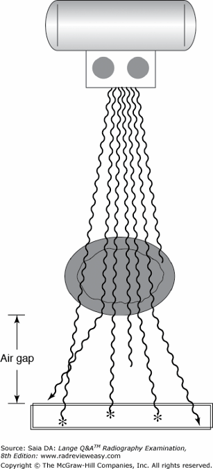
A Contrast would be increased.
-OID can affect contrast when it is used as an air gap. If a 6-in. air gap (OID) is introduced between the part and IR, much of the scattered radiation emitted from the body will not reach the IR, as shown in Figure 7–20. The OID thus is acting as a low-ratio grid and increasing image contrast.
An analog x-ray exposure of a particular part is made and restricted to a 14 × 17 in. field size. The same exposure is repeated, but the x-ray beam is restricted to a 4 × 4 in. field. Compared with the first image, the second image will demonstrate
- less receptor exposure
- more contrast
- more receptor exposure
A 1 only
B 1 and 2 only
C 3 only
D 2 and 3 only
B 1 and 2 only
-Less scattered radiation is generated within a part as the kilovoltage is decreased, as the size of the field is decreased, and as the thickness and density of tissue decrease. As the quantity of scattered radiation decreases from any of these sources, the less is the total receptor exposure.
Acceptable method(s) of minimizing motion unsharpness is (are)
- suspended respiration
- short exposure time
- patient instruction
A 1 only
B 1 and 2 only
C 1 and 3 only
D 1, 2, and 3
D 1, 2, and 3
-The shortest possible exposure time should be used to minimize motion unsharpness. Motion causes unsharpness that destroys spatial resolution. Careful and accurate patient instruction is essential for minimizing voluntary motion. Suspended respiration eliminates respiratory motion. Using the shortest possible exposure time is essential for decreasing involuntary motion. Immobilization is also very useful in eliminating motion unsharpness.
Factor(s) that impact receptor exposure include
1.milliamperage.
2.exposure time.
3.kilovoltage.
A 1 only
B 2 only
C 1 and 2 only
D 1, 2, and 3
D 1, 2, and 3
-Factors that regulate the number of x-ray photons produced at the target determine receptor exposure, namely milliamperage and exposure time (mAs). Receptor exposure is directly proportional to mAs; if the mAs is cut in half, the receptor exposure will decrease by one-half. Although kilovoltage is usually considered to regulate radiographic contrast (in analog imaging), it may also be used to impact receptor exposure in variable-kVp techniques, according to the 15% rule.
Grid cutoff due to off-centering would result in
A overall loss of receptor exposure
B both sides of the image being underexposed
C overexposure under the anode end
D underexposure under the anode en
A overall loss of receptor exposure
-Grids are composed of alternate strips of lead and interspace material and are used to trap scattered radiation after it emerges from the patient and before it reaches the IR. Accurate centering of the x-ray tube is required. If the x-ray tube is off-center but within the recommended focusing distance, there usually will be an overall loss of receptor exposure. Over- or under-exposure under the anode is usually the result of exceeding the focusing distance limits in addition to being off-center.
An exposure was made using 600 mA, 0.04 sec exposure, and 85 kVp. Each of the following changes will serve to reduce the receptor exposure by one-half except change to
A 1/50 sec exposure
B 72 kVp
C 18 mAs
D 300 mA
C 18 mAs
-Receptor exposure is directly proportional to milliampere-seconds. 600 mA with 0.04 sec = 24 mAs. It is desired to reduce receptor exposure to 12 mAs, or its near equivalent. If exposure time is halved from 0.04 sec to 0.02 (1/50) sec, receptor exposure will be cut in half. Changing to 300 mA also will halve the milliampere-seconds, effectively halving the receptor exposure. If the kilovoltage is decreased by 15%, from 85 to 72 kVp, receptor exposure will be halved according to the 15% rule. To cut the receptor exposure in half, the milliampere-seconds must be reduced to 12 mAs (not 18 mAs).
Spatial resolution is directly related to
- SID.
- tube current.
- focal-spot size.
A 1 only
B 1 and 2 only
C 2 and 3 only
D 1, 2, and 3
A 1 only
-As SID increases, so does spatial resolution because magnification is decreased - a direct relationship. Therefore, SID is directly related to spatial resolution. As focal spot size increases, spatial resolution decreases because more penumbral blur is produced. Focal spot size is thus inversely related to spatial resolution - as FSS increases,resolution decreases. Tube current affects receptor exposure and is unrelated to spatial resolution.
Central ray angulation may be required for
- magnification of anatomic structures
- foreshortening or self-superimposition
- superimposition of overlying structures
A 1 only
B 1 and 2 only
C 2 and 3 only
D 1, 2, and 3
C 2 and 3 only
-If structures are overlying or underlying the area to be demonstrated (e.g., the medial femoral condyle obscuring the joint space in the lateral knee projection), CR angulation is used (e.g., 5-degree cephalad angulation to see the joint space in the lateral knee).
If structures are likely to be foreshortened or self-superimposed (e.g., the scaphoid in a PA wrist), CR angulation may be employed to place the structure more closely parallel with the IR.
Another example is the oblique cervical spine, where cephalad or caudad angulation is required to “open” the intervertebral foramina.
Magnification is controlled by object-to- image-receptor distance (OID) and SID; it is unrelated to CR angulation.
Types of shape distortion include
- magnification
- elongation
- foreshortening
A 1 only
B 1 and 2 only
C 2 and 3 only
D 1, 2, and 3
C 2 and 3 only
-Size distortion (magnification) is inversely proportional to SID and directly proportional to OID. Increasing the SID and decreasing the OID decreases size distortion. Aligning the tube, part, and IR so that they are parallel reduces shape distortion. There are two types of shape distortion—elongation and foreshortening. Angulation of the part with relation to the IR results in foreshortening of the object. Tube angulation causes elongation of the object.
Which of the following examinations might require the use of 70 kV?
- AP abdomen
- Chest radiograph
- Barium-filled stomach
A 1 only
B 2 only
C 1 and 2 only
D 2 and 3 only
A 1 only
-It is appropriate to perform an AP abdomen radiograph with lower kilovoltage because it has such low subject contrast. Abdominal tissue densities are so similar that it takes high- or short-scale contrast (using low kilovoltage) to emphasize the little difference there is between tissues. However, high-kilovoltage factors are used frequently to even out densities in anatomic parts having high tissue contrast (e.g., the chest). However, since high kilovoltage produces added scattered radiation, it generally must be used with a grid. Barium-filled structures frequently are radiographed using 120 kV or more to penetrate the barium—to see through to posterior structures
In which of the following ways might higher image contrast be obtained in abdominal radiography?
1.By using lower kilovoltage
2.By using a contrast medium
3.By limiting the field size
A 1 only
B 1 and 2 only
C 1 and 3 only
D 1, 2, and 3
D 1, 2, and 3
-Higher/shorter scale contrast has few shades of gray between white and black. It is partly a result of lower energy photons (lower kVp). High contrast is also more likely when imaging parts having high tissue contrast, such as the chest. The abdomen has low tissue contrast, and abdominal radiographs can exhibit very low contrast unless technical factors are selected to increase contrast. To produce higher contrast in abdominal radiography, lower kVp can be used. To better demonstrate high contrast within a viscus, a contrast medium such as barium, iodine, or air can be used. Restricting the size of the field will also function to increase contrast because less scattered radiation will be generated.
Spatial resolution is directly related to
1.source-image distance (SID)
2.tube current.
3.focal spot size.
A 1 only
B 1 and 2 only
C 2 and 3 only
D 1, 2, and 3
A 1 only
-As SID increases, so does spatial resolution, because magnification is decreased. Therefore, SID is directly related to spatial resolution. As focal spot size increases, spatial resolution decreases because more blur/penumbra is produced. Focal spot size is thus inversely related to spatial resolution. Tube current affects receptor exposure and is unrelated to spatial resolution.
When involuntary motion must be considered, the exposure time may be cut in half if the kilovoltage is
A doubled
B increased by 15%
C increased by 25%
D increased by 35%
B increased by 15%
-If the exposure time is cut in half, one normally would double the milliamperage to maintain the same milliampere-seconds value and, consequently, the same receptor exposure. However, increasing the kilovoltage by 15% has a similar effect. For example, if the original kilovoltage were 85 kV, 15% of this is 13, and therefore, the new kilovoltage would be 98 kV. The same percentage value would be used to cut the receptor exposure in half (reduce kilovoltage by 15%).
Spatial resolution can be improved by decreasing
- the SID
- the OID
- patient/part motion
A 1 only
B 3 only
C 2 and 3 only
D 1, 2, and 3
C 2 and 3 only
-Motion, voluntary or involuntary, is most detrimental to good spatial resolution. Even if all other factors are adjusted to maximize detail, if motion occurs during exposure, resolution is lost. The most important ways to reduce the possibility of motion are using the shortest possible exposure time, careful patient instruction (for suspended respiration), and adequate immobilization when necessary. Minimizing magnification through the use of increased SID and decreased OID functions to improve spatial resolution.
An increase in kilovoltage will have which of the following effects?
- More scattered radiation will be produced.
- The exposure rate will increase.
- Radiographic contrast will increase.
A 1 only
B 1 and 2 only
C 2 and 3 only
D 1, 2, and 3
B 1 and 2 only
-An increase in kilovoltage (photon energy) will result in a greater number (i.e., exposure rate) of scattered photons (Compton interaction). These scattered photons carry no useful information and contribute to radiation fog, thus decreasing radiographic contrast.
A grid is usually employed
1.when radiographing a large or dense body part.
2.when using high kilovoltage.
3.when less patient dose is required.
A 1 only
B 3 only
C 1 and 2 only
D 1, 2, and 3
C 1 and 2 only
-Significant scattered radiation is produced when radiographing large or dense body parts and when using high kilovoltage. A radiographic grid is made of alternating lead strips and interspace material; it is placed between the patient and the IR to absorb energetic scatter emerging from the patient. Although a grid prevents much scattered radiation fog from reaching the radiograph, its use does necessitate a significant increase in patient exposure.
The absorption of useful radiation by a grid is called
A grid selectivity.
B grid cleanup.
C grid cutoff.
D latitude.
C grid cutoff.
-Grids are used in radiography to absorb scattered radiation before it reaches the IR (grid “cleanup”), thus improving radiographic contrast. Contrast obtained with a grid compared with contrast without a grid is termed contrast-improvement factor. The greater the percentage of scattered radiation absorbed compared with absorbed primary radiation, the greater is the “selectivity” of the grid. If a grid absorbs an abnormally large amount of useful radiation as a result of improper centering, tube angle, or tube distance, grid cutoff occurs.
A satisfactory radiograph of the abdomen was made at a 42-inch SID using 300 mA, 0.06-second exposure, and 80 kVp. If the distance is changed to 38 inches, what new exposure time would be required?
A 0.02 second
B 0.05 second
C 0.12 second
D 0.15 second

B 0.05 second
-According to the inverse square law of radiation, as the distance between the radiation source and the IR decreases, the exposure rate increases. Therefore, a decrease in technical factors is indicated. Theexposure maintenance formula is used to determine new mAs values when changing distance:
Then, to determine the new exposure time (mA × s = mAs),
300x = 14.7
x = 0.049 second at 300 mA
An exposure was made using 300 mA and 50 ms. If the exposure time is changed to 22 ms, which of the following milliamperage selections would most closely approximate the original receptor exposure?
A 300 mA
B 400 mA
C 600 mA
D 700 mA
D 700 mA
-Since 50 ms is equal to 0.050 s, and since mA × time mAs, the original milliampere-seconds value was 15 mAs. Now, it is only necessary to determine what milliamperage must be used with 22 ms to provide the same 15 mAs (and thus the same receptor exposure). Because mA × time = mAs
Which of the following can affect histogram appearance?
- Centering accuracy
- Positioning accuracy
- Processing algorithm accuracy
A 1 only
B 1 and 2 only
C 2 and 3 only
D 1, 2, and 3
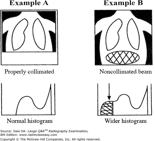
D 1, 2, and 3
-The computed radiography (CR) laser scanner recognizes the various tissue-density values and constructs a representative grayscale histogram. A histogram is a graphic representation showing the distribution of pixel values. Histogram analysis and use of the appropriate LUT together function to produce predictable image quality in CR. Histogram appearance can be affected by a number of things. Degree of accuracy in positioning and centering can have a significant effect on histogram appearance (as well as patient dose). Change is effected in average exposure level and exposure latitude; these changes will be reflected in the images informational numbers (i.e., S number and exposure index). Other factors affecting histogram appearance, and therefore these informational numbers, include selection of the correct processing algorithm (e.g., chest vs. femur vs. cervical spine) and changes in scatter, SID, OID, and collimation. Figure 7–21 illustrates the effect of incorrect collimation on histogram appearance—in short, anything that affects scatter and/or dose.
All the following have an impact on radiographic contrast except
A photon energy
B grid ratio
C OID
D focal-spot size
D focal-spot size
-As photon energy increases, more penetration and greater production of scattered radiation occur, producing a longer scale of contrast. As grid ratio increases, more scattered radiation is absorbed, producing a shorter scale of contrast. As OID increases, the distance between the part and the IR acts as a grid, and consequently, less scattered radiation reaches the IR, producing a shorter scale of contrast. Focal-spot size is related only to spatial resolution.
The use of which of the following is (are) essential in magnification radiography?
- High-ratio grid
- Fractional focal spot
- Direct exposure technique
A 1 only
B 2 only
C 1 and 3 only
D 1, 2, and 3
B 2 only
-Magnification radiography is used to enlarge details to a more perceptible degree. Hairline fractures and minute blood vessels are candidates for magnification radiography. The problem of magnification unsharpness is overcome by using a fractional focal spot; larger focal-spot sizes will produce excessive blurring unsharpness. Grids are usually unnecessary in magnification radiography because of the air-gap effect produced by the OID. Direct-exposure technique probably would not be used because of the excessive exposure required.
What type of x-ray imaging uses an area beam and a photostimulable phosphor as the IR?
A Film radiography
B Computed radiography
C Digital radiography
D Cineradiography
B Computed radiography
-Film radiography used an area x-ray beam, but the IR was film emulsion sandwiched between intensifying screens in a cassette. Computed radiography (CR) also uses an area x-ray beam, but the IR is a photostimulable phosphor such as europium-activated barium fluorohalide coated on an image plate. Digital radiography (DR) can use an area x-ray beam detected by a direct-capture solid-state device. DR can also use a fan-shaped x-ray beam. The fan-shaped beam is “read” by a linear array of detectors.
OID is related to spatial resolution in which of the following ways?
A Spatial resolution is directly related to OID.
B Spatial resolution is inversely related to OID.
C As OID increases, so does spatial resolution.
D OID is unrelated to spatial resolution.
B Spatial resolution is inversely related to OID.
-As the distance from the object to the IR (OID) increases, so does magnification distortion, thereby decreasing spatial resolution. Some magnification is inevitable in radiography because it is not possible to place anatomic structures directly on the IR. However, our understanding of how to minimize magnification distortion is an important part of our everyday work.
Shape distortion is influenced by the relationship between the
- x-ray tube and the part to be imaged.
- body part to be imaged and the IR.
- IR and the x-ray tube.
A 1 and 2 only
B 1 and 3 only
C 2 and 3 only
D 1, 2, and 3
D 1, 2, and 3
-Shape distortion is caused by misalignment of the x-ray tube, the body part to be radiographed, and the IR. An object can be falsely imaged (foreshortened or elongated) as a result of incorrect placement of the tube, the part, or the IR. Only one of the three need be misaligned for distortion to occur.
Which of the following factors is/are related to grid efficiency?
- Grid ratio
- Number of lead strips per inch
- Amount of scatter transmitted through the grid
A 1 only
B 2 only
C 1 and 2 only
D 1, 2, and 3
D 1, 2, and 3
-Grid ratio is defined as the ratio of the height of the lead strips to the width of the interspace material; the higher the lead strips, the more scattered radiation they will trap and the greater is the grid's efficiency. The greater the number of lead strips per inch, the thinner and less visible they will be on the finished radiograph. The function of a grid is to absorb scattered radiation in order to improve radiographic contrast. The selectivity of a grid is determined by the amount of primary radiation transmitted through the grid divided by the amount of scattered radiation transmitted through the grid.
If a radiograph, made using AEC, is overexposed because an exposure shorter than the minimum response time was required, the radiographer generally should
A decrease the milliamperage
B increase the milliamperage
C increase the kilovoltage
D decrease the kilovoltage
A decrease the milliamperage
-Decreasing or increasing the kilovoltage will produce a change in radiographic contrast. The image was overexposed (from excessive exposure time) because the AEC wasn't capable of producing the required extremely short exposure time. To bring the required exposure to an exposure time the AEC is capable of, the mA should be decreased (thus requiring a longer exposure time, within the capability of the AEC).
Which of the following is (are) characteristic(s) of a 16:1 grid?
- It absorbs more useful radiation than an 8:1 grid.
- It has more centering latitude than an 8:1 grid.
- It is used with higher-kilovoltage exposures than an 8:1 grid.
A 1 only
B 1 and 3 only
C 2 and 3 only
D 1, 2, and 3
B 1 and 3 only
-High-kilovoltage exposures produce large amounts of scattered radiation, and high-ratio grids are used often with high-kilovoltage techniques in an effort to absorb more of this scattered radiation. However, as more scattered radiation is absorbed, more primary radiation is absorbed as well. This accounts for the increase in milliampere-seconds required when changing from an 8:1 to a 16:1 grid. In addition, precise centering and positioning become more critical; a small degree of inaccuracy is more likely to cause grid cutoff in a high-ratio grid.
Exposure factors of 80 kVp and 8 mAs are used for a particular nongrid exposure. What should be the new milliampere-seconds value if an 8:1 grid is added?
A 16 mAs
B 24 mAs
C 32 mAs
D 40 mAs

C 32 mAs
-To change nongrid to grid exposure, or to adjust exposure when changing from one grid ratio to another, remember the factor for each grid ratio:
Therefore, to change from nongrid exposure to an 8:1 grid, multiply the original milliampere-seconds value by a factor of 4. Thus, a new setting of 32 mAs is required.
Which of the following units is (are) used to express resolution?
- Line-spread function
- Line pairs per millimeter
- Line-focus principle
A 1 only
B 1 and 2 only
C 2 and 3 only
D 1, 2, and 3
B 1 and 2 only
-Resolution describes how closely fine details may be associated and still be recognized as separate details before seeming to blend into each other and appear “as one.” The degree of resolution transferred to the IR is a function of the resolving power of each of the system components and can be expressed in line pairs per millimeter (lp/mm), line-spread function (LSP), or modulation transfer function(MTF). Lp/mm can be measured using a resolution test pattern; a number of resolution test tools are available. LSP is measured using a 10-μm x-ray beam; MTF measures the amount of information lost between the object and the IR. The effective focal spot is the foreshortened size of the actual focal spot as it is projected down toward the IR, that is, as it would be seen looking up into the emerging x-ray beam. This is called the line-focus principle and is not a unit used to express resolution.
What lies immediately under the phosphor layer of a PSP storage plate?
A Reflective layer
B Base
C Antistatic layer
D Lead foil
A Reflective layer
-The PSP storage plate within the IP has a layer of europium-activated barium fluorohalide (BaFX: Eu 2 +; X = halogen) mixed with a binder substance. This layer serves as the image receptor when exposed.
The barium fluorohalide is usually granular or turbid phosphors. Other examples of turbid phosphors are gadolinium oxysulfide and rubidium chloride. “Needle”-shaped, or columnar phosphors (usually cesium iodide), have the advantage of better x-ray absorption and less light diffusion.
Just under the barium fluorohalide layer is a reflective layer that helps direct emitted light up toward the CR reader. Below the reflective layer is the base, behind that is an antistatic layer, and then the lead foil to absorb backscatter. Over the top of the barium fluorohalide is a protective layer.
Terms that refer to size distortion include
- magnification
- attenuation
- elongation
A 1 only
B 1 and 2 only
C 1 and 3 only
D 1, 2, and 3
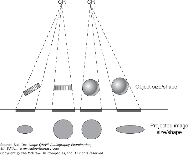
A 1 only
-Distortion is misrepresentation of the actual size or shape of the object being imaged. Size distortion is magnification. Shape distortion is a result of improper alignment of the x-ray tube, the part being radiographed, and the IR; the two types of shape distortion are foreshortening and elongation. The shapes of various structures can be misrepresented radiographically as a result of their position in the body, when the part is out of the central axis of the x-ray beam, or when the CR is angled (Figure 7–19). Parts sometimes are elongated intentionally for better visualization (e.g., sigmoid colon). Some body parts, because of their position in the body, are foreshortened, such as the carpal scaphoid. Attenuation refers to decreasing beam intensity and is unrelated to distortion.
Which of the following methods can be used effectively to decrease differential absorption, providing a longer scale of contrast in the diagnostic range?
- Using high peak kilovoltage and low milliampere-seconds factors
- Using compensating filtration
- Using factors that increase the photoelectric effect
A 1 only
B 1 and 2 only
C 2 and 3 only
D 1, 2, and 3
B 1 and 2 only
-When differences in absorption characteristics are decreased, body tissues absorb radiation more uniformly, and as a result, more grays are seen on the radiographic image. A longer scale of contrast is produced. High-kilovoltage and low-milliamperage factors achieve this. Compensating filtration is also used to “even out” densities in uneven anatomic parts, such as the thoracic spine. The photoelectric effect is the interaction between x-ray photons and matter that occurs at low-peak kilovoltage levels—levels that tend to produce short-scale contrast.
Disadvantages of using lower kilovoltage technical factors, with mAs compensated to maintain receptor exposure, include
- insufficient penetration
- increased patient dose
- diminished resolution
A 1 only
B 1 and 2 only
C 1 and 3 only
D 1, 2, and 3
B 1 and 2 only
-As the kilovoltage is decreased, x-ray-beam energy (i.e., penetration) is also decreased. Consequently, a shorter scale of contrast is obtained. As kilovoltage is reduced, the milliampere-seconds value must be increased accordingly to maintain adequate receptor exposure. This increase in milliampere-seconds results in greater patient dose. Resolution is not related to kV.
Focal-spot blur is greatest
A toward the anode end of the x-ray beam
B toward the cathode end of the x-ray beam
C directly along the course of the CR
D as the SID is increased
B toward the cathode end of the x-ray beam
-Focal-spot blur, or geometric blur, is caused by photons emerging from a large focal spot. Because the projected focal spot is greatest at the cathode end of the x-ray tube, geometric blur is also greatest at the corresponding part (cathode end) of the radiograph. The projected focal-spot size becomes progressively smaller toward the anode end of the x-ray tube.
Which of the following devices is used to overcome severe variation in patient anatomy or tissue density, providing more uniform radiographic density?
A Compensating filter
B Grid
C Collimator
D Protective filter
A Compensating filter
-A compensating filter is used when the part to be radiographed is of uneven thickness or tissue density (in the chest, mediastinum vs. lungs). The filter (made of aluminum or lead acrylic) is constructed in such a way that it will absorb much of the x-ray beam directed toward the low tissue-density area while not affecting the x-ray photons to directed toward the high tissue-density area. A collimator is used to decrease the production of scattered radiation by limiting the volume of tissue irradiated. The grid functions to trap scattered radiation before it reaches the IR, thus reducing scattered radiation fog.Protective filtration absorbs low energy x-ray photons that contribute only to patient (skin) dose and would never reach the image receptor.
An exposure was made using 300 mA, 40 ms exposure, and 85 kV. Each of the following changes will cut the receptor exposure in half except a change to
A 1/50 sec exposure
B 72 kV
C 10 mAs
D 150 mA
C 10 mAs
-Receptor exposure is directly proportional to milliampere-seconds. If exposure time is halved from 40 ms (0.04 or 1 /25) sec to 0.02 ( 1 / 50 ) sec, receptor exposure will be cut in half. Changing to 150 mA also will halve the milliampere-seconds, effectively halving the receptor exposure. If the kilovoltage is decreased by 15%, from 85 to 72 kV, receptor exposure will be halved according to the 15% rule. To cut the receptor exposure in half, the mAs value must be reduced to 6 mAs (rather than 10 mAs).
Which combination of exposure factors most likely will contribute to producing the shortest-scale contrast?
A mAs: 10; kV: 70; Grid ratio: 5:1; Field size: 14 × 17 in.
B mAs: 12; kV: 90; Grid ratio: 8:1; Field size: 14 × 17 in.
C mAs: 15; kV: 90; Grid ratio: 12:1; Field size: 11 × 14 in.
D mAs: 20; kV: 80; Grid ratio: 10:1; Field size: 8 × 10 in.
D mAs: 20; kV: 80; Grid ratio: 10:1; Field size: 8 × 10 in.
-Review the groups of factors. First, because the milliampere-seconds value has no effect on the scale of contrast produced, eliminate milliampere-seconds from consideration by drawing a line through the column. Then check the two entries in each column that are likely to produce shorter-scale contrast. For example, in the kilovoltage column, because lower kilovoltage can produce shorter-scale contrast, place checkmarks next to the 70 and 80 kV. Because higher-ratio grids permit less scattered radiation to reach the IR, the 10:1 and 12:1 grids can produce a shorter scale of contrast than the lower-ratio grids; check them. As the volume of irradiated tissue decreases, so does the amount of scattered radiation produced, and consequently, the shorter is the scale of radiographic contrast; therefore, check the 11 × 14 and 8 × 10 in. field sizes. An overview shows that the factors in groups (A) and (C) have more checkmarks, than the factors in group (D), indicating that group (D) is more likely to produce the shortest-scale contrast.
What is the single most important factor controlling size distortion?
A Tube, part, IR alignment
B IR dimensions
C SID
D OID
D OID
-Shape distortion (foreshortening, elongation) is caused by improper alignment of the tube, part, and image receptor. Size distortion, or magnification, is caused by too great an object–image distance or tooshort a source–image distance. OID is the primary factor influencing magnification, followed by SID.
A decrease from 200 to 100 mA will result in a decrease in which of the following?
- Wavelength
- Exposure rate
- Beam intensity
A 1 only
B 1 and 2 only
C 2 and 3 only
D 1, 2, and 3
C 2 and 3 only
-Technical factors can be expressed in terms of milliampere-seconds rather than milliamperes and time. The milliampere-seconds value is a quantitative factor because it regulates x-ray-beam intensity,exposure rate, quantity, or number of x-ray photons produced (the milliampere-seconds value is the single most important technical factor associated with receptor exposure and patient dose). The milliampere-seconds value is directly proportional to the intensity (i.e., exposure rate, number, and quantity) of x-ray photons produced and the resulting receptor exposure. If the milliampere-seconds value is doubled, twice the exposure rate and twice the receptor exposure occurs. If the milliampere-seconds value is cut in half, the receptor exposure and patient dose are cut in half. Kilovoltage is the qualitative exposure factor—it determines beam quality by regulating photon energy (i.e., wavelength).
Factors that determine AEC exposure determination include
- the thickness and density of the object
- positioning of the object with respect to the photocell
- beam restriction
A 1 only
B 1 and 2 only
C 2 and 3 only
D 1, 2, and 3
D 1, 2, and 3
-The AEC automatically terminates the exposure when the appropriate receptor exposure has been received by the IR. The important advantage of the AEC (phototimer or ionization chamber) is that it can accurately duplicate receptor exposures. It is very useful in providing accurate comparison in follow-up examinations and in decreasing patient exposure dose by reducing the number of “retakes” needed because of improper exposure. The AEC automatically adjusts the exposure required for body parts with different thicknesses and tissue densities. However, proper functioning of the AEC depends on accurate positioning by the radiographer. The correct photocell(s) must be selected, and the anatomic part of interest must completely cover the photocell to achieve an accurate receptor exposure. If collimation is inadequate and a field size larger than the part is used, excessive scattered radiation from the body or tabletop can cause the AEC to terminate the exposure prematurely, resulting in an underexposed image.
Geometric unsharpness is most likely to be greater
A at long SIDs.
B at the anode end of the image.
C with small focal spots.
D at the cathode end of the image.
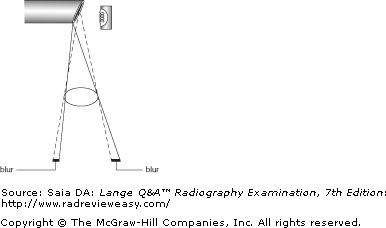
D at the cathode end of the image.
-The x-ray tube anode is designed according to the line focus principle, that is, with the focal track beveled (see the figure below). This allows a larger actual focal spot to project a smaller effective focal spot, resulting in improved spatial resolution with less blur. However, because of the target angle, penumbral blur varies along the longitudinal tube axis, being greater at the cathode end of the image and less at the anode end of the image.
In comparison with 60 kV, 80 kV will
- permit greater exposure latitude
- produce more scattered radiation
- produce shorter-scale contrast
A 1 only
B 2 only
C 1 and 2 only
D 2 and 3 only
C 1 and 2 only
-The higher the kilovoltage range, the greater is the exposure latitude (margin of error in exposure). Higher kilovoltage produces more energetic photons, is more penetrating, and produces more grays on the radiographic image, lengthening the scale of contrast. As kilovoltage increases, the percentage of scattered radiation also increases.
The component of a CR image plate (IP) that records the radiologic image is the
A emulsion
B helium–neon laser
C photostimulable phosphor
D scanner–reader
C photostimulable phosphor
-Inside the IP is the photostimulable phosphor (PSP). This PSP (or SPS—Storage Phosphor Screen), with its layer of europium-activated barium fluorohalide, serves as the IR because it is exposed in the traditional manner and receives the latent image. The PSP can store the latent image for several hours; after about 8 hours, noticeable image fading will occur. Once the IP is placed into the CR processor (scanner or reader), the PSP plate is removed automatically. The latent image on the PSP is changed to a manifest image as it is scanned by a narrow, high-intensity helium–neon laser to obtain the pixel data. As the PSP is scanned in the reader, it releases a violet light—a process referred to as photostimulated luminescence (PSL).
What pixel size has a 2,048 × 2,048 matrix with a 60-cm FOV?
A 0.3 mm
B 0.5 mm
C 0.15 mm
D 0.03 mm

A 0.3 mm
-In digital imaging, pixel size is determined by dividing the FOV by the matrix. In this case, the FOV is 60 cm; since the answer is expressed in millimeters, first change 60 cm to 600 mm. Then 600 divided by 2,048 equals 0.29 mm:
The FOV and matrix size are independent of one another; that is, either can be changed, and the other will remain unaffected. However, pixel size is affected by changes in either the FOV or matrix size. For example, if the matrix size is increased, pixel size decreases. If FOV increases, pixel size increases. Pixel size is inversely related to resolution. As pixel size increases, resolution decreases.
Which of the following is (are) directly related to photon energy?
- Kilovoltage
- Milliamperes
- Wavelength
A 1 only
B 1 and 2 only
C 1 and 3 only
D 1, 2, and 3
A 1 only
-Kilovoltage is the qualitative regulating factor; it has a direct effect on photon energy. That is, as kilovoltage is increased, photon energy increases. Photon energy is inversely related to wavelength. That is, as photon energy increases, wavelength decreases. Photon energy is unrelated to milliamperage.
A decrease in kilovoltage will result in
A a decrease in receptor exposure
B a decrease in image contrast
C a decrease in spatial resolution
D an increase in spatial resolution
A a decrease in receptor exposure
-As kilovoltage is increased, more electrons are driven to the anode with greater speed and energy. More high-energy electrons will result in production of more high-energy x-rays. Thus, kilovoltage affects both quantity and quality (energy) of the x-ray beam. However, although kilovoltage and receptor exposure are directly related, they are not directly proportional; that is, twice the radiographic receptor exposure is not achieved by doubling the kilovoltage. If it is desired to double the receptor exposure yet impossible to adjust the mAs, a similar effect can be achieved by increasing the kilovoltage by 15%. Conversely, the receptor exposure may be cut in half by decreasing the kilovoltage by 15%. Therefore, a decrease in kilovoltage will produce fewer x-ray photons, resulting in decreased receptor exposure. A decrease in kilovoltage will produce fewer shades of gray in analog imaging, that is, a shorter-scale, or higher/increased, contrast. Kilovoltage is unrelated to spatial resolution.
A satisfactory radiograph was made using a 36-in. SID, 12 mAs, and a 12:1 grid. If the examination will be repeated at a distance of 42 in. and using a 5:1 grid, what should be the new milliampere-seconds value to maintain the original receptor exposure?
A 5.6
B 6.5
C 9.7
D 13
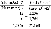
B 6.5
-According to the exposure-maintenance formula, if the SID is changed to 48 in., 16.33 mAs is required to maintain the original radiographic receptor exposure:
Thus, x = 16.33 mAs at 42 in. SID. Then, to compensate for changing from a 12:1 grid to a 5:1 grid, the milliampere-seconds value becomes 6.53 mAs:
Thus, x 6.53 mAs with 5:1 grid at 42 in. SID. Hence, 6.53 mAs is required to produce a receptor exposure similar to that of the original radiograph.
Recommended method(s) of minimizing motion unsharpness include
- suspended respiration
- short exposure time
- patient instruction
A 1 only
B 1 and 2 only
C 1 and 3 only
D 1, 2, and 3
D 1, 2, and 3
-The shortest possible exposure time should be used to minimize motion unsharpness. Motion causes unsharpness that destroys detail. Careful and accurate patient instruction is essential for minimizing voluntary motion. Suspended respiration eliminates respiratory motion. Using the shortest possible exposure time is essential to decreasing involuntary motion. Immobilization also can be useful in eliminating motion unsharpness.
Which of the following is (are) characteristic(s) of a 5:1 grid?
- It allows some positioning latitude.
- It is used with high-kilovoltage exposures.
- It absorbs a high percentage of scattered radiation.
A 1 only
B 1 and 3 only
C 2 and 3 only
D 1, 2, and 3
A 1 only
-Low-ratio grids, such as 5:1, 6:1, and 8:1, are used with moderate-kilovolt techniques and are not recommended for use beyond 85 kV. They are not able to clean up the amount of scatter produced at high kilovoltages, but their low ratio permits more positioning latitude than high-ratio grids. High-kilovoltage exposures produce large amounts of scattered radiation, and therefore, high-ratio grids are used in an effort to trap more of this scattered radiation. However, accurate centering and positioning become more critical to avoid grid cutoff
An exposure was made using 8 mAs and 60 kV. If the kilovoltage was changed to 70 to obtain longer-scale contrast, what new milliampere-seconds value is required to maintain receptor exposure?
A 2
B 4
C 16
D 32
B 4
-According to the 15% rule, if the kilovoltage is increased by 15%, receptor exposure will be doubled. Therefore, to compensate for this change and to maintain receptor exposure, the milliampere-seconds value should be reduced to 4 mAs
All the following statements regarding CR IPs are true except
A IPs have a thin lead foil backing.
B IPs can be placed in the Bucky tray.
C IPs must exclude all white light.
D IPs function to protect the PSP.
C IPs must exclude all white light.
-The image plate has a protective function for the flexible photostimulable storage phosphor within; it can be conveniently placed in a Bucky tray or under the anatomic part, and comes in a variety of sizes. The PSP within the IP is the image receptor/detector. IPs do not contain a light sensitive material and, therefore, do not need to be light-tight. The photostimulable PSP is not affected by light.
Which of the following groups of exposure factors would be most effective in eliminating prominent pulmonary vascular markings in the RAO position of the sternum?
A 500 mA, 1/30 s, 70 kV
B 200 mA, 0.04 second, 80 kV
C 300 mA, 1/10 s, 80 kV
D 25 mA, 7/10 s, 70 kV
D 25 mA, 7/10 s, 70 kV
-In the RAO position, the sternum must be visualized through the thorax and heart. Prominent pulmonary vascular markings can hinder good visualization. A method frequently used to overcome this problem is to use a milliampere-seconds value with a long exposure time. The patient is permitted to breathe normally during the (extended) exposure and by so doing blurs out the prominent vascularities.
In an AP abdomen taken at 105-cm SID during an IV urography series, one renal shadow measures 9 cm in width. If the OID is 18 cm, what is the actual width of the kidney?
A 5 cm
B 7.5 cm
C 11 cm
D 18 cm

B 7.5 cm
-As OID increases, magnification increases. Viscera and structures within the body will be varying distances from the image receptor, depending on their location within the body and the position used for the exposure. The size of a particular structure or image can be calculated using the following formula:
Substituting known quantities,
The relationship between SID, SOD, and OID and the equation for determining image or object size is illustrated in the figure below.
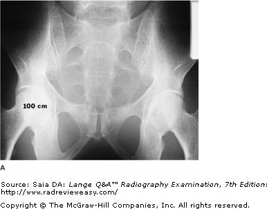
Both radiographic images seen in the figure below were made of the same subject using identical exposure factors. Which of the following statements correctly describes these images?
1.Image A illustrates less receptor exposure because a shorter SID was used.
2.Image A illustrates more receptor exposure because the subject was turned PA.
3.Image B illustrates more receptor exposure because a shorter SID was used.
A 1 only
B 2 only
C 3 only
D 1 and 2 only
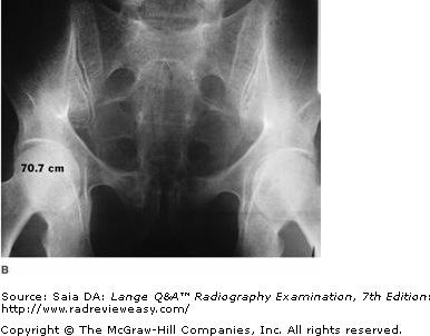
C 3 only
-In the figure shown, image B is darker because the receptor exposure was greater. Receptor exposure is significantly affected by mAs, SID, and exposure rate. In this case, there is a difference in SID between the two images. As SID decreases, exposure rate increases and receptor exposure increases. Image B is darker (received a greater exposure) than image A because image B was exposed at a shorter SID (and therefore a higher exposure rate).
Low-kilovoltage exposure factors usually are indicated for radiographic examinations using
- water-soluble, iodinated media
- a negative contrast agent
- barium sulfate
A 1 only
B 1 and 2 only
C 3 only
D 1 and 3 only
B 1 and 2 only
-Positive contrast medium is radiopaque; negative contrast material is radioparent. Barium sulfate (radiopaque, positive contrast material) is used most frequently for examinations of the intestinal tract, and high-kilovoltage exposure factors are used to penetrate (to see through and behind) the barium. Water-based iodinated contrast media (Conray, Amipaque) are also positive contrast agents. However, the K-edge binding energy of iodine prohibits the use of much greater than 70 kV with these materials. Higher kilovoltage values will obviate the effect of the contrast agent. Air is an example of a negative contrast agent, and high-kilovoltage factors are clearly not indicated.