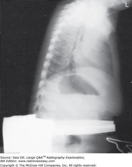
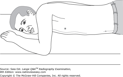
Which of the following statements is (are) true regarding the radiograph shown in Figure 6–16?
- The part is rotated.
- The patient is not shielded correctly.
- There is excessive receptor exposure.
A 1 only
B 2 only
C 1 and 2 only
D 1, 2, and 3
B 2 only
-The patient is well positioned; the spinous processes and sternum are seen clearly without superimposition. Adequate penetration and long-scale contrast are present without excessive receptor exposure. The patient had been shielded properly for the PA projection, but the shield was not moved to the correct location prior to the lateral exposure.
Posterior displacement of a tibial fracture would be best demonstrated in the
A AP projection.
B lateral projection.
C medial oblique projection.
D lateral oblique projection.
B lateral projection.
-A frontal projection (AP or PA) demonstrates the medial and lateral relationship of structures. A lateral projection demonstrates the anterior and posterior relationship of structures. Two views, at right angles to each other, are generally taken of most structures.
In which position of the shoulder is the outline of the greater tubercle superimposed on the humeral head?
A AP
B External rotation
C Internal rotation
D Neutral position
D Neutral position
-The external rotation position is the true AP position and places the greater tubercle in profile laterally and places the lesser tubercle anteriorly. The internal rotation position demonstrates the lesser tubercle in profile medially and places the humerus in a true lateral position; the greater tubercle is seen superimposed on the humeral head. The epicondyles should be superimposed and perpendicular to the IR. The neutral position places the epicondyles about 45 degrees to the IR and the outline of the greater tubercle superimposed on the humeral head.
With which of the following does the trapezium articulate?
A Fifth metacarpal
B First metacarpal
C Distal radius
D Distal ulna
B First metacarpal
-The first metacarpal, on the lateral side of the hand, articulates with the most lateral carpal of the distal carpal row, the greater multangular/trapezium. This articulation forms a rather unique and very versatile saddle joint named for the shape of its articulating surfaces.
Which of the following positions may be used to effectively demonstrate the hepatic flexure during radiographic examination of the large bowel?
1.RAO
2.LAO
3.LPO
A 1 only
B 1 and 2 only
C 1 and 3 only
D 2 and 3 only
C 1 and 3 only
-The hepatic and splenic flexures are not generally well demonstrated in the AP and PA projections. To "open" the flexures, oblique projections are required. The hepatic flexure is usually well demonstrated in the RAO (right PA oblique) and LPO (left AP oblique) positions. The LAO and RPO positions are used to demonstrate the splenic flexure.
Which of the following is (are) located on the anterior aspect of the femur?
- Patellar surface
- Intertrochanteric crest
- Linea aspera
A 1 only
B 1 and 2 only
C 2 and 3 only
D 1, 2, and 3
A 1 only
-The femur is the longest and strongest bone in the body. The femoral shaft is bowed slightly anteriorly and presents a long, narrow ridge posteriorly called the linea aspera. The proximal femur consists of a head that is received by the pelvic acetabulum. The femoral neck, which joins the head and shaft, normally angles upward about 120 degrees and forward (in anteversion) about 15 degrees. The greater and lesser trochanters are large processes on the posterior proximal femur. The intertrochanteric crest runs obliquely between the trochanters; the intertrochanteric line parallels the intertrochanteric crest on the anterior femoral surface. The intercondyloid fossa, a deep notch, is found on the distal posterior femur between the large femoral condyles, and the popliteal surface is a smooth surface just superior to the intercondyloid fossa. Just opposite the popliteal surface, on the distal anterior femur is the patellar surface—a smooth surface for patellar motion during flexion and extension of the knee.
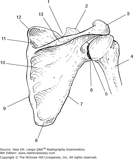
In Figure 2–29, which of the following is represented by the number 7?
A Medial border
B Lateral border
C Inferior angle
D Superior angle
B Lateral border
-Figure 2–29 depicts a posterior view of the right scapula and its articulation with the humerus (number 4). The scapula presents two borders—the lateral or axillary border (number 7) and the medial or vertebral border (number 9). It also presents three angles—the inferior angle (number 8), the superior angle (number 12), and the lateral angle (number 6). The processes of the scapula are the coracoid (number 2), the acromion (number 3), and the scapular spine (number 13). The scapula has a (supra) scapular notch (number 1), a supraspinatus fossa (number 11), and an infraspinatus fossa (number 10). Number 5 identifies the glenoid fossa—the articular surface for the humeral head, forming the glenohumeral articulation.
Which of the following is used to obtain a lateral projection of the upper humerus on patients who are unable to abduct their arm?
A Bicipital groove projection
B Superoinferior lateral
C Inferosuperior axial
D Transthoracic lateral
D Transthoracic lateral
-A transthoracic projection is used to obtain a lateral projection of the upper half to two-thirds of the humerus when the arm cannot be abducted. The affected arm is placed next to the upright Bucky, the unaffected arm rests on the head, and the CR is directed horizontally through the thorax, exiting the upper humerus. The superoinferior and inferosuperior projections of the shoulder both require abduction of the arm.
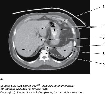
Which of the following statements is (are) true with regard to the two CT images seen below?
- Image A illustrates more superior structures.
- The images are sagittal reconstructions.
- The exam was performed without artificial contrast.
A 1 only
B 1 and 3 only
C 2 and 3 only
D 1, 2, and 3
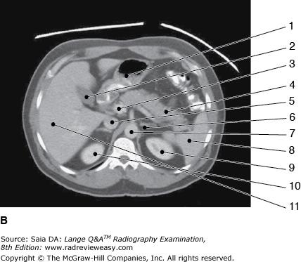
A 1 only
-The figures are axial CT images of the abdomen with contrast. In Figure A, the liver (number 1 left lobe; number 6 right lobe; number 3 caudate lobe), barium-filled stomach (number 2), spleen (number 5), and aorta (number 4) are seen in image A. In Figure B, more inferior structures such as the inferior vena cava (number 5) and kidneys (number 9 and number 10) are seen.
The sternal angle is at approximately the same level as the
A T2–3 interspace
B T9–10 interspace
C T5
D costal margin
C T5
-Surface landmarks, prominences, and depressions are very useful to the radiographer in locating anatomic structures that are not visible externally. The fifth thoracic vertebra is at approximately the same level as the sternal angle. The T2–3 interspace is about at the same level as the manubrial (suprasternal) notch. The costal margin is about the same level as L3.
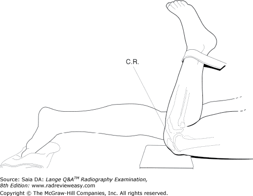
Which of the following positions is used to demonstrate vertical patellar fractures and the patellofemoral articulation?
A AP knee
B Lateral knee
C Tangential patella
D Tunnel view
C Tangential patella
-In the tangential (sunrise) projection of the patella, the CR is directed parallel to the longitudinal plane of the patella, thereby demonstrating a vertical fracture and providing the best view of the patellofemoral articulation. The AP knee projection could demonstrate a vertical fracture through the superimposed femur, but it does not demonstrate the patellofemoral articulation. The tunnel view of the knee is used to demonstrate the intercondyloid fossa.
AP stress studies of the ankle may be performed
- to demonstrate fractures of the distal tibia and fibula
- following inversion or eversion injuries
- to demonstrate a ligament tear
A 1 only
B 1 and 2 only
C 2 and 3 only
D 1, 2, and 3
C 2 and 3 only
-After forceful eversion or inversion injuries of the ankle, AP stress studies are valuable to confirm the presence of a ligament tear. Keeping the ankle in an AP position, the physician guides the ankle into inversion and eversion maneuvers. Characteristic changes in the relationship of the talus, tibia, and fibula will indicate ligament injury. Inversion stress demonstrates the lateral ligament, whereas eversion stress demonstrates the medial ligament. A fractured ankle would not be manipulated in this manner.
To demonstrate the glenoid fossa in profile, the patient is positioned
A 45 degrees oblique, affected side up.
B 45 degrees oblique, affected side down.
C 25 degrees oblique, affected side up.
D 25 degrees oblique, affected side down.
B 45 degrees oblique, affected side down.
-When viewing the glenoid fossa from the anterior, it is seen to angle posteriorly and laterally approximately 45 degrees. To view it in profile, then, it must be placed so that its surface is perpendicular to the IR. The patient is positioned in a 45-degree oblique, affected-side-down position, which places the glenoid fossa approximately perpendicular to the IR. The arm is abducted slightly, the elbow is flexed, and the hand and forearm are placed over the abdomen. The CR is directed perpendicular to the glenohumeral joint.
Which of the following criteria is (are) required for visualization of the greater tubercle in profile?
1.Epicondyles parallel to the IR
2.Arm in external rotation
3.Humerus in AP position
A 1 only
B 1 and 3 only
C 2 and 3 only
D 1, 2, and 3
D 1, 2, and 3
-The greater and lesser tubercles are prominences on the proximal humerus separated by the intertubercular (bicipital) groove. The AP projection of the humerus/shoulder places the epicondyles parallel to the IR and the shoulder in external rotation, and demonstrates the greater tubercle in profile.The lateral projection of the humerus places the shoulder in extreme internal rotation with the epicondyles perpendicular to the IR and demonstrates the lesser tubercle in profile.
Which projection of the foot will best demonstrate the longitudinal arch?
A Mediolateral
B Lateromedial
C Lateral weight-bearing
D 30-degree medial oblique
C Lateral weight-bearing
-The bones of the foot are arranged to form a number of longitudinal and transverse arches. The longitudinal arch facilitates walking and is evaluated radiographically in lateral weight-bearing (erect) projections. Recumbent laterals would not demonstrate any structural change that occurs when the individual is weight-bearing erect.
All elbow fat pads are best demonstrated in which position?
A AP
B Lateral
C Acute flexion
D AP partial flexion
B Lateral
-There are three important fat pads associated with the elbow. The anterior fat pad is located just anterior to the distal humerus. The posterior fat pad is located within the olecranon fossa at the distal posterior humerus. The supinator fat pad/stripe is located at the proximal radius just anterior to the head, neck, and tuberosity. The posterior fat pad is not visible radiographically in the normal elbow. All three fat pads can be demonstrated only in the lateral projection of the elbow.
A patient unable to extend his or her arm is seated at the end of the x-ray table, elbow flexed 90 degrees, with epicondyles perpendicular to IR. The CR is directed 45 degrees medially. Which of the following structures will be demonstrated best?
- Radial head
- Capitulum
- Coronoid process
A 1 only
B 1 and 2 only
C 2 and 3 only
D 1, 2, and 3
B 1 and 2 only
-The axial trauma lateral (Coyle) position is described. If routine elbow projections in extension are not possible because of limited part movement, this position can be used to demonstrate the coronoid process and/or radial head. With the elbow flexed 90 degrees, the epicondyles perpendicular to the IR, and the CR directed to the elbow joint at an angle of 45 degrees medially (i.e., toward the shoulder), the joint space between the radial head and capitulum should be revealed. With the elbow flexed 80 degrees and the CR directed to the elbow joint at an angle of 45 degrees laterally (i.e., from the shoulder toward the elbow), the elongated coronoid process will be visualized.
With which of the following does the lateral extremity of the clavicle articulate?
A Manubrium
B Coracoid process
C Coronoid process
D Acromion process
D Acromion process
-The S-shaped clavicle (“collar bone”) is usually the last bone to completely ossify, at about age 21, and is one of the most commonly fractured bones in young people. Its medial end articulates with the sternum to form the sternoclavicular joint; the clavicle articulates laterally with the scapula's acromion process, forming the acromioclavicular joint. Superior dislocation of the acromioclavicular joint is a common athletic injury.
Double-contrast examinations of the stomach or large bowel are performed to better visualize the
A position of the organ.
B size and shape of the organ.
C diverticula.
D gastric or bowel mucosa.
D gastric or bowel mucosa.
-Double-contrast studies of the stomach or large intestine involve coating the organ with a thin layer of barium sulfate, then introducing air. This permits seeing through the organ to structures behind it and, most especially, allows visualization of the mucosal lining of the organ. A barium-filled stomach or large bowel demonstrates the position, size, and shape of the organ and any lesion that projects out from its walls, such as diverticula. Polypoid lesions, which project inward from the wall of an organ, may go unnoticed unless a double-contrast examination is performed.

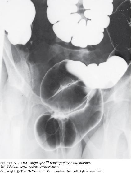
The structure indicated by the number 2 in Figure 6–11 is the
A ascending colon.
B descending colon.
C transverse colon.
D sigmoid colon.
B descending colon.
-The figure shown is a double-contrast BE, oblique position. Since the left colic/splenic flexure (number 1) is “open,” this is either a RPO or LAO position. Also demonstrated are the descending colon (number 2), and transverse colon (number 3). Barium has refluxed into the ileum (number 5).
Ingestion of barium sulfate is contraindicated in which of the following situations?
- Suspected perforation of a hollow viscus
- Suspected large bowel obstruction
- Preoperative patients
A 1 only
B 1 and 3 only
C 2 and 3 only
`D 1, 2, and 3
D 1, 2, and 3
-Barium sulfate suspension is the usual contrast medium of choice for investigation of the alimentary tract. There are, however, a few exceptions. Whenever there is the possibility of escape of contrast medium into the peritoneal cavity, barium sulfate is contraindicated, and a water-soluble iodinated medium is recommended because it is easily aspirated before surgery (or resorbed and excreted by the kidneys). Patients with a ruptured hollow viscus (e.g., perforated ulcer, diverticulitis, etc.), those with suspected large bowel obstruction, and those who are scheduled for surgery are examples of patients who should ingest only water-soluble iodinated media.
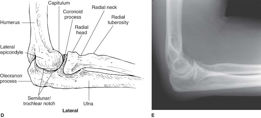
What portion of the humerus articulates with the ulna to help form the elbow joint?
A Semilunar/trochlear notch
B Radial head
C Capitulum
D Trochlea
D Trochlea
-The distal humerus articulates with the proximal radius and ulna to form the elbow joint. Specifically, the semilunar/trochlear notch of the proximal ulna articulates with the trochlea of the distal medial humerus. The capitulum is lateral to the trochlea and articulates with the radial head (Figure 2–50).
Which of the following is a vessel that does not carry oxygenated blood?
A Pulmonary vein
B Pulmonary artery
C Coronary artery
D Chordae tendineae
B Pulmonary artery
-Venous blood is returned to the right atrium of the heart via the superior (from the upper part of the body) and inferior (from the lower body) venae cavae and the coronary sinus (from the heart substance). During atrial systole, the blood passes through the tricuspid valve into the right ventricle. During ventricular systole, the blood is pumped through the pulmonary semilunar valve into the pulmonary artery (the only artery to carry unoxygenated blood) to the lungs for oxygenation. Blood is returned via the pulmonary veins (the only veins to carry oxygenated blood) to the left atrium. During atrial systole, blood passes through the mitral (bicuspid) valve into the left ventricle. During ventricular systole, the oxygenated blood is pumped through the aortic semilunar valve into the aorta. The coronary arteries supply oxygenated blood to the myocardium. The chordae tendineae are connective tissue fibers that help to limit the movement of valve flaps, preventing backflow of blood.
Which of the following examinations might require the use of 120 kVp?
1.AP abdomen
2.Chest radiograph
3.Barium-filled stomach
A 1 only
B 2 only
C 1 and 2 only
D 2 and 3 only
D 2 and 3 only
-High-kilovoltage factors are frequently used to even out densities in anatomic parts with high tissue contrast (eg, the chest). However, as high kilovoltage produces added scattered radiation, it generally must be used with a grid. It would be inappropriate to perform an AP abdomen with high kilovoltage because it has such low subject contrast. Barium-filled structures are frequently radiographed using 120 kV or more to penetrate the barium—to see through to structures behind.
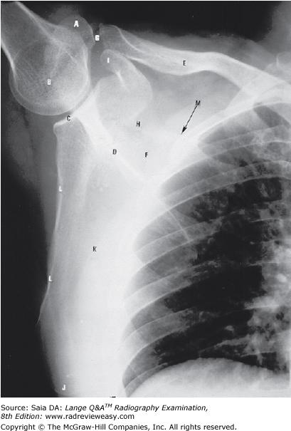
What is the structure indicated by the letter A in Figure 7–3?
A greater tubercle
B coronoid process
C coracoid process
D acromion process
D acromion process
-The radiograph illustrates an AP projection of the scapula; abduction of the arm moves the scapula away from the rib cage, revealing a greater portion of the scapula than would be visualized with the arm at the side. A number of bony structures are identified: the acromion process (A), the humeral head (B), glenoid fossa (C), scapular spine (D), clavicle (E), supraspinatus fossa (F), acromioclavicular joint (G), scapular notch (H), coracoid process (I), inferior angle/apex (j), body/costal surface (K), lateral/axillary border (L).
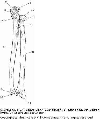
Which of the following correctly identifies the radial styloid process in the illustration in Figure A?
A Number 1
B Number 4
C Number 10
D Number 11
D Number 11
-An anterior view of the forearm is pictured. The proximal anterior surface of the ulna (number 8) presents a rather large pointed process at the anterior margin of the semilunar (trochlear) notch (5) called the coronoid process (6). The olecranon process is identified as number 4, and the radial notch of the ulna is number 7. Distally, the ulnar head is number 9, and the styloid process is labeled 10. The radius (number 12) is the lateral bone of the forearm. The radial head is number 3, the radial neck is number 2, and the radial tuberosity is number 1. Distally, the radial styloid process is labeled 11.
Which of the following should be demonstrated in a true AP projection of the clavicle?
- Clavicular body
- Acromioclavicular joint
- Sternocostal joint
A 1 only
B 1 and 2 only
C 2 and 3 only
D 1, 2, and 3
B 1 and 2 only
-The AP projection of the clavicle should demonstrate the clavicular body/shaft and its two extremities: the sternal extremity and its associated sternoclavicular articulation, and the acromial extremity and its associated acromioclavicular articulation. The sternocostal joint is the articulation between the sternum and rib and is not delineated in the AP clavicle image.
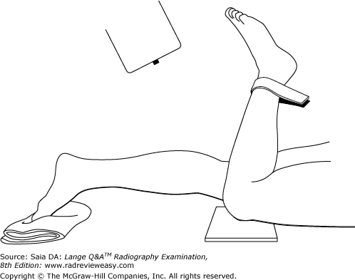
With the patient and the x-ray tube positioned as illustrated in Figure 2–2, which of the following will be visualized?
- Intercondyloid fossa
- Patellofemoral articulation
- Tangential patella
A 1 only
B 1 and 2 only
C 2 and 3 only
D 1, 2, and 3
C 2 and 3 only
-Note the relationship between the thigh, lower leg, patella, and CR. The CR is directed parallel to the plane of the patella, thereby providing a tangential projection of the patella (i.e., patella in profile) and an unobstructed view of the patellofemoral articulation (Figure 2–42). A tunnel view is required to demonstrate the intercondyloid fossa and the articulating surfaces of the tibia and femur.
All the following can be associated with the distal radius except
A head.
B styloid process.
C ulnar notch.
D radioulnar joint.
A head.
-The distal radius presents a styloid process laterally; the ulnar notch is located medially, helping to form the distal radioulnar articulation. The distal surface of the radius (carpal articular surface) is smooth for accommodating the scaphoid and lunate to form the radiocarpal (wrist) joint. The proximal radius has a cylindrical head with a medial surface that participates in the proximal radioulnar joint; its superior surface articulates with the capitulum of the humerus. Fracture of the distal radius is one of the most common skeletal fractures. Fractures of the radial head and neck frequently result from a fall onto an outstretched hand with the elbow partially flexed. Severe fractures often are accompanied by posterior dislocation of the elbow joint. Colles' fractures of the distal radius usually result from a fall onto an outstretched hand with the arm extended.
The following procedure can be employed to better demonstrate the carpal scaphoid:
1.elevate hand and wrist 20°.
2.place wrist in ulnar deviation.
3.angle CR 20° distally (toward fingers).
A 1 only
B 1 and 2 only
C 1 and 3 only
D 1, 2, and 3
B 1 and 2 only
-The carpal scaphoid is a curved, boat-shaped, bone, and is therefore superimposed on itself ("self-superimposition") in a routine PA projection. Since the scaphoid is the most frequently fractured carpal, special projections have been developed to help overcome self-superimposition. Stecher (in 1937) recommended elevating the hand and wrist 20° and using a perpendicular CR directed to the scaphoid. Effective variations of this position include employing ulnar deviation and angling the CR 20° proximally(toward the elbow). The 20° tube angulation would be used in place of the elevated hand/wrist.
All of the following bones are associated with condyles except the
A femur.
B tibia.
C fibula.
D mandible.

C fibula.
-The distal femur is associated with two large condyles; the deep depression separating them is the intercondyloid fossa (Fig. A). The proximal tibia has two condyles; their superior surfaces are smooth, forming the tibial plateau. The mandible has a condyle that articulates with the mandibular fossa of the temporal bone, forming the temporomandibular joint. The fibula has a proximal styloid process and a distal malleolus, but no condyle.

In which of the following positions was the radiograph in Figure A taken?
A RPO
B LPO
C AP axial
D Right lateral decubitus
B LPO
-The pictured radiograph is an oblique position of the large bowel, illustrating an "open" view of the hepatic/right colic flexure and ascending colon, with the splenic/left colic flexure superimposed on the descending colon. Therefore, the radiograph must have been made in either an RAO (if the patient was prone) or an LPO (if the patient was supine) position. The LAO and RPO positions are used to demonstrate the splenic/left colic flexure and descending colon free of self-superimposition. AP or PA axial is generally used to visualize the rectosigmoid colon.
The condition that allows blood to shunt between the right and left ventricles is called
A patent ductus arteriosus.
B coarctation of the aorta.
C atrial septal defect.
D ventricular septal defect.
D ventricular septal defect
-Ventricular septal defect is a congenital heart condition characterized by a hole in the interventricular septum that allows oxygenated and unoxygenated blood to mix. Some interventricular septal defects are small and close spontaneously; others require surgery. Coarctation of the aorta is a narrowing or constriction of the aorta. Atrial septal defect is a small hole (the remnant of the fetal foramen ovale) in the interatrial septum. It usually closes spontaneously in the first months of life; if it persists or is unusually large, surgical repair is necessary. The ductus arteriosus is a short fetal blood vessel connecting the aorta and pulmonary artery that usually closes within 10 to 15 hours after birth. A patent ductus arteriosus is one that persists and requires surgical closure.
Using the PA projection, which of the following tube angle and direction combinations is correct for an axial projection of the clavicle?
A 5 to 15 degrees caudad
B 5 to 15 degrees cephalad
C 15 to 30 degrees cephalad
D 15 to 30 degrees caudad
D 15 to 30 degrees caudad
-When the clavicle is examined in the PA axial projection, the CR must be directed 15 to 30 degrees caudad to project most of the clavicle's length above the ribs. The direction of the CR is reversed when examining the patient in the AP position.
Which of the anatomic structures listed below is seen most anteriorly in a lateral projection of the chest?
A Esophagus
B Trachea
C Cardiac apex
D Superimposed scapular borders
C Cardiac apex
-The relationship of these three structures is easily appreciated in a lateral projection of the chest. The heart is seen in the anterior half of the thoracic cavity, with its apex extending inferior and anterior. The air-filled trachea can be seen in about the center of the chest, and the air-filled esophagus is seen just posterior to the trachea (Figure 2–48). The superimposed vertebral and axillary borders of the scapulae would be seen most posteriorly.
A spontaneous fracture most likely would be associated with
A pathology.
B crepitus.
C trauma.
D metabolism.
A pathology.
-Spontaneous fractures most often affect bone weakened by a pathologic condition, for example, metastatic bone disease. The spontaneous fracture occurs suddenly, without trauma. One measure of a good radiographer is his or her ability to be cautious and resourceful when examining injured or debilitated patients having pathologic or traumatic conditions such as metastatic bone disease, arthritis, or bone fractures. Crepitus refers to a crackling sound made by a body part—such as the sound of fractured ends of bones rubbing together. Metabolism refers to the numerous energy and material transformations that take place in the body and is not associated with spontaneous fractures.
The ileocecal valve normally is located in which of the following body regions?
A Right iliac
B Left iliac
C Right lumbar
D Hypogastric
A Right iliac
-The abdomen is divided into nine regions. The upper lateral regions are the left and right hypochondriac, with the epigastric separating them. The middle lateral regions are the left and right lumbar, with the umbilical region between them. The lower lateral regions are the left and right iliac, with the hypogastric region between them. The ileocecal valve, cecum, and appendix (if present) are located in the lower right abdomen—therefore, the right iliac region
The manubrial notch is at approximately the same level as the
A fifth thoracic vertebra.
B T2–3 interspace.
C T4–5 interspace.
D costal margin.
B T2–3 interspace.
-Surface landmarks, prominences, and depressions are very useful to the radiographer in locating anatomic structures that are not visible externally. The fifth thoracic vertebra is at approximately the same level as the sternal angle. The T2–3 interspace is about at the same level as the manubrial (suprasternal) notch. The costal margin is about the same level as L3.
The AP Trendelenburg position is often used during an upper GI examination to demonstrate
A the duodenal loop
B filling of the duodenal bulb
C hiatal hernia
D hypertrophic pyloric stenosis
C hiatal hernia
-Placing the patient in a 20- to 30-degree AP Trendelenburg position during an upper GI examination helps to demonstrate the presence of a hiatal hernia. A 10- to 15-degree Trendelenburg position with the patient rotated slightly to the right also will help demonstrate regurgitation and hiatal hernia. Filling of the duodenal bulb and demonstration of the duodenal loop are best seen in the RAO position.Congenital hypertrophic pyloric stenosis is caused by excessive thickening of the pyloric sphincter. It is noted in infancy and characterized by projectile vomiting. The pyloric valve will let very little pass through, and as a result, the stomach becomes enlarged (hypertrophied).
The position illustrated in the figure below can be used successfully to demonstrate the
- PA oblique sternum
- barium-filled pylorus and duodenum
- left anterior axillary ribs
A 1 only
B 1 and 2 only
C 2 and 3 only
D 1, 2, and 3
D 1, 2, and 3
-The RAO position is shown. The barium-filled pylorus and duodenum are well demonstrated in this position, and the esophagus can be projected between the vertebrae and heart in this position. This RAO position is also used to superimpose the sternum onto the heart shadow to provide uniform receptor exposure throughout the sternum. The degree of obliquity depends on the patient's body habitus—greater obliquity is required for thinner chests. The RAO position is also used to see left anterior (axillary) ribs; in the anterior oblique positions, the affected side is away from the IR.
A near-frontal (AP/PA) view of the sternum is best accomplished in which of the following positions?
A AP
B PA
C RAO
D LAO
C RAO
-Because the sternum and vertebrae would be superimposed in a direct PA or AP projection, a slight oblique (just enough to separate the sternum from superimposition on the vertebrae) is used instead of a direct frontal (PA or AP) projection. In the RAO position, the heart superimposes a homogeneous tissue density over the sternum, thereby providing more clear radiographic visualization of its bony structure. If the LAO position were used to project the sternum to the right of the thoracic vertebrae, the posterior ribs and pulmonary markings would cast confusing shadows over the sternum because of their differing tissue densities.
The best projection to demonstrate the articular surfaces of the femoropatellar articulation is the
A AP knee.
B PA knee.
C tangential (“sunrise”) projection.
D tunnel view.
C tangential (“sunrise”) projection.
-The tangential (“sunrise”) projection is used to demonstrate the articular surfaces of the femur and patella. It is also used to demonstrate vertical fractures of the patella. The AP, PA, and oblique projections of the knee are used primarily to evaluate the joint space and articulating structures. The tunnel view is used to demonstrate the intercondyloid fossa.
Which of the following projections is most likely to demonstrate the carpal pisiform free of superimposition?
A Radial flexion/deviation
B Ulnar flexion/deviation
C AP (medial) oblique
D AP (lateral) oblique
C AP (medial) oblique
-In the direct PA projection of the wrist, the carpal pisiform is superimposed on the carpal triquetrum. The AP oblique projection (medial surface adjacent to the IR) separates the pisiform and triquetrum and projects the pisiform as a separate structure. The pisiform is the smallest and most palpable carpal.
The AP axial projection of the chest for pulmonary apices
- requires 15 to 20 degrees of cephalad angulation
- projects the apices above the clavicles
- should demonstrate the medial ends of the clavicles equidistant from the vertebral column
A 1 only
B 1 and 2 only
C 1 and 3 only
D 1, 2, and 3
C 1 and 3 only
-The AP axial projection is used to prevent the clavicles from superimposition on the pulmonary apices. A 15- to 20-degree cephalad angle projects the clavicles above the apices. The routine PA chest radiograph is evaluated for rotation by checking the distance between the medial ends of the clavicles and the lateral border of the vertebral column.
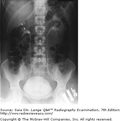
The number 1 in the radiograph in Figure A represents which of the following renal structures?
A Vesicoureteral junction
B Renal pelvis
C Minor calyx
D Major calyx
B Renal pelvis
-The pictured radiograph is one of a series of IVU (IU) images. It was done prone at 20 minutes after injection of the contrast medium. The urinary collecting system is well demonstrated. The renal pelvis(number 1) is the proximal expanded end of the ureter lying within the renal sinus. The minor calyces(number 3) receive urine from the collecting tubules of the renal pyramids and convey it to the major calyces (number 2), which empty into the renal pelvis. Urine is carried down the ureters by peristaltic waves.
Which of the following criteria are used to evaluate a PA projection of the chest?
1.Ten posterior ribs should be visualized.
2.Sternoclavicular joints should be symmetrical.
3.The scapulae should be lateral to the lung fields.
A 1 and 2 only
B 1 and 3 only
C 2 and 3 only
D 1, 2, and 3
D 1, 2, and 3
-To evaluate sufficient inspiration and lung expansion, 10 posterior ribs should be visualized. The sternoclavicular joints should be symmetrical; any loss of symmetry indicates rotation. To visualize maximum lung area, the shoulders are rolled forward to move the scapulae laterally from the lung fields.
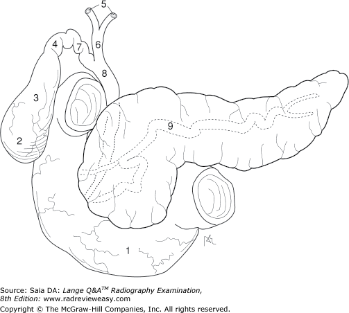
What is the structure indicated by the number 7 in Figure 2–18?
A Common hepatic duct
B Common bile duct
C Cystic duct
D Pancreatic duct
C Cystic duct
-Figure 2–18 illustrates a portion of the biliary system. Bile leaves the liver through the right and left hepatic ducts (number 5), which join to form the common hepatic duct (number 6). Bile enters the gallbladder through the cystic duct (number 7). The neck of the gallbladder is indicated by the number 4, its body by the number 3, and its fundus by the number 2. The gallbladder stores and concentrates bile, and when it contracts, bile flows out through the cystic duct and down the common bile duct (number 8). The common bile duct and pancreatic duct (number 9) unite to form the short hepatopancreatic ampulla (of Vater), which empties into the duodenum (number 1).
In the AP projection of the ankle, the
- plantar surface of the foot is vertical.
- fibula projects more distally than the tibia.
- calcaneus is well visualized.
A 1 only
B 1 and 2 only
C 2 and 3 only
D 1, 2, and 3
B 1 and 2 only
-To demonstrate the ankle joint space to best advantage, the plantar surface of the foot should be vertical in the AP projection of the ankle. Note that the fibula is the more distal of the two long bones of the lower leg and forms the lateral malleolus. The calcaneus is not well visualized in this projection because of superimposition with other tarsals.
Which of the following may be used to evaluate the glenohumeral joint?
1.Scapular Y projection
2.Inferosuperior axial
3.Transthoracic lateral
A 1 only
B 1 and 2 only
C 2 and 3 only
D 1, 2, and 3
D 1, 2, and 3
-The scapular Y projection is an oblique projection of the shoulder and is used to demonstrate anterior or posterior shoulder dislocation. The inferosuperior axial projection may be used to evaluate the glenohumeral joint when the patient is able to abduct the arm. The transthoracic lateral projection is used to evaluate the glenohumeral joint and upper humerus when the patient is unable to abduct the arm.

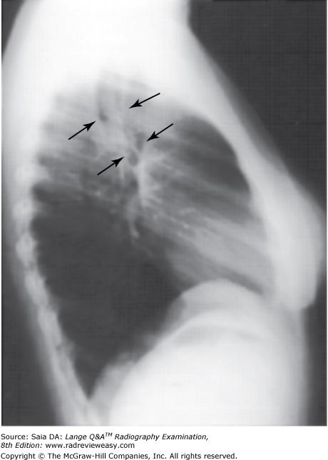
Which of the following statements is/are true regarding Figure A?
1.The radiograph was made in the LAO position.
2.The central ray should enter more inferiorly.
3.The sternum is projected onto the left side of the thorax.
A 1 only
B 2 only
C 2 and 3 only
D 1, 2, and 3
C 2 and 3 only
-The pictured radiograph is an RAO position of the sternum. The sternum is projected to the left side of the thorax, over the heart and other mediastinal structures in the RAO position, thus promoting more uniform receptor exposure. Although the upper limits of the sternum are well demonstrated in the figure, not all of the xiphoid process is seen, because the central ray was directed somewhat too superiorly. The central ray should be directed midway between the jugular (manubrial) notch and the xiphoid process.
Which of the following is most useful for bone age evaluation?
A Lateral skull
B PA chest
C AP pelvis
D PA hand
D PA hand
-A PA projection of the left hand and wrist is obtained most often to evaluate skeletal maturation. These images are compared with standard normal images for the age and sex of the child. Additional supplemental images may be requested.
The uppermost portion of the iliac crest is at approximately the same level as the
A costal margin
B umbilicus
C xiphoid tip
D fourth lumbar vertebra
D fourth lumbar vertebra
-Surface landmarks, prominences, and depressions are very useful to the radiographer in locating anatomic structures that are not visible externally. The costal margin is at about the same level as L3. The umbilicus is at approximately the same level as the L3–4 interspace; its location can be variable especially as body habitus varies. Bony landmarks are generally more reliable than soft tissue landmarks. The xiphoid tip is at about the same level as T10. The fourth lumbar vertebra is at approximately the same level as the iliac crest.
To demonstrate esophageal varices, the patient must be examined in
A the recumbent position
B the erect position
C the anatomic position
D the Fowler position
A the recumbent position
-Esophageal varices are tortuous dilatations of the esophageal veins. They are much less pronounced in the erect position and always must be examined with the patient recumbent. The recumbent position affords more complete filling of the veins because blood flows against gravity.
Knee arthrography may be performed to demonstrate a
1.torn meniscus.
2.Baker's cyst.
3.torn rotator cuff.
A 1 and 2 only
B 1 and 3 only
C 2 and 3 only
D 1, 2, and 3
A 1 and 2 only
-Knee arthrography may be performed to demonstrate torn meniscus (cartilage), Baker's cyst, loose bodies, and ligament damage. A torn rotator cuff would be demonstrated on a shoulder, not a knee arthrogram.
A patient in a recumbent position with the head lower than the feet is said to be in which of the following positions?
A Trendelenburg
B Fowler
C Sims
D Stenver
A Trendelenburg
-The patient is said to be in the Trendelenburg position when the head is positioned lower than the feet. This position is helpful in several radiographic procedures, such as separating redundant bowel loops and demonstration of hiatal hernias. It is also used in treating shock. In the Fowler position, the head is higher than the feet. The Sims position is the left anterior oblique (LAO) position with the right leg flexed up for insertion of the enema tip. The Stenver position is a radiographic position for radiographing the mastoids.
Which of the following projections of the abdomen may be used to demonstrate air or fluid levels?
1.Dorsal decubitus
2.Lateral decubitus
3.AP Trendelenburg
A 1 only
B 1 and 2 only
C 1 and 3 only
D 1, 2, and 3
B 1 and 2 only
-Air or fluid levels will be clearly demonstrated only if the central ray is directed parallel to them. Therefore, to demonstrate air or fluid levels, the erect or decubitus position should be used. Small amounts of fluid are best demonstrated in the lateral decubitus position, affected side down. Small amounts of air are best demonstrated in the lateral decubitus position, affected side up. Dorsal and ventral decubitus positions made with a horizontal x-ray beam can also be used to demonstrate air or fluid levels.
Which of the following views would best demonstrate arthritic changes in the knees?
A AP recumbent
B Lateral recumbent
C AP erect
D Medial oblique
C AP erect
-Arthritic changes in the knee result in changes in the joint bony relationships. These bony relationships are best evaluated in the AP position. Narrowing of the joint spaces is readily detected more on AP weight-bearing projections than on recumbent projections.
Which of the following statements regarding knee x-ray arthrography is (are) true?
- Ligament tears can be demonstrated.
- Sterile technique is observed.
- MRI can follow x-ray.
A 1 and 2 only
B 1 and 3 only
C 2 and 3 only
D 1, 2, and 3
D 1, 2, and 3
-X-ray arthrography requires the use of local anesthesia; sterile technique must be observed to avoid introducing infection into the joint. Fluoroscopy is used for proper placement of the needle and to obtain images immediately after the introduction of contrast medium. Many physicians follow up the x-ray arthrogram with an magnetic resonance (MR) arthrogram to visualize additional soft tissue structures. Arthrography is performed to detect compromised knee capsule structures, meniscal damage, ligament tears, and Baker cysts.
Particulate matter entering the respiratory bronchi can cause
A emphysema.
B empyema.
C pneumothorax.
D pneumoconiosis.
D pneumoconiosis.
-Pneumoconiosis is a condition of the lungs characterized by particulate matter having been deposited in lung tissue; it sometimes results in emphysema. Overdistension of the alveoli with air is emphysema.The condition is often a result of many years of smoking and is characterized by dyspnea, especially when recumbent. Empyema is pus in the thoracic cavity; pneumothorax is air or gas in the pleural cavity.
High-kilovoltage exposure factors are usually required for radiographic examinations using
1.water-soluble, iodinated media.
2.a negative contrast agent.
3.barium sulfate.
A 1 only
B 2 only
C 3 only
D 1 and 3 only
C 3 only
-Positive-contrast medium is radiopaque; negative-contrast material is radioparent. Barium sulfate (radiopaque, positive-contrast material) is most frequently used for examinations of the intestinal tract, and high-kVp exposure factors are used to penetrate (to see through and behind) the barium. Water-based iodinated contrast media (Conray, Amipaque) are also positive-contrast agents. However, the K-edge binding energy of iodine prohibits the use of much greater than 70 kVp with these materials. Higher kVp values will obviate the effect of the contrast agent. Air is an example of a negative-contrast agent, and high-kVp factors are clearly not indicated.
Esophageal varices are best demonstrated in which of the following positions?
A Erect
B Recumbent
C Fowler
D Sims
B Recumbent
-Esophageal varices are best demonstrated when there is increased venous pressure and when blood is flowing against gravity. Therefore, to demonstrate the twisted, dilated condition of venous varicosities, esophagograms must be performed in the recumbent position. In the erect position, the veins appear more smooth and normal. The Fowler position describes a position in which the patient's head is higher than the feet, and the Sims position is preferred for insertion of the enema tip.
Which of the following projections or positions will best demonstrate subacromial or subcoracoid dislocation?
A Tangential
B AP axial
C Transthoracic lateral
D PA oblique scapular Y
D PA oblique scapular Y
-The scapular Y refers to the characteristic Y formed by the humerus, acromion, and coracoid processes. The patient is placed in a PA oblique position—an RAO or LAO position depending on which is the affected side. The midcoronal plane is adjusted approximately 60 degrees to the IR, and the affected arm remains relaxed at the patient's side. The scapular Y position is employed to demonstrate anterior (subcoracoid) or posterior (subacromial) humeral dislocation. The humerus normally is superimposed on the scapula in this position; any deviation from this may indicate dislocation.
Which of the following projections is most likely to demonstrate the carpal pisiform free of superimposition?
A Radial flexion/deviation
B Ulnar flexion/deviation
C AP (medial) oblique
D AP (lateral) oblique
C AP (medial) oblique
-In the direct PA projection of the wrist, the carpal pisiform is superimposed on the carpal triquetrum. The AP oblique projection (medial surface adjacent to the IR) separates the pisiform and triquetrum and projects the pisiform as a separate structure. The pisiform is the smallest and most palpable carpal.
During an air-contrast BE, in what part of the colon is air most likely to be visualized with the body in the AP recumbent position?
A Transverse colon
B Descending colon
C Ascending colon
D Left and right colic flexures
A Transverse colon
-During a double-contrast BE, barium and air will distribute themselves according to the position of parts of the colon within the body and according to body position. When the body is in the AP recumbent position, the most anterior structures will be air filled. Anterior structures include the transverse colon and a portion of the sigmoid colon. Both flexures would be air filled in the erect position.
The pain experienced by an individual whose coronary arteries are not conveying sufficient blood to the heart is called
A tachycardia.
B bradycardia.
C angina pectoris.
D syncope.
C angina pectoris
-An individual whose coronary arteries are not carrying enough blood to the heart muscle (myocardium) as a result of partial or complete blockage of a cardiac vessel experiences crushing pain in the chest, frequently radiating to the left jaw and arm. This is termed angina pectoris. It may be relieved by the drug nitroglycerin, which dilates the coronary arteries, thus facilitating circulation. Tachycardia refers to rapid heart rate, and bradycardia, to slow heart rate. Syncope is fainting.
The functions of which body system include mineral homeostasis, protection, and triglyceride storage?
A Endocrine
B Integumentary
C Skeletal
D Muscular
C Skeletal
-The skeleton's design functions to protect vital internal organs such as the heart and lungs. Bone stores important minerals (e.g., calcium and phosphorus) and releases them into the blood as needed. Yellow bone marrow is composed mainly of fat cells and stores triglycerides for use as an energy reserve. The endocrine system is associated with hormone production; the integumentary system includes the skin that is important in protection and excretion; the muscular system is responsible for movement and heat production.
The secondary center of ossification in long bones is the
A periosteum.
B endosteum.
C epiphysis.
D diaphysis.
C epiphysis.
-Bones are classified as long, short, flat, and irregular. Many of the bones making up the extremities are long bones. Long bones have a shaft and two extremities (ends). The shaft (or diaphysis) of long bones is the primary ossification center during bone development. It is composed of compact tissue and covered with a membrane called periosteum. Within the shaft is the medullary cavity, which contains bone marrow and is lined by the membrane called endosteum. In the adult, yellow marrow occupies the shaft, and red marrow is found within the proximal and distal extremities of long bones. The secondary ossification center, the epiphysis, is separated from the diaphysis in early life by a layer of cartilage, the epiphyseal plate. As bone growth takes place, the epiphysis becomes part of the larger portion of bone and the epiphyseal plate disappears, but a characteristic line remains and is thereafter recognizable as the epiphyseal line.
To best visualize the lower ribs, the exposure should be made
A on normal inspiration
B on inspiration, second breath
C on expiration
D during shallow breathing
C on expiration
-Full or forced expiration is used to elevate the diaphragm and demonstrate the ribs below the diaphragm to best advantage (with exposure adjustment). Deep inspiration is used to depress the diaphragm and demonstrate as many ribs above the diaphragm as possible. Shallow breathing is used occasionally to visualize the ribs above the diaphragm while obliterating pulmonary vascular markings.
In which of the following positions/projections will the talocalcaneal joint be visualized?
A Dorsoplantar projection of the foot
B Plantodorsal projection of the calcaneus
C Medial oblique position of the foot
D Lateral foot
B Plantodorsal projection of the calcaneus
-The talocalcaneal, or subtalar, joint is a three-faceted articulation formed by the talus and the os calcis (calcaneus). The plantodorsal and dorsoplantar projections of the os calcis should exhibit sufficient receptor exposure to visualize the talocalcaneal joint (Figure 2–60). This is the only “routine” projection that will demonstrate the talocalcaneal joint. If evaluation of the talocalcaneal joint is desired, special views (such as the Broden and Isherwood methods) are required.
Which of the following projections/positions would best demonstrate structure number 6 seen in Figure 7–7?
A PA projection
B Lateral projection
C AP external oblique
D AP internal oblique
B Lateral projection
-The figure shows a posterior view of the elbow. The distal posterior humerus (number 1) is seen, as well as the proximal posterior radius (number 4) and ulna (number 3). Additional structures identified are the medial epicondyle (number 2), the olecranon fossa (number 5), olecranon process (number 6), lateral epicondyle (number 7), and radial head (number 8) The olecranon process (number 6) can best be demonstrated in the lateral projection; it can also be demonstrated in the acute flexion position. The AP internal oblique will demonstrate the coronoid process; the AP external oblique will demonstrate the radial head free of superimposition.
Which of the following positions is required to demonstrate small amounts of fluid in the pleural cavity?
A Lateral decubitus, affected side up
B Lateral decubitus, affected side down
C AP Trendelenburg
D AP supine
B Lateral decubitus, affected side down
-Air or fluid levels will be clearly delineated only if the central ray is directed parallel to them. Therefore, to demonstrate air or fluid levels, the erect or decubitus position should be used. Small amounts of fluid within the pleural space are best demonstrated in the lateral decubitus position, affected side down. Small amounts of air within the pleural space are best demonstrated in the lateral decubitus position, affected side up.
Correct preparation for a patient scheduled for a lower GI series is most likely to be
A iodinated contrast evening before examination; water only in the morning.
B NPO after midnight.
C cathartics and cleansing enemas.
D NPO after midnight, cleansing enemas, and filled bladder.
C cathartics and cleansing enemas.
-Diagnostic x-ray examinations that require contrast agents include upper GI series, lower GI series (BE), IVU, and the occasional GB series. Patient preparation is somewhat different for each of these examinations. An iodinated contrast agent, usually in the form of several pills, is taken by the patient the evening before a scheduled GB examination, and only water is allowed the morning of the examination. The patient scheduled for an upper GI series must receive NPO (nothing by mouth) after midnight. A lower GI series (BE) requires that the large bowel be very clean prior to the administration of barium; this requires the administration of cathartics (laxatives) and cleansing enemas.
Preparation for an IVU requires that the patient be NPO after midnight; some institutions also require that the large bowel be cleansed of gas and fecal material. Aftercare for barium examinations is very important. Patients typically are instructed to take milk of magnesia, increase their intake of fiber, drink plenty of water, and expect changes in stool color until all barium is evacuated and to call their physician if they do not have a bowel movement within 24 hours. Because water is removed from the barium sulfate suspension in the large bowel, it is essential to make patients understand the importance of these instructions to avoid barium impaction in the large bowel. The use of barium sulfate suspensions is contraindicated when ruling out visceral perforation.
To demonstrate esophageal varices, the patient must be examined in
A the recumbent position.
B the erect position.
C the anatomic position.
D Fowler's position.
A the recumbent position.
-Esophageal varices are tortuous dilatations of the esophageal veins. They are much less pronounced in the erect position and must always be examined with the patient recumbent. The recumbent position affords more complete filling of the veins, as blood flows against gravity.
Abnormal accumulation of air in pulmonary tissues, resulting in overdistention of the alveolar spaces, is
A emphysema.
B empyema.
C pneumothorax.
D pneumoconiosis.
A emphysema
-Overdistention of the alveoli with air is emphysema. The condition is often a result of many years of smoking and is characterized by dyspnea, especially when recumbent. Empyema is pus in the thoracic cavity; pneumothorax is air or gas in the pleural cavity. Pneumoconiosis is a condition of the lungs characterized by particulate matter having been deposited in lung tissue; it sometimes results in emphysema.
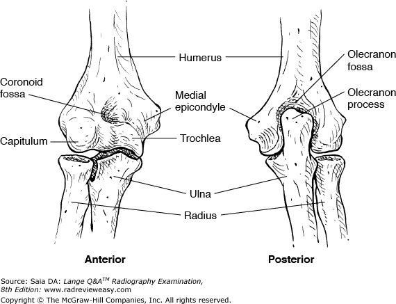
Which of the following is (are) located on the distal aspect of the humerus?
1.Capitulum
2.Intertubercular groove
3.Coronoid fossa
A 1 only
B 1 and 2 only
C 1 and 3 only
D 1, 2, and 3
C 1 and 3 only
-The distal humerus articulates with the radius and ulna to form the elbow joint. The lateral aspect of the distal humerus presents a raised, smooth, rounded surface, the capitulum, that articulates with the superior surface of the radial head. The trochlea is on the medial aspect of the distal humerus and articulates with the semilunar notch of the ulna. Just proximal to the capitulum and the trochlea are the lateral and medial epicondyles; the medial is more prominent and palpable. The coronoid fossa is found on the anterior distal humerus and functions to accommodate the coronoid process with the elbow in flexion. The intertubercular (bicipital) groove is located on the proximal humerus.
An acute infection of the lungs is called
A atelectasis.
B pneumothorax.
C pneumonia.
D COPD.
C pneumonia
-Pneumonia is an acute infection of the lung parenchyma characterized by productive cough, chest pain, fever, and chills and frequently accompanied by rales. Atelectasis is partial or complete collapse of a lung or lobe of a lung. Pneumothorax is the condition of air or gas in the pleural space. COPD (chronic obstructive pulmonary disease) is the name given to a number of disease processes that decrease the lung's ability to perform its function of ventilation.
During an intravenous urogram (IVU), the RPO position is used to demonstrate the
- left kidney parallel to the IR
- right kidney parallel to the IR
- right kidney perpendicular to the IR
A 1 only
B 2 only
C 1 and 2 only
D 1 and 3 only
D 1 and 3 only
-Since the kidneys do not lie parallel to the IR in the AP position, the oblique positions are used during IVU to visualize them better. With the AP oblique projections (i.e., RPO and LPO), the kidney that is farther away is placed parallel to the IR, and the kidney that is closer is placed perpendicular to the IR. Therefore, in the RPO position, the right kidney, being closer, is perpendicular to the IR. The left kidney, the one farther away, is placed parallel to the IR.
Conditions in which there is a lack of normal bone calcification include
- rickets.
- osteomalacia.
- osteoarthritis.
A 1 only
B 1 and 2 only
C 2 and 3 only
D 1, 2, and 3
B 1 and 2 only
-Rickets and osteomalacia are disorders in which there is softening of bone. Rickets results from a deficiency of vitamin D and usually is found affecting the growing bones of young children. The body's weight on the soft bones of the legs results in bowed and misshapen legs. Osteomalacia is an adult condition in which new bone fails to calcify. It is a painful condition and can result in easily fractured bones, especially in the lower extremities. Osteoarthritis is seen often in the elderly and is characterized by degeneration of articular cartilage in adjacent bones. The resulting rubbing of bone against bone results in pain and deterioration.
In which of the following examinations is exposure on full expiration required?
A PA chest
B Below diaphragm ribs
C AP lordotic chest
D Lateral thoracic spine
B Below diaphragm ribs
-Full or forced expiration is used to elevate the diaphragm and demonstrate the ribs below the diaphragm to best advantage. Deep inspiration is used to depress the diaphragm and demonstrate as many ribs above the diaphragm as possible. Shallow breathing is used occasionally to visualize the ribs above the diaphragm while obliterating pulmonary vascular markings. Shallow breathing is also used during exposure of the lateral thoracic spine and functions to blur prominent vascular markings. Posteroanterior (PA) and apical lordotic chest radiographs require full inspiration.
To evaluate the interphalangeal joints in the oblique and lateral positions, the fingers
A rest on the cassette for immobilization
B must be supported parallel to the IR
C are radiographed in natural flexion
D are radiographed in palmar flexion
B must be supported parallel to the IR
-The fingers must be supported parallel to the IR (e.g., on a finger sponge) in order that the joint spaces parallel the x-ray beam. When the fingers are flexed or resting on the cassette, the relationship between the joint spaces and the IR changes, and the joints appear “closed.”
Which of the following pathologic conditions would require an increase in exposure factors?
A Pneumoperitoneum
B Obstructed bowel
C Renal colic
D Ascites
D Ascites
-Because pneumoperitoneum is an abnormal accumulation of air or gas in the peritoneal cavity, it would require a decrease in exposure factors. Obstructed bowel usually involves distended, air- or gas-filled bowel loops, again requiring a decrease in exposure factors. With ascites, there is an abnormal accumulation of fluid in the abdominal cavity, necessitating an increase in exposure factors. Renal colic is the pain associated with the passage of renal calculi; no change from the normal exposure factors is usually required.
The position illustrated in the radiograph in Figure 2–28 may be obtained with the patient
- supine and the CR angled 30 degrees caudad.
- supine and the CR angled 30 degrees cephalad.
- prone and the CR angled 30 degrees caudad.
A 1 only
B 2 only
C 1 and 3 only
D 2 and 3 only
D 2 and 3 only
-A double-contrast examination of the large bowel is performed to see through the bowel to its posterior wall and to visualize any intraluminal lesions or masses. Oblique projections are used to “open up” the flexures—the RAO for the hepatic flexure and the LAO for the splenic flexure. The redundant S-shaped sigmoid can be demonstrated with the patient supine or prone. In the AP recumbent position the CR is directed 30 to 40 degrees cephalad. In the PA recumbent position the CR is directed 30 to 40 degrees caudal.
Evaluation criteria for a lateral projection of the humerus include
- epicondyles parallel to the IR
- lesser tubercle in profile
- superimposed epicondyles
A 1 only
B 1 and 3 only
C 2 and 3 only
D 1, 2, and 3
C 2 and 3 only
-The greater and lesser tubercles are prominences on the proximal humerus separated by the intertubercular (bicipital) groove. The lateral projection of the humerus places the shoulder in extreme internal rotation with the epicondyles perpendicular to the IR and superimposed. The lateral projection of the humerus should demonstrate the lesser tubercle in profile. The AP projection of the humerus/shoulder places the epicondyles parallel to the IR and the shoulder in external rotation and demonstrates the greater tubercle in profile.
Which of the following statements regarding the Norgaard method, “Ball-Catcher's position,” is (are) correct?
- Bilateral AP oblique hands are obtained.
- It is used for early detection of rheumatoid arthritis.
- The hands are obliqued about 45 degrees, palm up.
A 1 only
B 1 and 2 only
C 2 and 3 only
D 1, 2, and 3
D 1, 2, and 3
-Bilateral AP oblique hands are obtained using the Norgaard method or “Ball-Catcher position.” The method is used to detect early rheumatoid arthritis changes or fracture to the base of the fifth metacarpal. The hands are positioned and supported in a 45-degree oblique, palm-up position. The CR is directed to the level of the fifth metacarpophalangeal joint (MPJ) midway between the hands—both hands are exposed simultaneously.
A lateral projection of the hand in extension is often recommended to evaluate
- a fracture
- a foreign body
- soft tissue
A 1 only
B 2 only
C 2 and 3 only
D 1 and 3 only
C 2 and 3 only
-The lateral hand in extension, with appropriate technique adjustment, is recommended to evaluate foreign-body location in soft tissue. A small lead marker frequently is taped to the spot thought to be the point of entry. The physician then uses this external marker and the radiograph to determine the exact foreign-body location. Extension of the hand in the presence of a fracture would cause additional and unnecessary pain and possibly additional injury.
Which of the following projection(s) require(s) that the shoulder be placed in internal rotation?
1.AP humerus
2.AP thumb
3.Lateral humerus
A 1 only
B 1 and 2 only
C 2 and 3 only
D 1, 2, and 3
C 2 and 3 only
-When the arm is placed in the AP position, the epicondyles are parallel to the plane of the IR and the shoulder is placed in external rotation. In this position, an AP projection of the humerus, elbow, and forearm can be obtained; it places the greater tubercle of the humerus in profile. For the lateral projection of the humerus, the arm is internally rotated, elbow somewhat flexed, with the back of the hand against the thigh and the epicondyles superimposed and perpendicular to the IR. The AP projection of the thumb requires that the arm extended and internally rotated, placing the posterior surface of the thumb on the IR. The lateral projections of the humerus, elbow, and forearm all require that the epicondyles be perpendicular to the plane of the IR.
Which of the following positions can be used to effectively demonstrate the left colic flexure during radiographic examination of the large bowel?
- RAO
- LAO
- RPO
A 1 only
B 1 and 2 only
C 1 and 3 only
D 2 and 3 only
D 2 and 3 only
-The hepatic (right colic) and splenic (left colic) flexures are not generally well demonstrated in the AP and PA projections. To “open” the flexures, oblique projections are required. The left anterior oblique (LAO, left PA oblique) and right posterior oblique (RPO, right AP oblique) positions are used to demonstrate the splenic flexure. The hepatic flexure is usually well demonstrated in the RAO and LPO positions.
The esophagus commences at about the level of
A C3.
B C6.
C T1.
D T11.
B C6.
-The esophagus is a musculomembranous tube commencing at about the level of the cricoid cartilage, that is, C5–6. It is located posterior to the larynx and trachea and extends to about the level of T11, where it joins with the proximal stomach.
Gas-producing powder or crystals usually are ingested for which of the following examinations?
A Double-contrast barium enema (BE)
B Double-contrast gastrointestinal (GI) series
C Oral cholecystogram
D IV urogram (IVU)
B Double-contrast gastrointestinal (GI) series
-A double-contrast GI examination requires that the patient ingest gas-producing powder, crystals (eg. calcium and magnesium citrate), pills, or beverage followed by a small amount of high-density barium. The patient then may be asked to roll in the recumbent position in order to coat the gastric mucosa while the carbon dioxide expands. This procedure provides optimal visualization of the gastric walls. Although a double-contrast BE uses a negative contrast agent, it is not ingested but rather is delivered rectally. An oral cholecystogram can be performed approximately 3 hours after ingestion of special ipodate calcium granules. An IVU requires an IV injection of iodinated contrast medium.
The tarsals and metatarsals are arranged to form the
1.transverse arch.
2.longitudinal arch.
3.oblique arch.
A 1 only
B 1 and 2 only
C 2 and 3 only
D 1, 2, and 3
B 1 and 2 only
-The tarsals and metatarsals of the foot are arranged so as to form two arches: the transverse and the longitudinal (which has two parts—lateral and medial). The arches function to support and distribute the body's weight over the body. The ball of the foot usually accommodates about 40 percent of the body's weight, and the heel about 60 percent.
All the following positions are used frequently to demonstrate the sternoclavicular articulations except
A weight-bearing
B RAO
C LAO
D PA
A weight-bearing
-Sternoclavicular articulations may be examined with the patient PA, either bilaterally with the patient's head resting on the chin or unilaterally with the patient's head turned toward the side being examined. The sternoclavicular articulations also may be examined in the oblique position, with either the patient rotated slightly or the CR angled slightly medialward. Weight-bearing positions are used frequently for evaluation of acromioclavicular joints.
When a GI series has been requested on a patient with a suspected perforated ulcer, the type of contrast medium that should be used is
A a thin barium sulfate suspension.
B a thick barium sulfate suspension.
C water-soluble iodinated media.
D oil-based iodinated media.
C water-soluble iodinated media.
-Whenever a perforation of the GI tract is suspected, a water-soluble contrast agent (such as Gastrografin or oral Hypaque) should be used because it is easily absorbed from within the peritoneal cavity. Leakage of barium sulfate into the peritoneal cavity can have serious consequences. Water-soluble contrast agents also may be used in place of barium sulfate when the possibility of barium impaction exists. Oil-based contrast agents are used rarely today.
What process is best seen using a perpendicular CR with the elbow in acute flexion and with the posterior aspect of the humerus adjacent to the image receptor?
A Coracoid
B Coronoid
C Olecranon
D Glenoid
C Olecranon
-When the elbow is placed in acute flexion with the posterior aspect of the humerus adjacent to the image receptor and a perpendicular CR is used, the olecranon process of the ulna is seen in profile. The coronoid process is best visualized in the medial oblique position. The coracoid and glenoid are associated with the scapula
Compared with that of the hypersthenic and sthenic body types, the gallbladder of an asthenic patient is most likely to be located
A higher and more medial
B lower and more medial
C higher and more lateral
D lower and more lateral
B lower and more medial
-The four types of body habitus describe differences in visceral shape, position, tone, and motility. One body type is hypersthenic, the very large individual with short, wide heart and lungs, high transverse stomach and gallbladder, and peripheral colon. The sthenic individual is the average, athletic, most predominant type. The hyposthenic patient is somewhat thinner and a little more frail, with organs positioned somewhat lower. The asthenic type is smaller in the extreme, with a long thorax, a very long, almost pelvic stomach, and a low medial gallbladder. The asthenic colon is medial and redundant.
Which of the following is (are) accurate positioning or evaluation criteria for an AP projection of the normal knee?
- Femorotibial interspaces equal bilaterally.
- Patella superimposed on distal tibia.
- CR enters ½ in. distal to base of patella.
A 1 only
B 1 and 2 only
C 1 and 3 only
D 1, 2, and 3
A 1 only
-In the AP projection of the normal knee, the space between the tibial plateau and the femoral condyles is equal bilaterally. It is, therefore, important that there be no pelvic rotation that could change the appearance of an otherwise normal relationship. The AP projection of the knee superimposes the patella and femur. The CR should enter at the knee joint, located ½ in. distal to the patellar apex.
All of the following statements regarding respiratory structures are true except
A the right lung has two lobes.
B the uppermost portion of the lung is the apex.
C each lung is enclosed in pleura.
D the trachea bifurcates into mainstem bronchi.
A the right lung has two lobes.
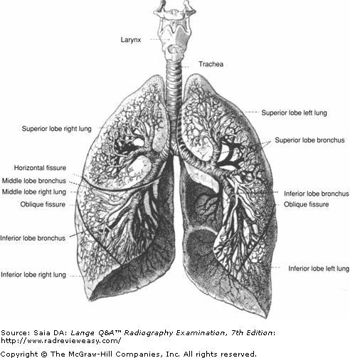
-The trachea (windpipe) bifurcates into left and right mainstem bronchi, each entering its respective lung hilum. The left bronchus divides into two portions, one for each lobe of the left lung. The right bronchus divides into three portions, one for each lobe of the right lung (Fig. A). The lungs are conical in shape, consisting of upper pointed portions, termed the apices (plural of apex), and broad lower portions (orbases). The lungs are enclosed in a double-walled serous membrane called the pleura
Which surface of the forearm must be adjacent to the IR to obtain a lateral projection of the fourth finger with optimal spatial resolution?
A Anterior
B Posterior
C Medial
D Lateral
C Medial
-A lateral projection of the fourth finger is best obtained if the finger is positioned so that there is as little OID as possible. Therefore, with only the fourth finger extended in the lateral position, the arm is positioned on the ulnar (medial) surface. This places the finger closer to the IR than if it were positioned radial side down. Excessive magnification distortion is avoided, and better spatial resolution is obtained.
The first carpometacarpal joint is formed by the articulation of the base of the first metacarpal and the
A distal radius.
B distal ulna.
C scaphoid.
D trapezium.
D trapezium.
-The bases of the proximal row of phalanges articulate with the heads of the metacarpals to form the (condyloid) metacarpophalangeal joints, which permit flexion and extension, abduction and adduction, and circumduction. The bases of the metacarpals articulate with each other and the distal row of carpals at the carpometacarpal joints. The first carpometacarpal joint (thumb) is a saddle joint, permitting flexion and extension, abduction and adduction, and circumduction; it is formed by the articulation of the base of the first metacarpal and the trapezium.
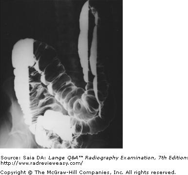
The radiograph pictured in Figure A may be used to evaluate
1.polypoid lesions.
2.the lateral wall of the descending colon.
3.the posterior wall of the rectum.
A 1 only
B 1 and 2 only
C 2 and 3 only
D 1, 2, and 3
B 1 and 2 only
-The pictured radiograph was made in the right lateral decubitus position. It is part of a series of radiographs made during an air-contrast (double-contrast) BE examination. A double-contrast examination of the large bowel is performed to see through the bowel to its posterior wall and to visualize any intraluminal (eg, polypoid) lesions or masses. Various body positions are used to redistribute the barium and air. To demonstrate the medial and lateral walls of the bowel, decubitus positions are performed. The radiograph presents a right lateral decubitus position, because the barium has gravitated to the right side (the side of the hepatic flexure). The air rises and delineates the medial side of the ascending colon and the lateral side of the descending colon. The posterior wall of the rectum could be visualized using the ventral decubitus position and a horizontal beam lateral of the rectum.
What projection of the calcaneus is obtained with the leg extended, the plantar surface of the foot vertical and perpendicular to the IR, and the CR directed 40 degrees cephalad?
A Axial plantodorsal projection
B Axial dorsoplantar projection
C Lateral projection
D Weight-bearing lateral projection
A Axial plantodorsal projection
-The plantodorsal projection of the os calsis/calcaneus is described. It is performed supine and requires cephalad angulation. The CR enters the plantar surface and exits the dorsal surface. The axial dorsoplantar projection requires that the CR enter the dorsal surface of the foot and exit the plantar surface.
Which of the following articulates with the base of the first metatarsal?
A First cuneiform
B Third cuneiform
C Navicular
D Cuboi
A First cuneiform
-The base of the first metatarsal articulates with the first (medial) cuneiform. The base of the second metatarsal articulates with the second (intermediate) cuneiform; the third base of the metatarsal articulates with the third (lateral) cuneiform. The bases of the fourth and fifth metatarsals articulate with the cuboid. The navicular articulates with the first and second cuneiforms anteriorly and the talus posteriorly.
Which of the following projections will best demonstrate the tarsal navicular free of superimposition?
A AP oblique, medial rotation
B AP oblique, lateral rotation
C Mediolateral
D Lateral weight-bearing
A AP oblique, medial rotation
-The medial oblique projection requires that the leg be rotated medially until the plantar surface of the foot forms a 30-degree angle with the IR. This position demonstrates the navicular with minimal bony superimposition. The lateral oblique projection of the foot superimposes much of the navicular on the cuboid. The navicular is also superimposed on the cuboid in lateral projections.
Which of the following is recommended to better demonstrate the tarsometatarsal joints in a dorsoplantar projection of the foot?
A Invert the foot.
B Evert the foot.
C Angle the CR 10 degrees posteriorly.
D Angle the CR 10 degrees anteriorly.
C Angle the CR 10 degrees posteriorly.
-In the dorsoplantar projection of the foot, the CR may be directed perpendicularly or angled 10 degrees posteriorly. Angulation serves to “open” the tarsometatarsal joints that are not well visualized on the dorsoplantar projection with perpendicular ray. Inversion and eversion of the foot do not affect the tarsometatarsal joints.

Which of the following statements is (are) true regarding the radiograph in Figure 2–12?
- The patient is placed in an RAO position.
- The midcoronal plane is about 60 degrees to the IR.
- The acromion process is free of superimposition.
A 1 only
B 1 and 2 only
C 2 and 3 only
D 1, 2, and 3
D 1, 2, and 3
-A right scapular Y is illustrated; this refers to the characteristic Y formed by the clearly visible humerus, acromion, and coracoid. The patient is positioned in a PA oblique position—in this case, an RAO projection to demonstrate the right side. The MCP is adjusted to approximately 60 degrees to the IR, and the affected arm is left relaxed at the patient's side. The scapular Y position is employed to demonstrate anterior or posterior humeral dislocation. The humerus is superimposed on the scapula in this position; any deviation from this may indicate dislocation.
Correct preparation for a patient scheduled for an upper gastrointestinal (GI) series is most likely to be
A iodinated contrast administration evening before examination; water only in the morning
B NPO after midnight
C cathartics and cleansing enemas
D NPO after midnight, cleansing enemas, and empty bladder before scout film
B NPO after midnight
-Diagnostic x-ray examinations that require contrast agents include upper gastrointestinal (GI) series, lower GI series (BE), intravenous urogram (IVU), and the occasional gallbladder (GB) series. Patient preparation is somewhat different for each of these examinations. An iodinated contrast agent, usually in the form of several pills, is taken by the patient the evening before a scheduled GB examination, and only water is allowed the morning of the examination. The patient scheduled for an upper GI series must receive NPO (nothing by mouth) after midnight. A lower GI series (BE) requires that the large bowel be very clean prior to the administration of barium; this requires the administration of cathartics(laxatives) and cleansing enemas. Preparation for an IVU requires that the patient be NPO after midnight; some institutions also require that the large bowel be cleansed of gas and fecal material. Aftercare for barium examinations is very important. Patients typically are instructed to take milk of magnesia, increase their intake of fiber, drink plenty of water, and expect changes in stool color until all barium is evacuated and to call their physician if they do not have a bowel movement within 24 hours. Because water is removed from the barium sulfate suspension in the large bowel, it is essential to make patients understand the importance of these instructions to avoid barium impaction in the large bowel. The use of barium sulfate suspensions is contraindicated when ruling out visceral perforation.
Which of the following statements is (are) correct with respect to postoperative cholangiography?
- A T-tube is in place in the common bile duct.
- Water-soluble contrast material is injected.
- The patency of biliary ducts is evaluated.
A 1 only
B 1 and 2 only
C 2 and 3 only
D 1, 2, and 3
D 1, 2, and 3
-Postoperative, or T-tube, cholangiography frequently is performed to evaluate the patency of the biliary ducts and to identify any previously undetected stones. Following surgery, a T-tube is left in place within the common bile duct, with the vertical portion of the T extending outside the body. Water-soluble iodinated contrast medium is injected, and fluoroscopic examination is carried out.
Shoulder arthrography is performed to
- evaluate humeral luxation
- demonstrate complete or partial rotator cuff tear
- evaluate the glenoid labrum
A 1 only
B 1 and 2 only
C 2 and 3 only
D 1, 2, and 3
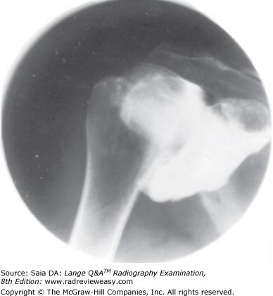
C 2 and 3 only
-Shoulder arthrograms (Figure 2–64) are used to evaluate rotator cuff tear, glenoid labrum (a ring of fibrocartilaginous tissue around the glenoid fossa), and frozen shoulder. Routine radiographs demonstrate arthritis, and the addition of a transthoracic humerus or scapular Y projection would be used to demonstrate luxation (dislocation).
In the AP knee projection of an asthenic patient who measures less than 19 cm from the anterior superior iliac spine (ASIS) to tabletop, the CR should be directed
A perpendicularly
B 5 degrees medially
C 5 degrees cephalad
D 5 degrees caudad
D 5 degrees caudad
-In the AP projection of the knee, the position of the joint space is significantly affected by the patient's overall body habitus and the distance between the ASIS and tabletop. When the patient is of sthenic habitus with a distance of 19 to 24 cm between the ASIS and tabletop, the CR is directed perpendicularly. When the patient is of asthenic habitus with a distance of less than 19 cm between the ASIS and tabletop, the CR is directed 5 degrees caudad. With a patient with a hypersthenic habitus and an ASIS-to-table measurement of greater than 24 cm, the CR is directed 5 degrees cephalad.
Demonstration of which anatomic structures require(s) ingestion of barium sulfate suspension?
- Duodenum
- Pylorus
- Ilium
A 1 only
B 1 and 2 only
C 2 and 3 only
D 1, 2, and 3
B 1 and 2 only
-Oral administration of barium sulfate is used to demonstrate the upper digestive system—the esophagus, fundus, body, and pylorus of the stomach—and barium progression through the small bowel. The small bowel includes the duodenum, jejunum, and ileum (ilium is part of the pelvis).
The patient usually is required to drink barium sulfate suspension in order to demonstrate which of the following structures?
- Descending duodenum
- Ilium
- Splenic flexure
A 1 only
B 1 and 3 only
C 2 and 3 only
D 3 only
A 1 only
-Oral administration of barium sulfate is used to demonstrate the upper digestive system, esophagus, fundus, and body and pylorus of the stomach and barium progression through the small bowel/intestine. The small intestine is composed of the duodenum, jejunum, and ileum. The duodenum is the shortest portion, beginning just beyond the pyloric sphincter and dividing into four portions—the duodenal cap or bulb, the descending duodenum, the transverse duodenum, and the ascending duodenum. These portions form a C-shaped loop that is occupied by the head of the pancreas. The descending portion receives the hepatopancreatic ampulla and duodenal papilla. The ascending portion terminates at the duodenojejunal flexure (angle of Treitz). Whereas the position of the short (9 in.) duodenum is fixed, the jejunum (9 ft) and ileum (13 ft) are very mobile. The large bowel, including the cecum, usually is demonstrated via rectal administration of barium. The approximately 5-ft-long large intestine (colon) begins at the terminus of the small intestine; its first portion is the dilated saclike cecum, located inferior to the ileocecal valve. Projecting posteromedially from the cecum is the short vermiform appendix. The ascending colon is continuous with the cecum, bending and forming the right colic (hepatic) flexure. The colon traverses the abdomen as the transverse colon and bends to form the left colic (splenic) flexure. The descending colon continues down the left side and moves medially to form the S-shaped sigmoid colon. The rectum is that part of the large intestine, approximately 5 in. in length, between the sigmoid and the anal canal. The ilium is the bony pelvis, whereas the ileum is the small bowel—which would be demonstrated by oral administration of barium.
The radiograph shown in Figure 2–15 demonstrates the articulation between the
- talus and the calcaneus
- calcaneus and the cuboid
- talus and the navicular
A 1 only
B 1 and 2 only
C 2 and 3 only
D 1, 2, and 3
C 2 and 3 only
-The radiograph shown is that of a medial oblique foot. With the foot rotated medially so that the plantar surface forms a 30-degree oblique with the IR, the sinus tarsi, the tuberosity of the fifth metatarsal, and several articulations should be demonstrated—the articulations between the talus and the navicular, between the calcaneus and the cuboid, between the cuboid and the bases of the fourth and fifth metatarsals, and between the cuboid and the lateral (third) cuneiform.
Abdominal viscera located in the retroperitoneum include the
- kidneys.
- duodenum.
- ascending and descending colon.
A 1 only
B 1 and 2 only
C 2 and 3 only
D 1, 2, and 3
D 1, 2, and 3
-Structures located behind the parietal peritoneum are referred to as retroperitoneal. Retroperitoneal structures include the kidneys, adrenal glands, pancreas, duodenum, ascending and descending colon, portions of the aorta, and the inferior vena cava.
Important considerations for radiographic examinations of traumatic injuries to the upper extremity include
- only the joint closest to the injured site must be supported during movement.
- both joints must be included in long bone studies.
- two views, at 90 degrees to each other, are required.
A 1 only
B 1 and 2 only
C 2 and 3 only
D 1, 2, and 3
C 2 and 3 only
-All traumatic injuries require the radiographer to be particularly alert and observant. Patient status must be observed and monitored continually. The radiographer must speak calmly to the patient,explaining the procedure even if the patient appears unconscious or unresponsive. In the case of an injured limb, both joints must be supported if any movement is required. Both joints also must be included when examining long bones. The injured limb need not be placed in exact AP and lateral positions, but any two views of the part at right angles to each other must be obtained.
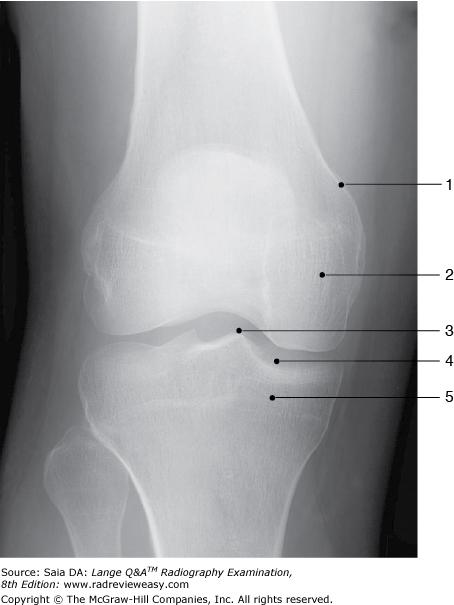
Identify the structure labeled 1 in the AP projection of the knee shown in Figure 2–16.
A Lateral condyle
B Lateral epicondyle
C Medial condyle
D Medial epicondyle
D Medial epicondyle
-Figure 2–16 shows an AP projection of the knee. The distal femur and proximal tibia and fibula are seen. The femorotibial joint space is open, and the tibial articular facets of the tibial plateau (number 4) are demonstrated. The intercondylar eminence (number 3) is seen. Number 2 is the medial femoral condyle; number 1 is the medial femoral epicondyle; and number 5 is the medial tibial condyle.
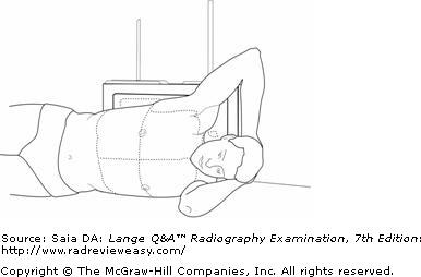
The position shown in Figure A is known as
A ventral decubitus.
B dorsal decubitus.
C left lateral decubitus.
D right lateral decubitus.
C left lateral decubitus.
-The illustration shows the patient positioned on his left side, with the cassette behind his back. This is a left lateral decubitus position. The x-ray beam is directed horizontally in decubitus positions to demonstrate air-fluid levels. Air or fluid levels will be clearly delineated only if the central ray is directed parallel to them. If the patient were lying on the right side, it would be a right lateral decubitus position. If the patient were lying on his or her back with a horizontal x-ray beam, it would be a dorsal decubitus position. Lying prone with a horizontal x-ray beam is termed a ventral decubitus position.
The type of ileus characterized by cessation of peristalsis is termed
A mechanical
B paralytic
C asymptomatic
D sterile
B paralytic
-Obstruction of the small bowel is termed ileus; there are two types of ileus—paralytic/adynamic and mechanical. Paralytic or adynamic ileus is characterized by an absence of peristalsis. This can be caused by infection (e.g., appendicitis or peritonitis) or postoperative difficulty. Mechanical ileus is caused by some sort of physical obstruction such as tumor or adhesions.

The scapula shown in Figure 2–29 demonstrates
- its posterior aspect
- its costal surface
- its sternal articular surface
A 1 only
B 1 and 2 only
C 1 and 3 only
D 1, 2, and 3
A 1 only
-Visualization of the scapular spine (number 13) indicates that this is a view of the posterior aspect of the scapula. The scapula's anterior, or costal, surface is that which is adjacent to the ribs. The scapula has no sternal articulation.
To better demonstrate the interphalangeal joints of the toes, which of the following procedures may be employed?
- Angle the CR 15 degrees caudad.
- Angle the CR 15 degrees cephalad.
- Place a sponge wedge under the foot with the toes elevated 15 degrees.
A 1 only
B 1 and 2 only
C 1 and 3 only
D 2 and 3 only
D 2 and 3 only
-Because the toes curve naturally downward, the interphalangeal joints are not well demonstrated in the AP (dorsoplantar) projection. To “open” the interphalangeal joints, the CR should be directed 15 degrees cephalad. Another method is to place a 15-degree foam sponge wedge under the foot, elevating the toes 15 degrees from the IR; the CR then would be directed perpendicularly.
Which of the following positions is most likely to place the right kidney parallel to the IR?
A AP
B PA
C RPO
D LPO
D LPO
-Since the kidneys do not lie parallel to the IR in the AP position, the oblique positions are used during IVU to visualize them better. With the AP oblique projections (RPO and LPO positions), the kidney that is farther away is placed parallel to the IR, and the kidney that is closer is placed perpendicular to the IR. Therefore, in the LPO position, the left kidney, being closer, is perpendicular to the IR. The right kidney,the one farther away, is placed parallel to the IR.
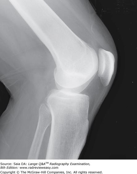
The mediolateral projection of the knee shown in Figure 6–1 could best be improved by
A rotating the patient forward
B rotating the patient backward
C angling the central ray (CR) about 5 degrees caudad
D angling the CR about 5 degrees cephalad
D angling the CR about 5 degrees cephalad
-The knee is formed by the proximal tibia, the patella, and the distal femur, which articulate to form the femorotibial and femoropatellar joints. The distal posterior femur presents two large medial and lateral condyles separated by the deep intercondyloid fossa. Because the medial femoral condyle is further from the IR, it is magnified and will obscure the femorotibial joint space, as seen in the figure. If the CR is angled about 5 degrees cephalad, the medial femoral condyle will be projected superiorly and superimposed on the lateral femoral condyle, thus opening the joint space. The patient should lie on the affected side with the patella perpendicular to the tabletop and the knee flexed 20 to 30 degrees. Rotating the part forward or backward will affect visualization of the femoropatellar joint.
Which of the following is an important consideration to avoid excessive metacarpal joint overlap in the oblique projection of the hand?
A Oblique the hand no more than 45 degrees.
B Use a support sponge for the phalanges.
C Clench the fist to bring the carpals closer to the IR.
D Use ulnar flexion.
A Oblique the hand no more than 45 degrees.
-The oblique projection of the hand should demonstrate minimal overlap of the third, fourth, and fifth metacarpals. Excessive overlap of these metacarpals is caused by obliquing the hand more than 45 degrees. The use of a 45-degree foam wedge ensures that the fingers will be extended and parallel to the IR, thus permitting visualization of the interphalangeal joints and avoiding foreshortening of the phalanges. Clenching of the fist and ulnar flexion are maneuvers used to better demonstrate the carpal scaphoid.
Which of the following positions would be the best choice for a right shoulder examination to rule out fracture?
A Internal and external rotation
B AP and tangential
C AP and AP axial
D AP and scapular Y
D AP and scapular Y
-The AP projection will give a general survey and show mediolateral and inferosuperior joint relationships. The scapular Y position (LAO or RAO) is employed to demonstrate anterior (subcoracoid) or posterior (subacromial) humeral dislocation. The humerus normally is superimposed on the scapula in this position; any deviation from this may indicate dislocation. Rotational views must be avoided in cases of suspected fracture. The AP and scapular Y combination is the closest to two views at right angles to each other.

The PA chest radiograph shown in the figure below demonstrates
- rotation
- scapula superimposed on lung fields
- adequate inspiration
A 1 only
B 1 and 3 only
C 2 and 3 only
D 1, 2, and 3
D 1, 2, and 3
-A PA projection of the chest is shown. Adequate inspiration is demonstrated by visualization of 10 posterior ribs above the diaphragm. The shoulders are rolled forward insufficiently, not completely removing the scapulae (especially the left) from the lung fields. Rotation of the chest is demonstrated by the unequal distance between the sternum and medial extremities of the clavicles. Pulmonary apices and costophrenic angles are demonstrated adequately. An air-filled trachea is seen in the lower cervical and upper thoracic region as a midline area of decreased brightness. Adequate long-scale contrast is evident, as indicated by visualization of pulmonary vascular markings.
Which of the following radiographic examinations require(s) the patient to be NPO 8–10 hours prior to examination for proper patient preparation?
- Abdominal survey
- Upper GI series
- BE
A 1 and 2 only
B 1 and 3 only
C 2 and 3 only
D 1, 2, and 3
C 2 and 3 only
-There is no preparation required for an abdominal survey. For an upper GI series and a lower GI series (BE), the patient should be NPO, or have nothing by mouth, for 8 to 10 hours prior to the examination. In addition, a low-residue diet may be imposed, fluid intake may be increased, and cleansing enemas and laxatives may be prescribed to rid the colon of fecal matter.
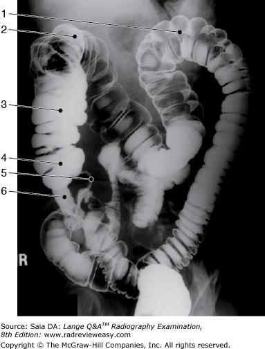
The structure indicated as number 4 in Figure 2–9 is the
A terminal ileum
B appendix
C cecum
D sigmoid
C cecum
-The radiograph shown is an oblique position of a double contrast study of the large bowel, illustrating an "open" view of the splenic/left colic flexure (number 1) and descending colon, with the hepatic/right colic flexure (number 2) somewhat superimposed on the transverse and ascending (number 3) colon. Therefore, the radiograph must have been made in either an RPO (if the patient was supine) or an LAO (if the patient was prone) position. The LPO and RAO positions are used to demonstrate the hepatic flexure and ascending colon free of self-superimposition. The distal ileum is well visualized (number 6), as well as the commencement of the large bowel—the cecum (number 4)—and its vermiform appendix (number 5). The AP or PA axial position generally is used to visualize the rectosigmoid colon.
Which of the following is (are) true regarding radiographic examination of the acromioclavicular joints?
- The procedure is performed in the erect position.
- Use of weights can improve demonstration of the joints.
- The procedure should be avoided if dislocation or separation is suspected.
A 1 only
B 1 and 2 only
C 1 and 3 only
D 2 and 3 only
B 1 and 2 only
-Evaluation of the acromioclavicular joints requires bilateral AP or PA erect projections with and without the use of weights. Weights are used to emphasize the minute changes within a joint caused by separation or dislocation. Weights should be anchored from the patient's wrists rather than held in the patient's hands because this encourages tightening of the shoulder muscles and obliteration of any small separation.
The plane that passes vertically through the body, dividing it into anterior and posterior halves, is termed the
A median sagittal plane (MSP)
B midcoronal plane
C sagittal plane
D transverse plane
B midcoronal plane
-he median sagittal, or midsagittal, plane (MSP) passes vertically through the midline of the body, dividing it into left and right halves. Any plane parallel to the MSP is termed a sagittal plane. The midcoronal plane is perpendicular to the MSP and divides the body into anterior and posterior halves. A transverse plane passes through the body at right angles to a sagittal plane. These planes, especially the MSP, are very important reference points in radiographic positioning (Figure 2–59).
Which of the following can be used to demonstrate the intercondyloid fossa?
- Prone, knee flexed 40 degrees, CR directed caudad 40 degrees to the popliteal fossa
- Supine, IR under flexed knee, CR directed cephalad to knee, perpendicular to tibia
- Prone, patella parallel to IR, heel rotated 5 to 10 degrees lateral, CR perpendicular to knee joint
A 1 only
B 1 and 2 only
C 2 and 3 only
D 1, 2, and 3
B 1 and 2 only
-Statement number 1 describes the PA axial projection (Camp-Coventry method) for demonstration of the intercondyloid fossa. Statement number 2 describes the AP axial projection (Béclère method) for demonstration of the intercondyloid fossa. The positions are actually the reverse of each other. Statement number 3 describes the method of obtaining a PA projection of the patella.
Which of the following statements is (are) correct with respect to evaluation criteria for a PA projection of the chest for lungs?
- Sternal extremities of clavicles are equidistant from vertebral borders.
- Ten posterior ribs are demonstrated above the diaphragm.
- The esophagus is visible in the midline.
A 1 only
B 1 and 2 only
C 2 and 3 only
D 1, 2, and 3
B 1 and 2 only
-In a PA projection of the chest, there should be no rotation, as evidenced by symmetry of sternal extremities of clavicles equidistant from vertebral borders. The shoulders are rolled forward to remove the scapulae from the lung fields. Inspiration should be adequate to demonstrate 10 posterior ribs above the diaphragm. The air-filled trachea should be seen midline; the esophagus is unlikely to be visualized without a contrast agent.
Blood is returned to the left atrium, from the lungs, via the
A aorta.
B superior vena cava.
C pulmonary veins.
D pulmonary artery
C pulmonary veins.
-Deoxygenated blood is collected by the superior and inferior vena cava and emptied into the right atrium.
Pulmonary circulation conveys deoxygenated blood from the right ventricle, through the pulmonary semilunar valve, into the pulmonary artery (the only artery that carries deoxygenated blood), and into the lungs where the blood becomes oxygenated. The oxygenated blood from the lungs is carried via the four pulmonary veins (the only veins that carry oxygenated blood) and emptied into the left atrium.
Which of the following is (are) distal to the tibial plateau?
- Intercondyloid fossa
- Tibial condyles
- Tibial tuberosity
A 1 only
B 1 and 2 only
C 2 and 3 only
D 1, 2, and 3
C 2 and 3 only
-The knee joint is formed by the femur and tibia. The most superior aspect of the tibia is the tibial plateau—formed by the tibial condyles just distal to it. The proximal tibia also presents the tibial tuberosity on its anterior surface, just distal to the condyles. Proximal to the tibial plateau, and articulating with it, are the femoral condyles—the deep notch separating them is the intercondyloid fossa.The term proximal refers to a part located closer to the point of attachment; the term distal refers to a part located farther away from the point of attachment.
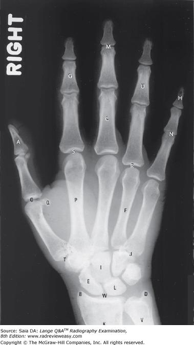
Which of the following correctly identifies the letter L in the radiograph shown in Figure 7–13?
A Hamate
B Lunate
C Scaphoid
D Trapezium
B Lunate
-The eight carpal bones are well visualized in this PA projection of the hand and wrist. The letters E(scaphoid) and L (lunate) are in the proximal carpal row. The capitate (I) is seen in the distal carpal row; just lateral to the capitate is the carpal trapezium, seen articulating with the base of the first metacarpal. The PA projection of the hand provides an oblique projection of the first finger (thumb).
In which of the following positions can the sesamoid bones of the foot be demonstrated to be free of superimposition with the metatarsals or phalanges?
A Dorsoplantar metatarsals/toes
B Tangential metatarsals/toes
C 30-degree medial oblique foot
D 30-degree lateral oblique foot
B Tangential metatarsals/toes
-The tangential projection projects the sesamoid bones separate from adjacent structures. The patient is best examined in the prone position because this places the parts of interest closest to the IR. The affected foot is dorsiflexed so as to place its plantar surface 15 to 20 degrees with the vertical. The CR is directed perpendicular to the posterior surface of the foot (near the metatarsophalangeal joints). The dorsoplantar and oblique projections of the foot will demonstrate the sesamoid bones superimposed on adjacent bony structures
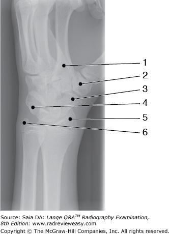
What is the structure labeled number 2 in Figure 2–37?
A Base of the 2nd metacarpal
B Pisiform
C Trapezium
D Trapezoid
C Trapezium
-The image illustrates a semipronation oblique of the wrist. This projection best demonstrates the lateral carpals. An oblique of the proximal metacarpals and distal radius and ulna are also visualized. The base of the second metacarpal is number 1. Just lateral, is seen the first carpometacarpal joint—the trapezium (lateral carpal, distal row) is labeled number 2. The scaphoid (lateral carpal, proximal row) is number 3. The pisiform is labeled number 4. The radial styloid process is number 5 and the ulnar styloid process is number 6.
In the lateral projection of the foot, the
- plantar surface should be perpendicular to the IR.
- metatarsals are superimposed.
- talofibular joint should be visualized.
A 1 only
B 1 and 2 only
C 2 and 3 only
D 1, 2, and 3
B 1 and 2 only
-When the foot is positioned for a lateral projection, the plantar surface should be perpendicular to the IR so as to superimpose the metatarsals. This may be accomplished with the patient lying on either the affected or the unaffected side (usually the affected), that is, mediolateral or lateromedial. The talofibular articulation is best demonstrated in the medial oblique projection of the ankle.
Which of the following statements is (are) correct, with respect to a left lateral projection of the chest?
- The MSP must be perfectly vertical and parallel to the IR.
- The right posterior ribs will be projected slightly posterior to the left posterior ribs.
- Arms must be raised high to prevent upper-arm soft-tissue superimposition on lung field.
A 1 only
B 1 and 2 only
C 1 and 3 only
D 1, 2, and 3
D 1, 2, and 3
-The chest should be examined in the upright position whenever possible to demonstrate any air–fluid levels. For the lateral projection, the patient elevates the arms well enough to avoid upper-arm soft-tissue superimposition on the lung fields. In the left lateral position, the right posterior ribs, being remote from the IR, will be somewhat magnified and very slightly posterior to the left posterior ribs. The MSP must remain vertical to avoid “tilt” distortion, and the coronal plane must be vertical to avoid rotation distortion.
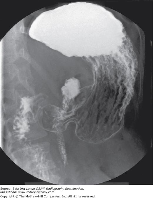
The image shown in Figure 7–4 was made in the following recumbent position
A RAO
B Lateral
C LPO
D PA
C LPO
-The stomach is generally not parallel with the long axis of the body—its fundus/superior portion lies more posterior, and the pylorus/inferior portion lies more anterior. Consequently, in the recumbent position, barium will gravitate to the fundus when the patient is supine and to the pylorus when the patient is prone. Since the image shows a barium-filled fundus, it must have been made in the AP projection or LPO position. A look at the vertebrae indicates that the body is somewhat obliqued, indicating an LPO position. Note the double-contrast demonstration of rugal folds in the body of the stomach and double-contrast delineation of the pylorus and duodenal bulb
In which of the following procedures is quiet, shallow breathing recommended during the exposure to obliterate prominent pulmonary vascular markings?
- RAO sternum
- Lateral thoracic spine
- AP scapula
A 1 only
B 1 and 2 only
C 2 and 3 only
D 1, 2, and 3
D 1, 2, and 3
-Pulmonary vascular markings often are prominent in the elderly and in smokers. Quiet, shallow breathing may be used during a long exposure (with a compensating low milliamperage) to blur them out. Oblique sternum, AP scapula, and lateral thoracic spine projections are examinations in which this technique is useful.
Which of the following conditions require(s) a decrease in technical factors?
- Emphysema
- Osteomalacia
- Atelectasis
A 1 only
B 1 and 2 only
C 2 and 3 only
D 1, 2, and 3
B 1 and 2 only
-Subcutaneous emphysema is a pathologic distension of tissues with air; pulmonary emphysema is a chronic disease characterized by overdistension of the alveoli with air. Osteomalacia is a softening of bone so that it becomes flexible, brittle, and deformed. All three of these conditions involve a decrease in tissue density and, therefore, require a decrease in technical factors. Atelectasis is a collapsed or airless lung; it requires an increase in technical factors.
The act of inspiration will cause elevation of the
- sternum.
- ribs.
- diaphragm.
A 1 only
B 1 and 2 only
C 2 and 3 only
D 1, 2, and 3
B 1 and 2 only
-The diaphragm is the major muscle of respiration. On inspiration/inhalation, the diaphragm and abdominal viscera are depressed, enabling filling and expansion of the lungs, accompanied by upward movement of the sternum and ribs. During expiration/exhalation, air leaves the lungs, and they deflate while the diaphragm relaxes and moves to a more superior position along with the abdominal viscera. As the diaphragm relaxes and moves up, the sternum and ribs move inferiorly.
Which of the following is proximal to the carpal bones?
A Distal interphalangeal joints
B Proximal interphalangeal joints
C Metacarpals
D Radial styloid process
D Radial styloid process
-The term proximal refers to structures closer to the point of attachment. For example, the elbow is described as being proximal to the wrist; that is, the elbow is closer to the point of attachment (the shoulder) than is the wrist. Referring to the question, then, the interphalangeal joints (both proximal and distal) and the metacarpals are both distal to the carpal bones. The radial styloid process is proximal to the carpals.
In which of the following positions was the radiograph in Figure 2–9 taken?
A RPO
B LPO
C AP axial
D Right lateral decubitus
A RPO
-The radiograph shown is an oblique position of a double contrast study of the large bowel, illustrating an “open” view of the splenic/left colic flexure (number 1) and descending colon, with the hepatic/right colic flexure (number 2) somewhat superimposed on the transverse and ascending (number 3) colon. Therefore, the radiograph must have been made in either an RPO (if the patient was supine) or an LAO(if the patient was prone) position. The LPO and RAO positions are used to demonstrate the hepatic flexure and ascending colon free of self-superimposition. The distal ilium is well visualized (number 6), as well as the commencement of the large bowel—the cecum (number 4)—and its vermiform appendix (number 5). The AP or PA axial position generally is used to visualize the rectosigmoid colon.
An axial projection of the clavicle is often helpful in demonstrating a fracture that is not visualized using a perpendicular CR. When examining the clavicle in the PA axial projection, how should the Central Ray directed?
A Cephalad
B Caudad
C Medially
D Laterally
B Caudad
-With the patient positioned erect or supine, the CR is directed cephalad 25 to 30°. This serves to project the clavicle away from the pulmonary apices and ribs, projecting most of the clavicle above the thorax. The reverse is true when the patient is examined for the PA axial projection, i.e. the CR is directed caudally. The PA and PA axial projections can be useful for better resolution because of the reduced OID.
Which of the following positions will separate the radial head, neck, and tuberosity from superimposition on the ulna?
A AP
B Lateral
C Medial oblique
D Lateral oblique
D Lateral oblique
-In the AP projection of the elbow, the proximal radius and ulna are partially superimposed. In the lateral projection, the radial head is partially superimposed on the coronoid process, facing anteriorly. In the medial oblique projection, there is even greater superimposition. The lateral oblique projection completely separates the proximal radius and ulna, projecting the radial head, neck, and tuberosity free of superimposition with the proximal ulna.
Which of the following sequences correctly describes the path of blood flow as it leaves the left ventricle?
A Arteries, arterioles, capillaries, venules, veins
B Arterioles, arteries, capillaries, veins, venules
C Veins, venules, capillaries, arteries, arterioles
D Venules, veins, capillaries, arterioles, arteries
A Arteries, arterioles, capillaries, venules, veins
-Blood is oxygenated in the lungs and carried to the left atrium by the four pulmonary veins. From the left atrium, blood flows through the bicuspid (mitral) valve into the left ventricle. Blood leaving the left ventricle is bright red, oxygenated blood that travels through the systemic circulation, which delivers oxygenated blood via arteries and returns deoxygenated blood to the lungs via veins. From the left ventricle, blood first goes through the largest arteries and then goes to progressively smaller arteries (arterioles), to the capillaries, to the smallest veins (venules), and on to progressively larger veins.
Which of the following are characteristics of the hypersthenic body type?
- Short, wide, transverse heart
- High and peripheral large bowel
- Diaphragm positioned low
A 1 and 2 only
B 1 and 3 only
C 2 and 3 only
D 1, 2, and 3
A 1 and 2 only
-The hypersthenic body type is large and heavy. The thoracic cavity is short, the lungs are short with broad bases, and the heart is usually in an almost transverse position. The diaphragm is high; the stomach and gallbladder are high and transverse. The large bowel is positioned high and peripheral (and often requires that 14 × 17 inch cassettes be placed cross-wise for imaging a BE).
Which of the following projections require(s) that the shoulder be placed in external rotation?
- AP humerus
- Lateral forearm
- Lateral humerus
A 1 only
B 1 and 2 only
C 2 and 3 only
D 1, 2, and 3
A 1 only
-When the arm is placed in the AP position, the epicondyles are parallel to the plane of the cassette, and the shoulder is placed in external rotation. In this position, an AP projection of the humerus, elbow, and forearm can be obtained. For the lateral projection of the humerus, elbow, or forearm, the epicondyles must be perpendicular to the plane of the cassette.
Which of the following projections of the ankle would best demonstrate the distal tibiofibular joint?
A Medial oblique 15° to 20°
B Lateral oblique 15° to 20°
C Medial oblique 45°
D Lateral oblique 45°
C Medial oblique 45°
-To best demonstrate the distal tibiofibular articulation, a 45° medial oblique projection of the ankle is required. The 15° medial oblique is used to demonstrate the ankle mortise (joint). Although the joint is well demonstrated in the 15° medial oblique, there is some superimposition of the distal tibia and fibula, and greater obliquity is required to separate the bones.
Which of the following groups of organs/structures are located in the left upper quadrant?
A Left kidney, left suprarenal gland, and gastric fundus
B Left suprarenal gland, pylorus, and duodenal bulb
C Hepatic flexure, cecum, and pancreas
D Gastric fundus, liver, and cecum
A Left kidney, left suprarenal gland, and gastric fundus
-The abdomen is divided anatomically into nine regions and four quadrants. The region designation usually is used for anatomic studies, whereas the quadrant designation is used most often to describe the location of a lesion, pain, tumor, or other abnormality. Some of the structures found in the left upper quadrant (LUQ) are the fundus of the stomach, the left kidney and suprarenal gland, and the splenic flexure.
The image shown in Figure 2–34 was made in what position?
A AP or PA erect
B Dorsal decubitus
C Left lateral decubitus
D Right lateral decubitus
D Right lateral decubitus
-The radiograph shown was made in the right lateral decubitus position. Although there is no visible marker, the more superior flexure is the left/splenic and the inferior flexure is the right/hepatic. It is part of a series of images made during an air-contrast (double-contrast) BE examination. A double-contrast examination of the large bowel is performed to see through the bowel to its posterior wall and to visualize any intraluminal (e.g., polypoid) lesions or masses. Various body positions are used to redistribute the barium and air. To demonstrate the medial and lateral walls of the bowel, decubitus positions are used. The radiograph shows a right lateral decubitus position because the barium has gravitated to the right side (the side of the hepatic flexure). The air rises and delineates the medial side of the ascending colon and the lateral side of the descending colon. The posterior wall of the rectum could be visualized using the ventral decubitus position and a horizontal beam lateral to the rectum.
An increase in exposure factors usually is required in which of the following circumstances?
- Edema
- Ascites
- Acromegaly
A 1 only
B 1 and 2 only
C 1 and 3 only
D 1, 2, and 3
D 1, 2, and 3
-An increase in exposure factors will be required when imaging pathologic conditions that cause greater attenuation of the x-ray beam. The x-ray beam suffers more attenuation as the thickness and/or density of the tissues increases. Examples include conditions involving an increase in part size as a result of fluid accumulation (edema) following trauma, an accumulation of fluid in the abdomen (ascites), or an increase in bone size and density (acromegaly) as a result of an endocrine disorder. The radiographer needs a good working knowledge of pathologic conditions, their effect on the body, and the resulting modifications in technical factors required.
The fifth metacarpal is located on which aspect of the hand?
A Medial
B Lateral
C Radial
D Volar
A Medial
-The fifth metacarpal is located on the medial aspect of the hand. Remember to always view a part in its an atomic position. With the arm in the anatomic position, the fifth metacarpal and the ulna lie medially.
The patient's chin should be elevated during chest radiography to
A permit the diaphragm to move to its lowest position
B avoid superimposition on the apices
C assist in maintaining an upright position
D keep the MSP parallel
B avoid superimposition on the apices
-Chest positioning must be correct and accurate; thoracic structures are easily distorted. To avoid superimposition on the upper medial apices, the patient's chin should be sufficiently elevated. Movement of the diaphragm to its lowest position is a function of the erect position and of making the exposure after the second inspiration. The MSP is perpendicular to the IR in the PA projection and parallel to the IR in the lateral projection. The position of the chin has little to do with the MSP.

The structure labeled number 5 in Figure 2–41 is the
A sternoclavicular joint
B acromioclavicular joint
C glenohumeral joint
D acromiohumeral joint
C glenohumeral joint
-An AP, external rotation, projection of the shoulder is pictured. The hand is supinated, and the arm is in the anatomical position. Therefore, the greater tubercle (number 3) is well visualized. The greater portion of the clavicle is seen, the acromioclavicular joint (number 1), the acromion process (number 2), the coracoid process (number 4), and the glenohumeral joint (number 5). The coronoid process is located on the ulna.
Which of the following positions is required to demonstrate small amounts of air in the pleural cavity?
A AP supine
B AP Trendelenburg
C Lateral decubitus, affected side down
D Lateral decubitus, affected side up
D Lateral decubitus, affected side up
-Air or fluid levels will be clearly delineated only if the CR is directed parallel to them. Therefore, to demonstrate air or fluid levels, the erect or decubitus position should be used. Small amounts of fluid within the pleural space are best demonstrated in the lateral decubitus position, affected side down. Small amounts of air within the pleural space are best demonstrated in the lateral decubitus position,affected side up.
What could be done to improve the mediolateral projection of the knee seen in Figure 2–3?
A Rotate the pelvis slightly forward/anteriorly.
B Rotate the pelvis slightly backward/posteriorly.
C Angle the x-ray tube 5 degrees cephalad.
D Angle the x-ray tube 5 degrees caudad.
B Rotate the pelvis slightly backward/posteriorly.
-The figure illustrates a mediolateral projection of the knee. The femoral condyles are not superimposed posteriorly, indicating incorrect degree of forward (anterior)/backward (posterior) rotation. Because the magnified medial femoral condyle is obscuring the femoropatellar articulation, the radiographer should rotation the pelvis backward, i.e. posteriorly, a bit. This will superimpose the femoral condyles, place the patella perpendicular to the tabletop, and open the femoropatellar joint space.
Impingement on the wrist's median nerve causing pain and disability of the affected hand and wrist is known as
A carpal boss syndrome
B carpal tunnel syndrome
C carpopedal syndrome
D radioulnar syndrome
B carpal tunnel syndrome
-Carpal tunnel syndrome involves pain and numbness to some parts of the median nerve distribution (i.e., palmar surface of the thumb, index finger, and radial half of the fourth finger and palm). Carpal tunnel syndrome occurs frequently in those who continually use vibrating tools or machinery.Carpopedal spasm is spasm of the hands and feet, commonly encountered during hyperventilation.Carpal boss is a bony growth on the dorsal surface of the third metacarpophalangeal joint.
Ulnar deviation will best demonstrate which carpal(s)?
- Medial carpals
- Lateral carpals
- Scaphoid
A 1 only
B 1 and 2 only
C 2 and 3 only
D 1, 2, and 3
C 2 and 3 only
-The carpal scaphoid is somewhat curved and consequently foreshortened radiographically in the PA position. To better separate it from the adjacent carpals, the ulnar deviation maneuver is employed frequently. In addition to correcting foreshortening of the scaphoid, ulnar deviation opens the interspaces between adjacent lateral carpals. Radial deviation is used to better demonstrate medial carpals.
A dorsal decubitus position of the chest may be used to evaluate small amounts of
1.fluid in the posterior chest.
2.air in the posterior chest.
3.fluid in the anterior chest.
A 1 only
B 1 and 2 only
C 2 and 3 only
D 1, 2, and 3
A 1 only
-The dorsal decubitus position is obtained with the patient supine and the x-ray beam directed horizontally. The finished radiograph looks similar to a routine lateral projection of the chest. However, small amounts of fluid will gravitate posteriorly, and small amounts of air will rise anteriorly.
Which of the following will best demonstrate the size and shape of the liver and kidneys?
A Lateral abdomen
B AP abdomen
C Dorsal decubitus abdomen
D Ventral decubitus abdomen
B AP abdomen
-The AP projection provides a general survey of the abdomen showing the size and shape of the liver, spleen, and kidneys. When performed erect, it should demonstrate both hemidiaphragms. The lateral projection is sometimes requested and is useful for evaluating the prevertebral space occupied by the aorta. Ventral and dorsal decubitus positions provide a lateral view of the abdomen that is useful for demonstration of air–fluid levels.
Which cholangiographic procedure uses an indwelling drainage tube for contrast medium administration?
A Endoscopic retrograde cholangiographic pancreatography (ERCP)
B Operative cholangiography
C T-tube cholangiography
D Percutaneous transhepatic cholangiography
C T-tube cholangiography
-Contrast media may be administered in a variety of manners in cholangiography, including (1) an endoscope with a cannula placed in the hepatopancreatic ampulla (of Vater) for an ERCP, (2) a needle or small catheter placed directly in the common bile duct for an operative cholangiogram, (3) a very fine needle through the patient's side and into the liver for a percutaneous transhepatic cholangiogram, and (4) via an indwelling T-tube for a postoperative or T-tube cholangiogram.
How should a chest examination to rule out air–fluid levels be obtained on a patient having traumatic injuries?
A Perform the examination in the Trendelenburg position.
B Erect inspiration and expiration images should be obtained.
C Include a lateral chest examination performed in dorsal decubitus position.
D Perform the examination AP supine at 44 inches SID.
C Include a lateral chest examination performed in dorsal decubitus position.
-One of the most important principles in chest radiography is that it be performed, whenever possible, in the erect position. It is in this position that the diaphragm can descend to its lowest position during inspiration, and any air–fluid levels can be detected. However, patients having traumatic injuries frequently must be examined in the supine position. An AP supine chest is performed first. If the examination is also being performed to rule out air–fluid levels, this can be determined by performing the lateral projection in the dorsal decubitus position. The patient is lying supine, and a horizontal (cross-table) x-ray beam is used.
During an upper gastrointestinal (GI) examination, a stomach of average shape demonstrates a barium-filled fundus and double contrast of the pylorus and duodenal bulb. The position used is most likely
A AP erect
B PA
C RAO
D LPO
D LPO
-With the body in the AP recumbent position (or LPO position), barium flows easily into the fundus of the stomach (from the more distal portions of the stomach), displacing/drawing the stomach somewhat superiorly. The fundus, then, is filled with barium, whereas the air that had been in the fundus is now displaced into the gastric body, pylorus, and duodenum, illustrating them in double contrast. Double-contrast delineation of these structures allows us to see through the stomach to the retrogastric areas and structures. The RAO position demonstrates a barium-filled pylorus and duodenum. Anterior and posterior aspects of the stomach are visualized in the lateral position; medial and lateral aspects of the stomach are visualized in the AP projection.
In the lateral projection of the scapula, the
- vertebral and axillary borders are superimposed.
- acromion and coracoid processes are superimposed.
- inferior angle is superimposed on the ribs.
A 1 only
B 1 and 2 only
C 1 and 3 only
D 1, 2, and 3
A 1 only
-A lateral projection of the scapula superimposes its medial and lateral borders (vertebral and axillary, respectively). The coracoid and acromion processes should be readily identified separately (not superimposed) in the lateral projection. The entire scapula should be free of superimposition with the ribs. The erect position is probably the most comfortable position for a patient with scapular pain.
In which of the following tangential axial projections of the patella is complete relaxation of the quadriceps femoris required for an accurate diagnosis?
- Supine flexion 45 degrees (Merchant)
- Prone flexion 90 degrees (Settegast)
- Prone flexion 55 degrees (Hughston)
A 1 only
B 1 and 2 only
C 2 and 3 only
D 1, 2, and 3 only
A 1 only
-The tangential axial projections of the patella are also often referred to as sunrise or skyline views. The supine flexion 45-degree (Merchant) position requires a special apparatus, and the patellae can be examined bilaterally. This position also requires patient comfort without muscle tension—muscle tension can cause a subluxed patella to be pulled into the intercondyler sulcus, giving the appearance of a normal patella. The two prone positions differ according to the degree of flexion employed. The 90-degree flexion (Settegast) position must not be employed with suspected patellar fracture.
Tangential axial projections of the patella can be obtained in which of the following positions?
1.supine flexion 45° (Merchant)
2.prone flexion 90° (Settegast)
3.prone flexion 55° (Hughston)
A 1 only
B 1 and 2 only
C 2 and 3 only
D 1, 2, and 3 only
D 1, 2, and 3 only
-The tangential axial projections of the patella are also often referred to as "sunrise" or "skyline" views. The supine flexion 45° (Merchant) position requires a special apparatus, and the patellae can be examined bilaterally. This position also requires patient comfort without muscle tension—muscle tension can cause a subluxed patella to be pulled into the intercondyler sulcus, giving the appearance of a normal patella. The two prone positions differ according to the degree of flexion employed. The 90° flexion (Settegast) position must not be employed with suspected patellar fracture.
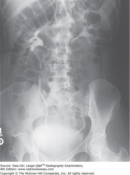
Which of the following statements regarding the image in Figure 2–4 is correct?
A The left kidney is more parallel to the IR.
B The image was made in the left posterior oblique position.
C The left ureter is better visualized.
D The image was made postvoid.
A The left kidney is more parallel to the IR.
-The image shown is one of a series of IVU images taken 15 minutes after injection of the contrast medium. The urinary collecting system is well demonstrated. An RPO position is illustrated, with the right marker indicating the right side; also, the right ilium is more “open” than the left. The RPO position places the left kidney and right ureter parallel to the IR. The urinary bladder contains considerable contrast, indicating that it is most likely a prevoid image.
During an upper gastrointestinal (GI) examination, the AP recumbent projection of a stomach of average shape will usually demonstrate
1.anterior and posterior aspects of the stomach.
2.barium-filled fundus.
3.double-contrast body and antral portions.
A 1 only
B 1 and 2 only
C 2 and 3 only
D 1, 2, and 3
C 2 and 3 only
-With the body in the AP recumbent position, barium flows easily into the fundus of the stomach, displacing the stomach somewhat superiorly. The fundus, then, is filled with barium, while the air that had been in the fundus is displaced into the gastric body, pylorus, and duodenum, illustrating them in double-contrast fashion. Air-contrast delineation of these structures allows us to see through the stomach to the retrogastric areas and structures. Anterior and posterior aspects of the stomach are visualized in the lateral position; medial and lateral aspects of the stomach are visualized in the AP projection.
Valid evaluation criteria for a lateral projection of the forearm requires that
- the epicondyles be parallel to the IR.
- the radius and ulna be superimposed distally.
- the radial tuberosity should face anteriorly.
A 1 only
B 1 and 2 only
C 2 and 3 only
D 1, 2, and 3
C 2 and 3 only
-To accurately position a lateral forearm, the elbow must form a 90-degree angle with the humeral epicondyles perpendicular to the IR and superimposed. The radius and ulna are superimposed distally. Proximally, the coronoid process and radial head are superimposed, and the radial head faces anteriorly. Failure of the elbow to form a 90-degree angle or the hand to be lateral results in a less than satisfactory lateral projection of the forearm.
To demonstrate the entire circumference of the radial head, the required exposure(s) must include
- epicondyles perpendicular to the IP
- hand pronated
- hand supinated as much as possible
A 1 only
B 1 and 2 only
C 1 and 3 only
D 1, 2, and 3
D 1, 2, and 3
- Although routine elbow projections may be essentially negative, conditions may exist (such as an elevated fat pad) that seem to indicate the presence of a small fracture of the radial head. To demonstrate the entire circumference of the radial head, four individual exposures are required with the elbow flexed 90 degrees and with the humeral epicondyles superimposed and perpendicular to the IP: one with the hand supinated as much as possible, one with the hand lateral, one with the hand pronated, and one with the hand in internal rotation, thumb down. Each maneuver changes the position of the radial head, and a different surface is presented for inspection. (Frank, Long, and Smith, 11th ed., vol. 1, pp. 152–153)
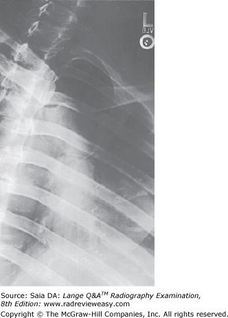
Which of the following statements is (are) true regarding the figure below?
- The image was made in the LAO position.
- The CR should enter more inferiorly.
- The sternum is projected onto the left side of the thorax.
A 1 only
B 2 only
C 2 and 3 only
D 1, 2, and 3
C 2 and 3 only
-The image demonstrates the sternum made in the RAO position. This position projects the sternum to the left side of the thorax. Superimposition of the sternum onto the heart and other mediastinal structures promotes more uniform receptor exposure. The CR for this image should have been directed more inferiorly, that is, midway between the jugular (manubrial) notch and the xiphoid process.
The medical term for congenital clubfoot is
A coxa plana.
B osteochondritis.
C talipes.
D muscular dystrophy.
C talipes.
-Talipes is the term used to describe congenital clubfoot. There are several types of talipes, generally characterized by a deformed talus and a shortened Achilles tendon, giving the foot a clubfoot appearance. Osteochondritis (Osgood–Schlatter disease) is a painful incomplete separation of the tibial tuberosity from the tibial shaft. It is often seen in active adolescent boys. Coxa plana (Legg–Calvé–Perthes disease) is ischemic necrosis leading to flattening of the femoral head. Muscular dystrophy is a congenital disorder characterized by wasting of skeletal muscles.
Which of the following projections of the elbow should demonstrate the radial head free of ulnar superimposition?
A AP
B Lateral
C Medial oblique
D Lateral oblique
D Lateral oblique
-On the AP projection of the elbow, the radial head and ulna normally are somewhat superimposed. The lateral oblique projection demonstrates the radial head free of ulnar superimposition. The lateral projection demonstrates the olecranon process in profile. The medial oblique projection demonstrates considerable overlap of the proximal radius and ulna but should clearly demonstrate the coronoid process free of superimposition and the olecranon process within the olecranon fossa.
What is the structure indicated by the number 8 in Figure 2–18?
A Common hepatic duct
B Common bile duct
C Cystic duct
D Pancreatic duct
B Common bile duct
-Figure 2–18 illustrates a portion of the biliary system. Bile leaves the liver through the right and left hepatic ducts (number 5), which join to form the common hepatic duct (number 6). Bile enters the gallbladder through the cystic duct (number 7). The neck of the gallbladder is indicated by the number 4, its body by the number 3, and its fundus by the number 2. The gallbladder stores and concentrates bile, and when it contracts, bile flows out through the cystic duct and down the common bile duct (number 8). The common bile duct and pancreatic duct (number 9) unite to form the short hepatopancreatic ampulla (of Vater), which empties into the duodenum (number 1)
To demonstrate a profile view of the glenoid fossa, the patient is AP recumbent and obliqued 45 degrees
A toward the affected side
B away from the affected side
C with the arm at the side in the anatomic position
D with the arm in external rotation
A toward the affected side
-In an AP projection of the shoulder, there is superimposition of the humeral head and glenoid fossa. With the patient obliqued 45 degrees toward the affected side, the glenohumeral joint is open, and the glenoid fossa is seen in profile. The patient's arm is abducted somewhat and placed in internal rotation.
Place the following anatomic structures in order from anterior to posterior:
1.Trachea
2.Apex of heart
3.Esophagus
A Trachea, esophagus, apex of heart
B Esophagus, trachea, apex of heart
C Apex of heart, trachea, esophagus
D Apex of heart, esophagus, trachea
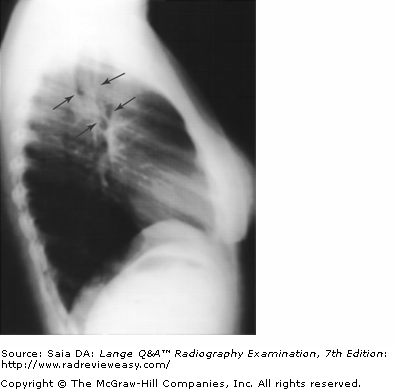
C Apex of heart, trachea, esophagus
-The relationship of these three structures can be appreciated in a lateral projection of the chest. The heart is seen in the anterior half of the thoracic cavity, with its apex extending inferior and anterior. The air-filled trachea can be seen in about the center of the chest, and the air-filled esophagus just posterior to the trachea (Fig. A).
Which of the following statements is (are) true regarding the CR image artifact seen in the erect PA projection of the chest shown in Figure 4–31?
A The object is located within the patient.
B The object is located within the IP.
C The object is located between the patient and the x-ray tube.
D The object is located between the patient and the IR.
B The object is located within the IP.
-The artifact shown in the figure has sharply delineated edges, indicating that it is located adjacent to the PSP within the IP. The farther the object is from the IR, the more blurred its edges will be as a result of magnification distortion.
A compression fracture of the posterolateral humeral head and associated with an anterior dislocation of the glenohumeral joint is called a(an)
A Hill-Sachs defect.
B Bankart lesion.
C rotator cuff tear.
D adhesive capsulitis.
A Hill-Sachs defect.
-A Hill-Sachs defect is a compression fracture of the posterolateral humeral head, usually associated with anterior dislocation of the shoulder joint. It can involve the cartilage of the humeral head, causing instability and predisposing the shoulder to subsequent dislocations. A Bankart lesion is a fracture of the anteroinferior portion of the rim of the glenoid fossa. A rotator cuff tear involves injury to one or more of the muscles participating in formation of that muscular structure. The supraspinatus, infraspinatus, subscapularis, and teres minor are the major muscles of the rotator cuff. Adhesive capsulitis, or "frozen shoulder," causes very diminished shoulder movement as a result of chronic joint inflammation.
The zygapophyseal articulations of the thoracic spine are demonstrated with the
A coronal plane 45° to the IR.
B midsagittal plane 45° to the IR.
C coronal plane 70° to the IR.
D midsagittal plane 70° to the IR.
C coronal plane 70° to the IR.
-The thoracic zygapophyseal joints are demonstrated by placing the patient in an oblique position with the coronal plane 70° to the IR (MSP 20° to the IR). This may be accomplished by first placing the patient lateral, then obliquing the patient 20° "off lateral." The zygapophyseal joints closest to the IR are demonstrated in the PA oblique, and those remote from the IR in the AP oblique. Comparable resolution is obtained using either method, because the OID is about the same.
The lesser tubercle of the humerus will be visualized in profile in the
A AP shoulder external rotation radiograph.
B AP shoulder internal rotation radiograph.
C AP elbow radiograph.
D Lateral elbow radiograph.
B AP shoulder internal rotation radiograph.
-The greater and lesser tubercles are prominences on the proximal humerus, separated by the bicipital groove. The AP projection of the humerus in external rotation demonstrates the greater tubercle in profile. With the arm placed in internal rotation, the humerus is placed in a true lateral position, and the lesser tubercle is demonstrated.
Characteristics of a patient with pulmonary emphysema include
- shoulder girdle elevation
- increased AP diameter of the chest
- hyperventilation
A 1 only
B 1 and 2 only
C 2 and 3 only
D 1, 2, and 3
B 1 and 2 only
-Emphysema is a chronic obstructive pulmonary disease (COPD) characterized by pathologic distension of the pulmonary alveoli with (destructive) changes in their walls, resulting in a loss of elasticity. Emphysema is seen occasionally following asthma or tuberculosis, but it is caused most frequently by cigarette smoking. Because the emphysematous patient's greatest difficulty is exhalation, it becomes a conscious, forced effort. Breathing is shallow and rapid. Forced and ineffective breathing results in expansion of the AP diameter of the chest and elevated shoulder girdle in established emphysema. Hyperventilation results from too frequent deep breaths in the anxious or tense individual, and is generally not related to pulmonary emphysema. Hyperventilation results in a feeling of dizziness and tingling of the extremities.
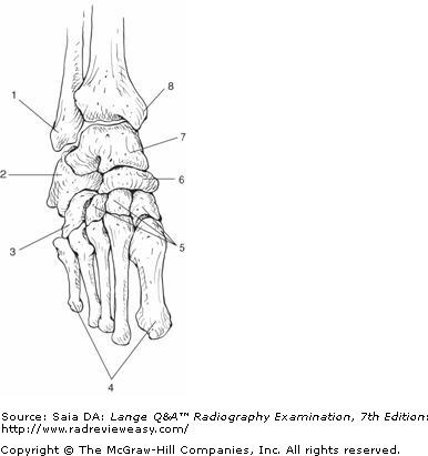
Which of the labeled bones in Figure A identifies the tarsal navicular?
A Number 2
B Number 3
C Number 6
D Number 7
C Number 6
-An anterior view of the foot and ankle bones is illustrated. The ankle joint is formed by the articulation of the tibia, fibula, and talus (7). The tibial (medial) malleolus is labeled 8; the fibular (lateral) malleolus is labeled 1. The talus articulates with the calcaneus (2) inferiorly and with the navicular (6) anteriorly. The cuboid (3) is seen anterior to the calcaneus, and the three cuneiforms (5) are anterior to the navicular.
During GI radiography, the position of the stomach may vary depending on
- the respiratory phase
- body habitus
- patient position
A 1 and 2 only
B 1 and 3 only
C 2 and 3 only
D 1, 2, and 3
D 1, 2, and 3
-During GI radiography, the position of the stomach may vary depending on the respiratory phase, the body habitus, and the patient position. Inspiration causes the lungs to fill with air and the diaphragm to descend, thereby pushing the abdominal contents downward. On expiration, the diaphragm will rise, allowing the abdominal organs to ascend. Body habitus is an important factor in determining the size and shape of the stomach. An asthenic patient may have a long, J-shaped stomach, whereas the stomach of a hypersthenic patient may be transverse. The body habitus is an important consideration in determining the positioning and placement of the cassette. The patient position also can alter the position of the stomach. If a patient turns from the RAO position into the AP position, the stomach will move into a more horizontal position. Although the cardiac sphincter and the pyloric sphincter are relatively fixed, the fundus is quite mobile and will vary in position
Which of the following may be used to evaluate the glenohumeral joint?
- Scapular Y projection
- Inferosuperior axial
- Transthoracic lateral
A 1 only
B 1 and 2 only
C 2 and 3 only
D 1, 2, and 3
D 1, 2, and 3
-The scapular Y projection is an oblique projection of the shoulder that is used to demonstrate anterior or posterior shoulder dislocation. The inferosuperior axial projection may be used to evaluate the glenohumeral joint when the patient is able to abduct the arm. The transthoracic lateral projection is used to evaluate the glenohumeral joint and upper humerus when the patient is unable to abduct the arm.
In which projection of the foot are the sinus tarsi, cuboid, and tuberosity of the fifth metatarsal best demonstrated?
A Lateral oblique foot
B Medial oblique foot
C Lateral foot
D Weight-bearing foot
B Medial oblique foot
-To best demonstrate most of the tarsals and intertarsal spaces (including the cuboid, sinus tarsi, and tuberosity of the fifth metatarsal), a medial oblique is required (plantar surface and IR form a 30° angle). The lateral oblique demonstrates the interspaces between the first and second metatarsals and between the first and second cuneiforms. Weight-bearing lateral feet are used to demonstrate the longitudinal arches.

What should be done to better demonstrate the coracoid process shown in Figure 2–22?
A Use a perpendicular CR.
B Angle the CR about 30 degrees cephalad.
C Angle the CR about 30 degrees caudad.
D Angle the MSP 15 degrees toward the affected side.
B Angle the CR about 30 degrees cephalad.
-The figure shows an AP projection of the shoulder. A plane passing through the epicondyles is parallel to the IR (and perpendicular to the CR). To project the coracoid process with less self-superimposition, the CR must be angled cephalad 15 degrees. The amount of cephalad angulation depends on the degree of thoracic kyphosis; the greater the degree of kyphosis, the greater is the degree of cephalad angulation required. A 30-degree angle is used for the average patient.
What are the positions most commonly employed for a radiographic examination of the sternum?
1.Lateral
2.RAO
3.LAO
A 1 and 2 only
B 1 and 3 only
C 2 and 3 only
D 1, 2, and 3
A 1 and 2 only
-Because the sternum and vertebrae would be superimposed in a direct PA or AP projection, a slight oblique (just enough to separate the sternum from superimposition on the vertebrae) is used instead of a direct frontal projection. In the RAO position, the heart superimposes its homogeneous density over the sternum, thereby providing clearer radiographic visualization of its bony structure. If the LAO position were used to project the sternum to the right of the thoracic vertebrae, the posterior ribs and pulmonary markings would cast confusing shadows over the sternum because of their differing tissue densities. The lateral projection requires that the shoulders be rolled back sufficiently to project the sternum completely anterior to the ribs. Prominent pulmonary vascular markings can be obliterated using a "breathing technique," that is, using an exposure time long enough (with appropriately low milliamperage) to equal at least a few respirations.
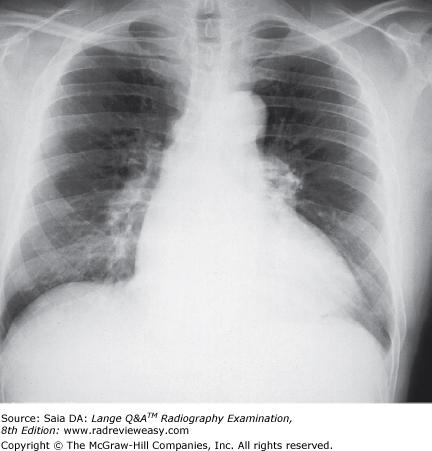
The PA chest analog image shown in the figure below demonstrates
- excessive receptor exposure
- insufficient kilovoltage
- underpenetration
A 1 only
B 1 and 2 only
C 2 and 3 only
D 1, 2, and 3
C 2 and 3 only
-The PA projection of the chest demonstrates high-, or short-scale, contrast. Additionally, faint visualization of the thoracic vertebrae through the heart is not demonstrated—indicating that there is inadequate penetration, that is, insufficient kilovoltage selected. Receptor exposure is adequate, but contrast and penetration are unacceptable for diagnostic purposes.
All the following procedures demonstrate renal function except
A IVP.
B descending urography.
C retrograde urography.
D infusion nephrotomography.
C retrograde urography.
-Retrograde urography is not considered a functional study of the urinary system. IVP, descending urography, and infusion nephrotomography are all considered functional urinary tract studies because the contrast medium is introduced intravenously and excreted by the kidneys. Retrograde urography involves introduction of contrast medium into the kidneys via catheter, thereby demonstrating their structure but not their function.
What is the most superior structure of the scapula?
A Apex
B Acromion process
C Coracoid process
D Superior angle
B Acromion process
-It is easy to determine the highest point of the scapula when it is viewed laterally. The coracoid process projects anteriorly and is quite superior. However, the acromion process, which is an anterior extension of the scapular spine, projects considerably more superior than the coracoid.

With the patient positioned as shown in Figure 6–13, how should the CR be directed to best demonstrate the intercondyloid fossa?
A Perpendicular to the popliteal depression
B 40 degrees caudad to the popliteal depression
C Perpendicular to the long axis of the femur
D 40 degrees cephalad to the popliteal depression

B 40 degrees caudad to the popliteal depression
-To demonstrate the intercondyloid fossa, the CR must be directed perpendicular to the long axis of the tibia (Figure 6–25). Because the knee is flexed so that the tibia forms a 40-degree angle with the IR, the CR must be directed 40 degrees caudad to place the CR perpendicular to the long axis of the tibia. Directing the CR to the popliteal depression aligns the CR parallel with the knee joint space.
What is the position/projection to demonstrate the longitudinal arch of the foot, while permitting the patient to keep weight equally distributed on both feet?
A Mediolateral
B Lateromedial
C Mediolateral weight-bearing lateral
D Lateromedial weight-bearing lateral
D Lateromedial weight-bearing lateral
-Weight-bearing lateral projections of the foot are requested often to evaluate the longitudinal arch structure of the foot. The patient stands on a small platform, weight equally distributed on feet. The IR is placed between the feet, in a slot provided on the platform, with the top of the IR against the medial aspect of the foot. The CR is directed to enter the lateral aspect of the foot perpendicular to the base of the fifth metatarsal and to exit the medial side of the foot. When the mediolateral projection is used, the patient must balance on the foot being imaged while bringing the other foot back.
A flat and upright abdomen is requested on an acutely ill patient, to demonstrate the presence of air-fluid levels. Because of the patient's condition, the x-ray table can be tilted upright only 70° (rather than the desired 90°). How should the central ray be directed?
A Perpendicular to the IR
B Parallel to the floor
C 20° caudad
D 20° cephalad
B Parallel to the floor
-Whenever a part is being radiographed for demonstration of air-fluid levels, the central ray must be directed parallel to the floor; horizontal beam is necessary for visualization of a air fluid levels. In this example, the patient was unable to tolerate the 90° tilt of the x-ray table. If the radiographer were to compensate for this by directing the central ray perpendicular to the IR (angling 20° caudad), it is very possible that any air-fluid level would be blurred and indistinct, and would go unrecognized. Remember that air or fluid always levels out parallel to the floor. Thus, if the air-fluid level needs to be demonstrated, the central ray must also be parallel to the floor.
Which of the following positions may be used to effectively demonstrate the right posterior axillary ribs?
A LAO
B RAO
C RPO
D LPO
C RPO
-To place the right posterior axillary ribs parallel to the IR, an RPO position is required. The LAO will also demonstrate the right axillary ribs, but primarily the anterior portion. The RAO position will demonstrate the left anterior axillary ribs, and the LPO will demonstrate the left posterior axillary ribs.
Which of the following anatomic structures is best demonstrated in the LPO position, in a positive-contrast exam?
A Hepatic flexure
B Splenic flexure
C Sigmoid colon
D Ileocecal valve
A Hepatic flexure
-The AP oblique positions (RPO and LPO) demonstrate the colonic structures farther from the IR. The LPO position will demonstrate the hepatic flexure and ascending colon, whereas the RPO position demonstrates the splenic flexure and descending colon. In the prone oblique positions (RAO and LAO), the flexure disclosed is the one closer to the IR. Therefore, the LAO position will “open up” the left colic, or splenic, flexure, and the RAO position will demonstrate the right colic, or hepatic, flexure.
All the following statements regarding respiratory structures are true except
A the right lung has three lobes.
B the inferior portion of the lung is the apex.
C each lung is enclosed in serous membrane.
D the main stem bronchi enter the lung hilum.
B the inferior portion of the lung is the apex.
-The trachea (windpipe) bifurcates into left and right main stem bronchi, each entering its respective lung hilum. The left bronchus divides into two portions, one for each lobe of the left lung. The right bronchus divides into three portions, one for each lobe of the right lung (Figure 2–43). The lungs are conical in shape, consisting of upper pointed portions, termed the apices (plural of apex), and broad lower portions (or bases). The lungs are enclosed in a double-walled serous membrane called the pleura.

Which of the following anatomic structures is indicated by the number 2 in Figure 2–7?
A medial epicondyle
B trochlea
C capitulum
D olecranon process
D olecranon process
-The image illustrates a medial oblique (internal rotation) projection of the elbow with epicondyles 45 degrees to the IR. An oblique view of the proximal radius and ulna and the distal humerus is obtained. This projection is particularly useful to demonstrate the coronoid process in profile (number 4), the trochlea (number 3), and the medial epicondyle (number 1). The olecranon process (number 2) fits into the olecranon fossa during extension of the elbow. A small portion of the radial head (number 5) not superimposed on the ulna can be seen. The external oblique (lateral rotation) projection demonstrates the entire radial head free of superimposition as well as the radial neck and the humeral capitulum.
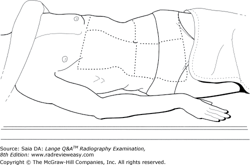
Which of the following statements is (are) true regarding the position illustrated in Figure 2–19?
- The left (elevated) ureter is parallel to the IR.
- The left (elevated) kidney is parallel to the IR.
- The degree of obliquity should be about 30 degrees.
A 1 only
B 1 and 2 only
C 2 and 3 only
D 1, 2, and 3
C 2 and 3 only
-An RPO position is illustrated. The oblique IVU projections should be approximately 30 degrees; this position significantly changes the position of the kidneys and ureters. When the abdomen is obliqued, the kidney of the “down” side is perpendicular to the IR and its ureter is parallel to the IR. The kidney of the “up” side is parallel to the IR.
Moderate hypertension can produce damage to which of the following organ(s)
1. Lungs
2. Kidneys
3. Brain
A 1 only
B 1 and 2 only
C 2 and 3 only
D 1, 2, and 3
D 1, 2, and 3
-Normal blood pressure is 110–140 mmHg systolic and 60–80 mmHg diastolic. High blood pressure (hypertension) is indicated by a systolic pressure higher than 140 mmHg and a diastolic pressure higher than 90 mmHg. Hypertension can be identified as extreme or moderate. Extreme hypertension can result in brain damage within just a few minutes. Moderate hypertension can cause damage to organs—the lungs, kidneys, brain, heart, etc. Various disease processes can produce hypertension as well as contributing factors such as medications, obesity, smoking, and stress.
The following instructions should be given to a patient following a barium sulfate contrast examination:
- Increase fluid and fiber intake for several days.
- Changes in stool color will occur until all barium has been evacuated.
- Contact a physician if no bowel movement occurs in 24 hours.
A 1 only
B 2 only
C 1 and 3 only
D 1, 2, and 3
D 1, 2, and 3
-Physicians often prescribe a mild laxative to aid in the elimination of barium sulfate. If a laxative is not given, the patient should be instructed to increase dietary fluid and fiber and to monitor bowel movements (the patient should have at least one within 24 hours). Patients should also be aware of the white appearance of their stool that will be present until all barium is expelled.

The structure labeled number 4 in Figure 2–41 is the
A acromion process
B coracoid process
C coronoid process
D glenoid process
B coracoid process
-An AP, external rotation, projection of the shoulder is pictured. The hand is supinated, and the arm is in the anatomical position. Therefore, the greater tubercle (number 3) is well visualized. The greater portion of the clavicle is seen, the acromioclavicular joint (number 1), the acromion process (number 2), the coracoid process (number 4), and the glenohumeral joint (number 5). The coronoid process is located on the ulna.
Muscles that contribute to the formation of the rotator cuff include the
1. subscapularis.
2. infraspinatus.
3. teres minor.
A 1 only
B 1 and 2 only
C 2 and 3 only
D 1, 2, and 3
D 1, 2, and 3
-The rotator cuff is a musculotendinous structure that includes the supraspinatus, infraspinatus subscapularis, and teres minor muscles. The muscles function to stabilize the humeral head in all arm motions and, together with the deltoid, function to abduct and rotate the arm. Weakness of the rotator cuff can lead to impingement syndrome and/or tendonitis. A tear of the cuff can result in subluxation; calcification can lead to shoulder immobilization.
Differences between body habitus types are likely to affect all the following except
A the size and shape of an organ.
B the position of an organ.
C the position of the diaphragm.
D the degree of bone porosity.
D the degree of bone porosity.
-The four types of body habitus are (from upper extreme to lower extreme) hypersthenic, sthenic, hyposthenic, and asthenic. The gallbladder and stomach are higher and more lateral and the large bowel more peripheral in the hypersthenic individual. The diaphragm is in a higher position in the hypersthenic individual. Recognition of a patient's body habitus and its characteristics is an important part of accurate radiography. Bone porosity generally is unrelated to body habitus.
Involuntary motion can be caused by
1.peristalsis.
2.severe pain.
3.heart muscle contraction.
A 1 only
B 2 only
C 1 and 2 only
D 1, 2, and 3
D 1, 2, and 3
-Patients who are able to cooperate are usually able to control voluntary motion. However, certain body functions and responses create involuntary motion that is not controllable by the patient. Severe pain, muscle spasm, and chills all cause involuntary movements. Peristaltic activity of the intestinal tract and motion caused by contraction of the heart muscle are other sources of involuntary motion.
During endoscopic retrograde cholangiopancreatography (ERCP) examination, contrast medium is injected into the
A hepatic duct
B cystic duct
C pancreatic duct
D common bile duct
D common bile duct
-Endoscopic retrograde cholangiopancreatography (ERCP) is performed to diagnose disease of the biliary and/or pancreatic organs. Fluoroscopic control is used to introduce the fiber-optic endoscope through the mouth and into the duodenum. The hepatopancreatic ampulla (of Vater) then is located and cannulated, and contrast medium is injected into the common bile duct.
Which of the following bones participate(s) in the formation of the knee joint?
- Femur
- Tibia
- Patella
A 1 and 2 only
B 1 and 3 only
C 2 and 3 only
D 1, 2, and 3
A 1 and 2 only
-The knee (tibiofemoral joint) is the largest joint of the body, formed by the articulation of the femur and tibia. There are three neighboring articulations—the patellofemoral joint, the lateral tibiofemoral joint (lateral femoral condyle with tibial plateau), and the medial tibiofemoral joint (medial femoral condyle with tibial plateau). Although the knee itself is classified as a synovial (diarthrotic) hinge-type joint, the patellofemoral joint actually is a gliding joint, and the medial and lateral tibiofemoral joints are hinge type.

What is the name of the structure indicated as number 5 in Figure 7–7?
A Coronoid fossa
B Coracoid fossa
C Olecranon fossa
D Trochlear/semilunar notch
C Olecranon fossa
-The figure shows a posterior view of the elbow. The distal posterior humerus (number 1) is seen, as well as the proximal posterior radius (number 4) and ulna (number 3). Additional structures identified are the medial epicondyle (number 2), the olecranon fossa (number 5), olecranon process (number 6), lateral epicondyle (number 7), and radial head (number 8) The olecranon process (number 6) can best be demonstrated in the lateral projection; it can also be demonstrated in the acute flexion position. The AP internal oblique will demonstrate the coronoid process; the AP external oblique will demonstrate the radial head free of superimposition.
In which projection of the foot are the interspaces between the first and second cuneiforms best demonstrated?
A Lateral oblique foot
B Medial oblique foot
C Lateral foot
D Weight-bearing foot
A Lateral oblique foot
-The lateral oblique demonstrates the interspaces between the first and second metatarsals and between the first and second cuneiforms. To best demonstrate most of the tarsals and intertarsal spaces (including the cuboid, sinus tarsi, and tuberosity of the fifth metatarsal), a medial oblique projection is required (plantar surface and IR form a 30-degree angle). A weight-bearing lateral projection of the feet is used to demonstrate the longitudinal arches.

What is the structure labeled number 5 in Figure 2–37?
A Trapezium
B Scaphoid
C Ulnar styloid
D Radial styloid
D Radial styloid
-The image illustrates a semipronation oblique of the wrist. This projection best demonstrates the lateral carpals. An oblique of the proximal metacarpals and distal radius and ulna are also visualized. The base of the second metacarpal is number 1. Just lateral, is seen the first carpometacarpal joint—the trapezium (lateral carpal, distal row) is labeled number 2. The scaphoid (lateral carpal, proximal row) is number 3. The pisiform is labeled number 4. The radial styloid process is number 5 and the ulnar styloid process is number 6.
Synovial fluid is associated with the
A brain.
B spinal canal.
C peritoneal cavity.
D bony articulations.
D bony articulations.
-Synovial fluid is associated with diarthrotic (freely movable) bony articulations. Other types of bony articulations are fibrous (synarthrotic/immovable) and cartilaginous (amphiarthrotic/partially movable).
Fluid associated with the brain and spinal canal is cerebrospinal fluid (CSF). The peritoneal and pleural cavities are associated with a lubricating serous fluid.
Types of inflammatory bowel disease include
- ulcerative colitis.
- Crohn's disease.
- intussusception.
A 1 only
B 1 and 2 only
C 2 and 3 only
D 1, 2, and 3
B 1 and 2 only
-The two most common types of chronic inflammation of the intestines are ulcerative colitis and Crohn's disease. The latter can attack any part of the GI tract and extends through all layers of the intestinal wall (therefore the possibility of forming fistulous tracks to contiguous structures). Ulcerative colitis attacks only the large bowel and only the mucosal layer of the intestinal wall. Curiously, cigarette smoking increases the risk for Crohn's disease and decreases the risk for ulcerative colitis. Intussusception is an obstructive disorder.
Which of the following is a radiologic procedure that functions to dilate a stenotic vessel?
A Percutaneous nephrolithotomy
B Percutaneous angioplasty
C Renal arteriography
D Surgical nephrostomy
B Percutaneous angioplasty
-Plaque deposited on arterial walls in cases of atherosclerosis causes arterial stenosis. Percutaneous transluminal angioplasty (PTA) is a procedure that uses a balloon catheter to permanently increase the size of the arterial lumen, thus reopening the vessel and restoring blood flow. A percutaneous nephrolithotomy is a procedure performed to remove a renal calculus from a kidney or proximal ureter.Renal arteriography is the radiologic investigation of the renal arteries. Nephrostomy is the surgical formation of an artificial opening into the kidney.
Which of the following are components of a trimalleolar fracture?
- Fractured lateral malleolus
- Fractured medial malleolus
- Fractured posterior tibia
A 1 only
B 1 and 3 only
C 2 and 3 only
D 1, 2, and 3
D 1, 2, and 3
-A trimalleolar fracture involves three separate fractures. The lateral malleolus is fractured in the “typical” fashion, but the medial malleolus is fractured on both its medial and posterior aspects. The trimalleolar fracture frequently is associated with subluxation of the articular surfaces.
In a lateral projection of the normal knee, the
- fibular head should be somewhat superimposed on the proximal tibia.
- patellofemoral joint should be visualized.
- femoral condyles should be superimposed.
A 1 only
B 2 only
C 1 and 3 only
D 1, 2, and 3
D 1, 2, and 3
-To better visualize the joint space in the lateral projection of the knee, 20 to 30 degrees of flexion is recommended. The femoral condyles are superimposed so as to demonstrate the patellofemoral joint and the articulation between the femur and the tibia. The head of the fibula will be slightly superimposed on the proximal tibia. The correct degree of forward or backward body rotation is responsible for visualization of the patellofemoral joint. Cephalad tube angulation of 5 to 7 degrees is responsible for demonstrating the articulation between the femur and the tibia (by removing the magnified medial femoral condyle from superimposition on the joint space).
The usual patient preparation for an upper GI examination is
A nothing by mouth (NPO) 8 hours before the examination.
B light breakfast only on the morning of the examination.
C clear fluids only on the morning of the examination.
D 2 oz of castor oil and enemas until clear.
A nothing by mouth (NPO) 8 hours before the examination.
-To obtain a diagnostic examination of the stomach, it must first be empty. The usual preparation is NPO (nothing by mouth) after midnight (approximately 8 hours before the examination). Any material in the stomach can simulate the appearance of disease.
What is the position of the gallbladder in an asthenic patient?
A Superior and medial
B Superior and lateral
C Inferior and medial
D Inferior and lateral
C Inferior and medial
-The position, shape, and motility of various organs can differ greatly from one body habitus to another. The hypersthenic individual is large and heavy; the lungs and heart are high, the stomach is high and transverse, the gallbladder is high and lateral, and the colon is high and peripheral. In contrast, the other habitus extreme is the asthenic individual. This patient is slender and light, and has a long and narrow thorax, a low and long stomach, a low and medial gallbladder, and a low medial and redundant colon. The radiographer must consider these characteristic differences when radiographing individuals of various body types.
The act of expiration will cause the
- diaphragm to move inferiorly
- sternum and ribs to move inferiorly
- diaphragm to move superiorly
A 1 only
B 1 and 2 only
C 2 and 3 only
D 1, 2, and 3
C 2 and 3 only
-The diaphragm is the major muscle of respiration. On inspiration/inhalation, the diaphragm and abdominal viscera are depressed, enabling the filling and expansion of the lungs, accompanied by upward movement of the sternum and ribs. During expiration/exhalation, air leaves the lungs and they deflate, and the diaphragm relaxes and moves to a more superior position along with the abdominal viscera. As the diaphragm relaxes and moves up, the sternum and ribs move inferiorly.
Another name for Hirschsprung's disease, the most common cause of lower GI obstruction in neonates, is
A intussusception.
B volvulus.
C congenital megacolon.
D pyloric stenosis.
C congenital megacolon.
-Hirschsprung disease, or congenital megacolon, is caused by the absence of some or all of the bowel ganglion cells—usually in the rectosigmoid area but occasionally more extensively. Hirschsprung disease is the most common cause of lower GI obstruction in neonates and is treated surgically by excision of the affected area followed by reanastomosis with the normal, healthy bowel. Hirschsprung disease is diagnosed by BE or, in mild cares, by rectal biopsy. Intussusception is “telescoping” of the bowel, causing (mechanical) obstruction. Volvulus is twisting of the bowel on itself causing (mechanical) obstruction. Pyloric stenosis is a condition of the upper GI tract.
Routine excretory urography usually includes a postmicturition radiograph of the bladder. This is done to demonstrate
- tumor masses.
- residual urine.
- prostatic enlargement.
A 2 only
B 1 and 3 only
C 2 and 3 only
D 1, 2, and 3
D 1, 2, and 3
-Variance from the normal bladder contour will be noted while the bladder is full of contrast medium. However, a postmicturition (postvoiding) radiograph is also an essential part of an IVU/IVP. The presence of residual urine may be an indication of small tumor masses or, in male patients, enlargement of the prostate gland.
Adult orthoroentgenography, or radiographic measurement of long bones of an upper or lower extremity, requires which of the following accessories?
- Bell-Thompson scale
- Bucky tray
- Cannula
A 1 only
B 1 and 2 only
C 1 and 3 only
D 1, 2, and 3
B 1 and 2 only
-Adult orthoroentgenography is the radiographic measurement of long-bone length. It can be required on adults or children having extremity length (especially leg) discrepancies. This can be performed most easily with the use of the metallic Bell-Thompson scale secured to the x-ray tabletop adjacent to the limb being examined (or between both limbs for simultaneous bilateral examination). A 14 × 17 inch cassette is in the Bucky tray (to permit movement of the cassette between exposures), and three well-collimated exposures are made—at the hip joint, the knee joint, and the ankle joint. A cannula is a tube placed in a cavity to introduce or withdraw material and is unrelated to orthoroentgenography.
All the following structures are associated with the posterior femur except
A popliteal surface
B intercondyloid fossa
C intertrochanteric line
D linea aspera
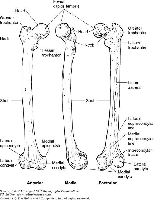
C intertrochanteric line
-The femur is the longest and strongest bone in the body. The femoral shaft is bowed slightly anteriorly and presents a long, narrow ridge posteriorly called the linea aspera. The distal femur is associated with two large condyles; the deep depression separating them posteriorly is the intercondyloid fossa (Figure 2–49). Just superior to the large condyles are the smaller medial and lateral epicondyles. The posterior distal femoral surface presents the popliteal surface, whereas the distal anterior surface presents the patellar surface. Proximally, the femur presents a head, neck, and greater and lesser trochanters. The intertrochanteric crest is a prominent ridge of bone between the trochanters posteriorly; anteriorly the intertrochanteric line is seen. The femoral head presents a roughened prominence, the fovea capitis femoris—ligaments attached here secure the femoral head to the acetabulum.
Fluoroscopic imaging of the ileocecal valve is generally part of a(n)
A esophagram.
B upper GI series.
C small-bowel series.
D ERCP.
C small-bowel series.
-The ileocecal valve is located at the terminal ileum, where it meets the first portion of the large bowel, the cecum. Most small-bowel examinations are performed following oral administration of barium sulfate suspension. The first small-bowel radiograph is taken 15 minutes after the first swallow of barium, with subsequent radiographs made every 15 to 30 minutes, depending on how quickly the barium is moving through the small bowel. Each image is shown to the radiologist, and a decision is made regarding the time of the next image. When the barium reaches the terminal ileum, fluoroscopy may be performed and compression spot images taken of the ileocecal valve.
In the lateral projection of the ankle, the
- talotibial joint is visualized.
- talofibular joint is visualized.
- tibia and fibula are superimposed.
A 1 only
B 1 and 2 only
C 1 and 3 only
D 1, 2, and 3
C 1 and 3 only
-In a lateral projection of the ankle, the tibia and fibula are superimposed, and the foot is somewhat dorsiflexed to better demonstrate the talotibial joint. The talofibular joint is not visualized because of superimposition with other bony structures. It may be well visualized in the medial oblique projection of the ankle.
Demonstration of the posterior fat pad on the lateral projection of the adult elbow can be caused by
- trauma or other pathology
- greater than 90-degree flexion
- less than 90-degree flexion
A 1 only
B 3 only
C 1 and 2 only
D 1 and 3 only
D 1 and 3 only
-There are three important fat pads associated with the elbow, best demonstrated in the true lateral projection. They cannot be demonstrated in the AP projection because of their superimposition on bony structures. The anterior fat pad is located just anterior to the distal humerus. The posterior fat pad is located within the olecranon fossa at the distal posterior humerus. The supinator fat pad/stripe is located at the proximal radius just anterior to the head, neck, and tuberosity. The posterior fat pad is not visible radiographically in the normal elbow. The posterior fat pad is visible in cases or trauma or other pathology and when the elbow is insufficiently flexed.
With the patient recumbent on the x-ray table with the head lower than the feet, the patient is said to be in the
A Trendelenburg position
B Fowler position
C decubitus position
D Sims position
A Trendelenburg position
-When the patient is recumbent with the head lower than the feet, he or she is said to be in the Trendelenburg position. The decubitus position is used to describe the patient who is recumbent (prone, supine, or lateral) with the CR directed horizontally. In the Fowler position, the patient's head is positioned higher than the feet. The Sim position is the (LAO) position assumed for enema tip insertion.
All the following statements regarding large bowel radiography are true except
A the large bowel must be completely empty prior to examination.
B retained fecal material can obscure pathology.
C single-contrast studies help to demonstrate intraluminal lesions.
D double-contrast studies help to demonstrate mucosal lesions.
C single-contrast studies help to demonstrate intraluminal lesions.
-Perhaps the most important prerequisite to a successful BE examination is a thoroughly clean large bowel. Any retained fecal material can simulate or obscure pathology. A single-contrast examination demonstrates the anatomy and contour of the large bowel, as well as anything that may project out from the bowel wall (e.g., diverticula). In a double-contrast examination, the bowel wall (mucosa) is coated with barium, and then the lumen is filled with air. This enables visualization of any intraluminal lesions such as polyps and tumor masses.
Which of the following is the most likely site for a lumbar puncture?
A S1–2
B L3–4
C L1–2
D C6–7
B L3–4
-The spinal cord is a column of nervous tissue about 17 in. (44 cm) in length. It is somewhat flattened anteroposteriorly and extends from the medulla oblongata of the brain to the level of L2 within the spinal canal. Because the adult spinal cord ends at the level of L2, a lumbar puncture usually is performed below that level—generally at the level of L3 to L4. A lumbar puncture may be performed for the removal of spinal fluid for diagnostic purposes or for the injection of medications.
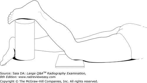
With the patient positioned as illustrated in Figure 2–20, which of the following structures is best demonstrated?
A Patella
B Patellofemoral articulation
C Intercondyloid fossa
D Tibial tuberosity
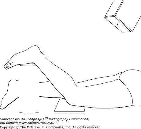
C Intercondyloid fossa
-The PA axial projection (Camp-Coventry method) of the intercondyloid fossa (tunnel view) is shown. The knee is flexed about 40 degrees, and the CR is directed caudally 40 degrees and perpendicular to the tibia (Figure 2–57). The patella and patellofemoral articulation are demonstrated in the axial/tangential view of the patella.

Which of the following statements is (are) true regarding the images shown in Figure 2–33?
- Image A is positioned in internal rotation.
- Image B is positioned in internal rotation.
- The greater tubercle is better demonstrated in image A.
A 1 only
B 2 only
C 1 and 3 only
D 2 and 3 only
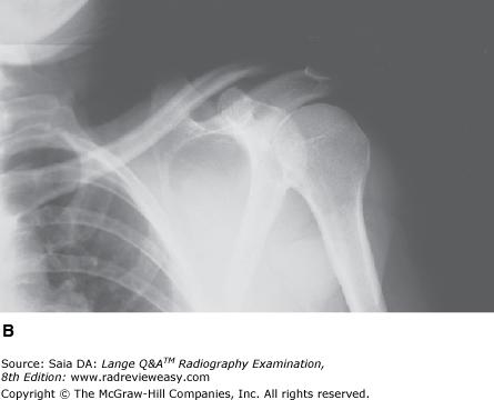
D 2 and 3 only
-When the shoulder is placed in internal rotation, a greater portion of the glenoid fossa is superimposed by the humeral head, and the lesser tubercle is visualized, as in image B. The external rotation position (image A) removes the humeral head from a large portion of the glenoid fossa and better demonstrates the greater tubercle.

Which of the following correctly identifies the letter T in the radiograph shown in Figure 7–13?
A Gliding joint
B Pivot joint
C Diarthrotic joint
D Amphiarthrotic joint
C Diarthrotic joint
-The radiograph is a PA projection of the hand and wrist; an oblique projection of the thumb is obtained. The letter T is pointing out the first carpometacarpal joint, formed by the base of the first metacarpal and the trapezium. This is classified as a saddle-type diarthrotic joint. Diarthrotic joints are freely movable joints and the most plentiful type of joint in the human body. Amphiarthrotic joints are partially movable; synarthrotic joints are immovable.
The AP axial projection of the pulmonary apices requires the CR to be directed
A 15 degrees cephalad
B 15 degrees caudad
C 30 degrees cephalad
D 30 degrees caudad
A 15 degrees cephalad
-It is occasionally necessary to view the lung apices free of superimposition with the clavicles. This objective can be achieved in an AP axial projection. The patient is positioned AP erect with the CR directed 15 degrees cephalad, entering the manubrium. An AP axial projection can also be obtained with the patient in the lordotic position. If sufficient lordosis can be assumed, the CR is directed perpendicular to the IR.
Double-contrast examinations of the stomach or large bowel are performed to better visualize the
A position of the organ
B size and shape of the organ
C diverticula
D gastric or bowel mucosa
D gastric or bowel mucosa
-Double-contrast studies of the stomach or large intestine involve coating the organ with a thin layer of barium sulfate and then introducing air. This allows the operator to see through the organ to structures behind it and, most especially, allows visualization of the mucosal lining of the organ. A barium-filled stomach or large bowel demonstrates the position, size, and shape of the organ and any lesion that projects out from its walls, such as diverticula. Polypoid lesions, which project inward from the wall of an organ, may go unnoticed unless a double-contrast examination is performed.
Which of the following procedures will best demonstrate the cephalic, basilic, and subclavian veins?
A Aortofemoral arteriogram
B Upper-limb venogram
C Lower-limb venogram
D Renal venogram
B Upper-limb venogram
-The cephalic, basilic, and subclavian veins should be demonstrated on an upper limb venogram. Venography of the upper limb usually is performed to rule out venous obstruction or thrombosis. The injection site is usually in the hand or wrist, and images should be obtained up to the area of the superior vena cava.
In the 15° medial oblique projection of the ankle, demonstrates the entire
1.talofibular joint.
2. tibiotalar joint.
3.ankle mortise.
A 1 only
B 1 and 2 only
C 2 and 3 only
D 1, 2, and 3
D 1, 2, and 3
-The medial oblique projection of the ankle can be performed either as a 15° to 20° oblique or as a 45° oblique. The 15° to 20° oblique demonstrates the entire ankle mortise, that is, the articulations between the talus, tibia, and fibula. The talofibular articulation is open, the tibiotalar articulation is open, and the entire mortise is visible. The 45° oblique opens the distal tibiofibular joint. It is often recommended that the plantar surface be vertical.
Which of the following articulations may be described as diarthrotic?
- Knee
- Intervertebral joints
- Temporomandibular joint (TMJ)
A 1 only
B 2 only
C 1 and 3 only
D 1, 2, and 3
C 1 and 3 only
-Diarthrotic, or synovial, joints, such as the knee and the TMJ, are freely movable. Most diarthrotic joints are associated with a joint capsule containing synovial fluid. Diarthrotic joints are the most numerous in the body and are subdivided according to type of movement. Amphiarthrotic joints are partially movable joints whose articular surfaces are connected by cartilage, such as intervertebral joints. Synarthrotic joints, such as the cranial sutures, are immovable.
All the following can be associated with the elbow joint except
A the capitulum.
B the trochlea.
C the tubercles.
D the epicondyles.
C the tubercles.
-The distal humerus articulates with the radius and ulna to form the elbow joint. The lateral aspect of the distal humerus presents a raised, smooth, rounded surface, the capitulum, that articulates with the superior surface of the radial head. The trochlea is on the medial aspect of the distal humerus and articulates with the semilunar/trochlear notch of the ulna. Just proximal to the capitulum and trochlea are the lateral and medial epicondyles; the medial is more prominent and palpable. Lateral epicondylitis("tennis elbow") is a painful condition caused by prolonged rotary motion of the forearm. The tubercles are prominences located at the proximal humerus and are anatomically remote from the elbow joint.
Which of the following is a major cause of bowel obstruction in children?
A Appendicitis
B Intussusception
C Regional enteritis
D Ulcerative colitis
B Intussusception
-Intussusception is the telescoping of one part of the intestinal tract into another. It is a major cause of bowel obstruction in children, usually in the region of the ileocecal valve, and is much less common in adults. Radiographically, intussusception appears as the classic “coil spring,” with barium trapped between folds of the telescoped bowel. The diagnostic BE procedure occasionally can reduce the intussusception, although care must be taken to avoid perforation of the bowel. Appendicitis occurs when an obstructed appendix becomes inflamed. Distension of the appendix occurs, and if the appendix is left untended, gangrene and perforation can result. Regional enteritis (Crohn disease) is a chronic granulomatous inflammatory disorder that can affect any part of the GI tract but generally involves the area of the terminal ileum. Ulceration and formation of fistulous tracts often occur.Ulcerative colitis occurs most often in young adults; its etiology is unknown, although psychogenic or autoimmune factors seem to be involved.
The AP oblique projection (medial rotation) of the elbow demonstrates which of the following?
1.Radial head free of superimposition
2.Olecranon process within the olecranon fossa
3.Coronoid process free of superimposition
A 1 only
B 1 and 2 only
C 2 and 3 only
D 1, 2, and 3
C 2 and 3 only
-The AP oblique projection (medial rotation) of the elbow superimposes the radial head and neck on the proximal ulna. It demonstrates the olecranon process within the olecranon fossa, and it projects the coronoid process free of superimposition. The radial head is projected free of superimposition in the AP oblique projection (lateral rotation) of the elbow.
The body habitus characterized by a long and narrow thoracic cavity and low midline stomach and gallbladder is the
A asthenic
B hyposthenic
C sthenic
D hypersthenic
A asthenic
-The four types of body habitus describe differences in visceral shape, position, tone, and motility. One body type is hypersthenic, characterized by the very large individual with short, wide heart and lungs, high transverse stomach and gallbladder, and peripheral colon. The sthenic individual is the average, athletic, most predominant type. The hyposthenic patient is somewhat thinner and a little frailer, with organs positioned somewhat lower. The asthenic type is smaller in the extreme, with a long thorax, a very long, almost pelvic stomach, and a low medial gallbladder. The colon is medial and redundant. Hypersthenic patients usually demonstrate the greatest motility.

What does the number 8 in Figure 6–14 identify?
A Medial malleolus
B Lateral malleolus
C Medial cuneiform
D Talus
A Medial malleolus
-An anterior view of the foot and ankle bones is shown. The ankle joint is formed by the articulation of the tibia, fibula, and talus (number 7). The tibial (medial) malleolus is labeled 8; the fibular (lateral) malleolus is labeled 1. The talus articulates with the calcaneus (number 2) inferiorly and with the navicular (number 6) anteriorly. The cuboid (number 3) is seen anterior to the calcaneus, and the three cuneiforms (number 5) are anterior to the navicular.
In myelography, the contrast medium generally is injected into the
A cisterna magna
B individual intervertebral disks
C subarachnoid space between the first and second lumbar vertebrae
D subarachnoid space between the third and fourth lumbar vertebrae
D subarachnoid space between the third and fourth lumbar vertebrae
-Generally, contrast medium is injected into the subarachnoid space between the third and fourth lumbar vertebrae (Figure 2–62). Because the spinal cord ends at the level of the first or second lumbar vertebra, this is considered to be a relatively safe injection site. The cisterna magna can be used, but the risk of contrast medium entering the ventricles and causing side effects increases. Diskography requires injection of contrast medium into the individual intervertebral disks.
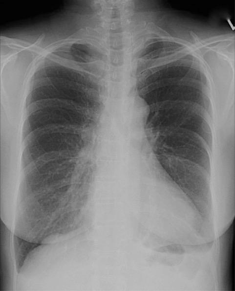
Which of the following statements with respect to the PA chest seen in Figure 2–11 is (are) correct?
- Adequate inspiration is demonstrated.
- The shoulders are rolled forward adequately.
- Rotation is demonstrated.
A 1 only
B 1 and 2 only
C 2 and 3 only
D 1, 2, and 3
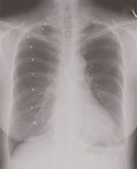
D 1, 2, and 3
-A PA projection of the chest is illustrated below. Adequate inspiration is present because the diaphragm is depressed enough to demonstrate 10 posterior ribs. The shoulders are rolled forward sufficiently; the scapulae are completely removed from superimposition on the lung fields. The major fault with this image is the obvious rotation, as evidenced by the asymmetry between the medial extremities of the clavicles and the vertebral borders. Note that the medial extremity of the right clavicle is completely visible, whereas the left is not seen at all.
Which of the following projections of the elbow should demonstrate the coronoid process free of superimposition and the olecranon process within the olecranon fossa?
A AP
B Lateral
C Medial oblique
D Lateral oblique
C Medial oblique
-On the AP projection of the elbow, the radial head and ulna are normally somewhat superimposed. The lateral oblique demonstrates the radial head free of ulnar superimposition. The lateral projection demonstrates the olecranon process in profile. The medial oblique demonstrates considerable overlap of the proximal radius and ulna, but should clearly demonstrate the coronoid process free of superimposition and the olecranon process within the olecranon fossa.
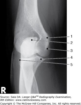
In which of the following projections was the image in Figure 2–7 made?
A AP
B medial oblique
C lateral oblique
D acute flexion
B medial oblique
-The image illustrates a medial oblique (internal rotation) projection of the elbow with epicondyles 45 degrees to the IR. An oblique view of the proximal radius and ulna and the distal humerus is obtained. This projection is particularly useful to demonstrate the coronoid process in profile, the trochlea, and the medial epicondyle. The external oblique (lateral rotation) projection demonstrates the radial head free of superimposition as well as the radial neck and the humeral capitulum. The acute flexion projection (Jones Method) of the elbow is a two-projection method demonstrating the elbow anatomy when the part cannot be extended for an AP projection.
Which of the following pathologic conditions probably will require a decrease in exposure factors?
A Osteomyelitis
B Osteoporosis
C Osteosclerosis
D Osteochondritis
B Osteoporosis
-Osteoporosis is a condition, often seen in the elderly, marked by increased porosity and softening of bone. The bones are much less dense, and thus a decrease in exposure is required. Osteomyelitis and osteochondritis are inflammatory conditions that usually increase bone density. Osteosclerosis is abnormal hardening of the bone, and an increase in exposure factors would be required.
The secondary center of ossification in long bones is the
A diaphysis
B epiphysis
C metaphysis
D apophysis
B epiphysis
-Long bones are composed of a shaft, or diaphysis, and two extremities. The diaphysis is referred to as the primary ossification center. In the growing bone, the cartilaginous epiphyseal plate (located at the extremities of long bones) is gradually replaced by bone. For this reason, the epiphyses are referred to as the secondary ossification centers. The ossified growth area of long bones is the metaphysis.An apophysis is a normal bony outgrowth arising from a separate ossification center which fuses in time. An apophysis is the site of ligament or tendon attachment (whereas an epiphysis contributes to joint formation).
With the patient seated at the end of the x-ray table, elbow flexed 80 degrees, and the CR directed 45 degrees laterally from the shoulder to the elbow joint, which of the following structures will be demonstrated best?
A Radial head
B Ulnar head
C Coronoid process
D Olecranon process
C Coronoid process
-The axial trauma lateral (Coyle) position is described. If routine elbow projections in extension are not possible because of limited part movement, these positions can be used to demonstrate the coronoid process and/or radial head. With the elbow flexed 90 degrees and the CR directed to the elbow joint at an angle of 45 degrees medially (i.e., toward the shoulder), the joint space between the radial head and capitulum should be revealed. With the elbow flexed 80 degrees and the CR directed to the elbow joint at an angle of 45 degrees laterally (i.e., from the shoulder toward the elbow), the elongated coronoid process will be visualized.
During studies of the soft tissue of the neck, the exposure can be made
1.during phonation before/after opacification.
2.during Valsalva maneuver.
3.at the height of swallowing motion with opacification.
A 1 only
B 1 and 2 only
C 2 and 3 only
D 1, 2, and 3
D 1, 2, and 3
-Soft tissue neck studies can be performed for a number of reasons including to determine presence of foreign body or to evaluate the swallowing mechanism following a stroke event. Phonation of various vowel sounds, with or without contrast media opacification, can help demonstrate the vocal cords. Performance of the Valsalva maneuver fills the larynx and trachea with air, which is then well demonstrated on soft tissue study. Pharyngeal structures are demonstrated during swallowing motion.
To radiograph an infant for suspected free air within the abdominal cavity, which of the following projections of the abdomen will demonstrate the condition with the least patient exposure?
A PA erect with grid
B Left lateral decubitus with grid
C Left lateral decubitus without grid
D Recumbent AP without grid
C Left lateral decubitus without grid
-Air–fluid levels are demonstrated in the erect or decubitus position. Grid radiography requires about a 3 to 4 times greater dose than nongrid radiography. A left lateral decubitus projection without a grid, then, would demonstrate fluid levels with a considerably smaller dose to the infant. PA erect with grid would be the next-best choice – the PA position reduces dose to radiosensitive future reproductive cells. A recumbent AP projection would not demonstrate air–fluid levels. Left lateral decubitus is preferred to right lateral decubitus because the liver moves down slightly in this position, creating a space between the liver and right hemidiaphragm (right subphrenic space). Any abdominal free air would then be visualized below the right hemidiaphragm in the subphrenic space along the lateral aspect of the liver.
Which of the following statements is (are) correct regarding the chest radiograph in Figure 6–19?
- Rotation of the chest is demonstrated.
- The pulmonary apices are not visualized.
- The costophrenic angles are demonstrated.
A 1 only
B 1 and 3 only
C 2 and 3 only
D 1, 2, and 3
B 1 and 3 only
-Rotation of the chest is evidenced in the following ways: The distance between the medial aspect of the clavicles and the lateral portion of the vertebral column is asymmetric, the air-filled trachea is off midline, and the scapulae and air-filled lungs are asymmetric. The exposure was made during reasonably good inspiration, as evidenced by visualization of eight ribs above the diaphragm. The upper and lateral aspects of the lungs, including the pulmonary apices and costophrenic angles, are demonstrated. Even minimal rotation of the chest introduces significant distortion of the heart.

Which of the labeled bones in Figure 6–14 identifies the tarsal navicular?
A Number 2
B Number 3
C Number 6
D Number 7
C Number 6
-An anterior view of the foot and ankle bones is shown. The ankle joint is formed by the articulation of the tibia, fibula, and talus (number 7). The tibial (medial) malleolus is labeled 8; the fibular (lateral) malleolus is labeled 1. The talus articulates with the calcaneus (number 2) inferiorly and with the navicular (number 6) anteriorly. The cuboid (number 3) is seen anterior to the calcaneus, and the three cuneiforms (number 5) are anterior to the navicular.
The relationship between the ends of fractured long bones is referred to as
A angulation
B apposition
C luxation
D sprain
B apposition
-Various terms are used to describe the position of fractured ends of long bones. The term apposition is used to describe the alignment, or misalignment, between the ends of fractured long bones. The termangulation describes the direction of misalignment. The term luxation refers to a dislocation. A sprain refers to a wrenched articulation with ligament injury.
Which of the following positions would best demonstrate the proximal tibiofibular articulation?
A AP
B 90 degrees mediolateral
C 45-degree internal rotation
D 45-degree external rotation
C 45-degree internal rotation
-In the AP projection, the proximal fibula is at least partially superimposed on the lateral tibial condyle.Medial rotation of 45 degrees will “open” the proximal tibiofibular articulation. Lateral rotation will obscure the articulation even more.
Moderate hypertension can produce damage to which of the following organs?
1. Lungs
2. Kidneys
3. Brain
A 1 only
B 1 and 2 only
C 2 and 3 only
D 1, 2, and 3
D 1, 2, and 3
-Normal blood pressure is 110–140 mmHg systolic and 60–80 mmHg diastolic. High blood pressure (hypertension) is indicated by a systolic pressure higher than 140 mmHg and a diastolic pressure higher than 90 mmHg. Hypertension can be identified as extreme or moderate. Extreme hypertension can result in brain damage within just a few minutes. Moderate hypertension can cause damage to organs—the lungs, kidneys, brain, heart, etc. Various disease processes can produce hypertension as well as contributing factors such as medications, obesity, smoking, and stress.
An AP oblique (lateral rotation) of the elbow demonstrates which of the following?
- Radial head free of superimposition
- Capitulum of the humerus
- Olecranon process within the olecranon fossa
A 1 only
B 1 and 2 only
C 2 and 3 only
D 1, 2, and 3
B 1 and 2 only
-The radial head and neck are projected free of superimposition in the AP oblique projection (lateral /external rotation) of the elbow. The humeral capitulum is also well demonstrated in this external oblique position. The AP oblique projection (medial /internal rotation) of the elbow superimposes the radial head and neck on the proximal ulna. The medial rotation demonstrates the olecranon process within the olecranon fossa, and it projects the coronoid process free of superimposition.
In Figure 2–29, which of the following is represented by the number 3?
A Acromion process
B Scapular spine
C Coracoid process
D Acromioclavicular joint
A Acromion process
-Figure 2–29 depicts a posterior view of the right scapula and its articulation with the humerus (number 4). The scapula presents two borders—the lateral or axillary border (number 7) and the medial or vertebral border (number 9). It also presents three angles—the inferior angle (number 8), the superior angle (number 12), and the lateral angle (number 6). The processes of the scapula are the coracoid (number 2), the acromion (number 3), and the scapular spine (number 13). The scapula has a (supra) scapular notch (number 1), a supraspinatus fossa (number 11), and an infraspinatus fossa (number 10). Number 5 identifies the glenoid fossa—the articular surface for the humeral head, forming the glenohumeral articulation.
The thoracic cavity is lined by
A parietal pleura.
B visceral pleura.
C parietal peritoneum.
D visceral peritoneum.
A parietal pleura.
-The thoracic and abdominal cavities are associated with serous membranes: the thoracic cavity with the pleura and the abdominal cavity with the peritoneum. The pleura and peritoneum each have two walls, a parietal (outer) wall and a visceral (inner) wall. The parietal pleura lines the thoracic cavity, while the visceral pleura is reflected over the surface of the lungs and projects between the fissures. The parietal peritoneum lines the abdominal cavity, and the visceral peritoneum invests the abdominal viscera.
In a posteroanterior (PA) projection of the chest being used for cardiac evaluation, the heart measures 14.7 cm between its widest points. If the magnification factor is known to be 1.2, what is the actual diameter of the heart?
A 10.4 cm
B 12.25 cm
C 13.5 cm
D 17.64 cm
B 12.25 cm
-The formula for magnification factor (MF) = image size/object size. In the stated problem, the anatomic measurement is 14.7 cm, and the magnification factor is known to be 1.2. Substituting the known factors in the appropriate equation
During chest radiography, the act of inspiration
- elevates the diaphragm
- raises the ribs
- depresses the abdominal viscera
A 1 only
B 1 and 2 only
C 2 and 3 only
D 1, 2, and 3
C 2 and 3 only
-With inspiration, the diaphragm moves inferiorly and depresses the abdominal viscera. The ribs and sternum are elevated. As the ribs are elevated, their angle is decreased. Receptor exposure can vary considerably in appearance depending on the phase of respiration during which the exposure is made.
Which of the following radiologic examinations can demonstrate ureteral reflux?
A IV urogram
B Retrograde pyelogram
C Voiding cystourethrogram
D Nephrotomogram
C Voiding cystourethrogram
-Ureteral reflux is best demonstrated during voiding. It can occur even when the bladder is only partially filled with a contrast medium. The vesicourethral orifice, as well as other sphincter muscles, relaxes during urination; however, the vesicoureteral orifices may also relax and cause reflux.
The condition in which pulmonary alveoli lose their elasticity and become permanently inflated, causing the patient to consciously exhale, is
A bronchial asthma
B bronchitis
C emphysema
D tuberculosis
C emphysema
-Emphysema is a progressive disorder caused by long-term irritation of the bronchial passages, such as by air pollution or cigarette smoking. Emphysema patients are unable to exhale normally because of the loss of elasticity of the alveolar walls. If emphysema patients receive oxygen, it is usually administered at a very slow rate because their respirations are controlled by the level of carbon dioxide in the blood.
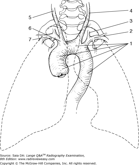
The structure labeled number 6 in Figure 2–39 is the
A left subclavian artery
B brachiocephalic artery
C right common carotid artery
D left vertebral artery
B brachiocephalic artery
-The figure illustrates the aortic arch (number 1) and its three main branches—the brachiocephalic artery (number 6), the left common carotid artery (number 4), and the left subclavian artery (number 2). The right common carotid artery (number 5) and the right subclavian artery (number 7) are branches of the brachiocephalic. The vertebral arteries are the first main branch of the subclavian arteries. The left vertebral artery is labeled number 3.
The term varus refers to
A turned outward
B turned inward
C rotated medially
D rotated laterally
B turned inward
-
The term varus refers to bent or
turned inward. In genu varus, the tibia or femur turns
inward causing bowlegged deformity; in talipes varus, the foot turns
inward (clubfoot deformity). The term valgus refers to a
part turned/deformed outward—as in hallux valgus and talipes
valgus. Hallux valgus is angulation of the great toe away from the
midline; talipes valgus is a foot deformity with the heel turned
outward—a component of clubfoot.
Which of the following positions will demonstrate the right axillary ribs?
1.RAO
2.LAO
3.RPO
A 1 only
B 1 and 2 only
C 2 and 3 only
D 1, 2, and 3
C 2 and 3 only
-The axillary portion of the ribs is best demonstrated in a 45° oblique position. The axillary ribs are demonstrated in the AP oblique projection with the affected side adjacent to the IR, and in the PA oblique projection with the affected side away from the IR. Therefore, the right axillary ribs would be demonstrated in the RPO (AP oblique with affected side adjacent to the IR) and LAO (PA oblique with affected side away from the IR) positions.
Which projection(s) of the abdomen would be used to demonstrate pneumoperitoneum?
1.Right lateral decubitus
2.Left lateral decubitus
3.Upright
A 2 only
B 1 and 3 only
C 2 and 3 only
D 1, 2, and 3
C 2 and 3 only
-An erect abdomen or left lateral decubitus should be performed for demonstration of air–fluid levels in the abdomen. The right lateral decubitus position is used to demonstrate the layering of gallstones. It will not show free air within the peritoneum because of the overlying gastric bubble on the elevated left side of the body.
Which of the following is (are) evaluation criteria for a PA chest radiograph of the heart and lungs?
- Ten posterior ribs should be seen above the diaphragm.
- The medial ends of the clavicles should be equidistant from the vertebral column.
- The scapulae should be seen through the upper lung fields.
A 1 only
B 1 and 2 only
C 2 and 3 only
D 1, 2, and 3
B 1 and 2 only
-Sufficient inspiration is demonstrated by the visualization of 10 posterior ribs projected above the diaphragm. Rotation of the chest is detected by asymmetry in the distance between the medial ends of the clavicles and the vertebral column. The scapulae should be free of superimposition with the lung fields; this is accomplished by rolling the shoulders forward while positioning for the PA projection.
What is the name of the plane indicated by the number 1 in Figure 6–17?
A Midcoronal plane
B Midsagittal plane
C Transverse plane
D Horizontal plane
A Midcoronal plane
-The midcoronal plane (number 1) divides the body into anterior and posterior halves. A coronal plane is any plane parallel to the midcoronal plane. The midsagittal plane (number 2) divides the body into left and right halves. A sagittal plane is any plane parallel to the midsagittal plane. A transverse or horizontal plane (number 3) is perpendicular to the midsagittal plane and midcoronal plane, dividing the body into superior and inferior portions.
Which of the following is the preferred scheduling sequence?
A Lower GI series, abdomen ultrasound, upper GI series
B Abdomen ultrasound, lower GI series, upper GI series
C Abdomen ultrasound, upper GI series, lower GI series
D Upper GI series, lower GI series, abdomen ultrasound
B Abdomen ultrasound, lower GI series, upper GI series
-Diagnostic imaging examinations must be scheduled appropriately. Retained barium sulfate contrast medium can obscure necessary anatomic details in x-ray or ultrasound studies that are scheduled later. Therefore, the ultrasound examination should come first, followed by the lower GI series (BE), and finally the upper GI series. Retained barium from the lower GI series probably will not obscure upper GI structures.
Which of the following equipment is necessary for ERCP?
- A fluoroscopic unit with imaging device and tilt-table capabilities
- A fiberoptic endoscope
- Polyethylene catheters
A 1 and 2 only
B 1 and 3 only
C 2 and 3 only
D 1, 2, and 3
D 1, 2, and 3
-A fluoroscopic unit with spot device and tilt table should be used for endoscopic retrograde pancreatography. The Trendelenburg position is sometimes necessary to fill the interhepatic ducts, and a semierect position may be necessary to fill the lower end of the common bile duct. Also necessary are a fiberoptic endoscope for locating the hepatopancreatic ampulla and polyethylene catheters for the introduction of contrast medium.
The primary center of ossification in long bones is the
A diaphysis.
B epiphysis.
C metaphysis.
D apophysis.
A diaphysis.
-Long bones are composed of a shaft, or diaphysis, and two extremities. The diaphysis is referred to as the primary ossification center. In the growing bone, the cartilaginous epiphyseal plate (located at the extremities of long bones) is gradually replaced by bone. For this reason, the epiphyses are referred to as the secondary ossification centers. The ossified growth area of long bones is the metaphysis. Apophysis refers to vertebral joints formed by articulation of superjacent articular facets.
Which of the following structures is (are) located in the right upper quadrant (RUQ)?
- Hepatic flexure
- Gallbladder
- Ileocecal valve
A 1 only
B 1 and 2 only
C 2 and 3 only
D 1, 2, and 3
B 1 and 2 only
-The gallbladder is located on the posterior surface of the liver in the right upper quadrant (RUQ). The hepatic (right colic) flexure, so named because of its close proximity to the liver, is also in the RUQ. The cecum, located in the right lower quadrant (RLQ), is continuous with the terminal ileum—forming the ileocecal valve
Which of the following is (are) essential to high-quality mammographic examinations?
- Small-focal-spot x-ray tube
- Short-scale contrast
- Use of a compression device
A 1 only
B 1 and 2 only
C 1 and 3 only
D 1, 2, and 3
D 1, 2, and 3
-Breast tissue has very low subject contrast, but it is imperative to visualize micro-calcifications and subtle tissue density differences. Fine detail is necessary to visualize any micro-calcifications; therefore, a small-focal-spot tube is essential. High, short-scale contrast (and, therefore, low kilovoltage) is needed to accentuate minute differences in tissue density. A compression device serves to even out differences in tissue thickness (thicker at the chest wall, thinner at the nipple) and decrease OID and helps to decrease the production of scattered radiation.
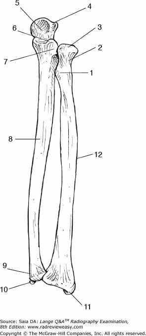
What is the anatomic structure indicated by the number 7 in Figure 6–22?
A Coracoid process
B Coronoid process
C Trochlear notch
D Radial notch
D Radial notch
-An anterior view of the forearm is shown. The proximal anterior surface of the ulna (number 8) presents a rather large pointed process at the anterior margin of the semilunar (trochlear) notch (number 5) called the coronoid process (number 6). The olecranon process is identified as number 4, and the radial notch of the ulna is number 7. Distally, the ulnar head is number 9, and the styloid process is labeled 10. The radius (number 12) is the lateral bone of the forearm. The radial head is number 3, the radial neck is number 2, and the radial tuberosity is number 1. Distally, the radial styloid process is labeled 11
Free air in the abdominal cavity is best demonstrated in which of the following positions?
A AP projection, left lateral decubitus position
B AP projection, right lateral decubitus position
C PA recumbent position
D AP recumbent position
A AP projection, left lateral decubitus position
-The erect position is employed most often to demonstrate air–fluid levels in the chest or abdomen or both. However, patients having traumatic injuries frequently must be examined in the recumbent position. The recumbent position will not demonstrate air–fluid levels unless it is a decubitus position. If free air is being questioned, we will look for that quantity of air on the “up” side because air rises. However, because liver tissue is so homogeneous, a small amount of air will be perceived more easily superimposed on it rather than on left-sided structures. Thus, an AP projection obtained in the left lateral decubitus position will best demonstrate a small amount of free air because that air will be superimposed on the liver.
Which of the following is (are) part of the bony thorax?
- Manubrium
- Clavicles
- 24 ribs
A 1 only
B 1 and 2 only
C 1 and 3 only
D 1, 2, and 3
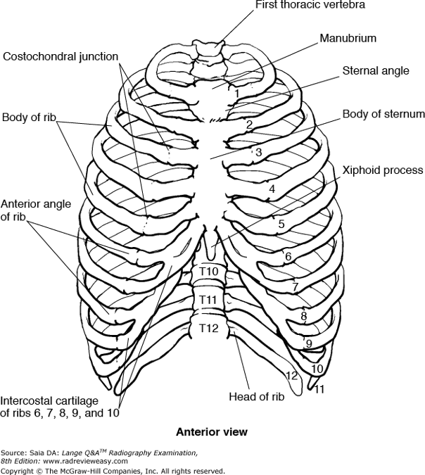
C 1 and 3 only
-The bony thorax consists of 12 pairs of ribs and the structures to which they are attached anteriorly and posteriorly: the sternum (consisting of manubrium, body/gladiolus, and xiphoid/ensiform process) and the 12 thoracic vertebrae (Figure 2–55). These structures form a bony cage that surrounds and protects the vital organs within (the heart, lungs, and great vessels). The scapulae, together with the clavicles, form the shoulder (pectoral) girdle of the upper extremity.
All of the following statements regarding the RAO position of the sternum are true, except
A the sternum is generally projected to the left of the vertebral column.
B shallow breathing during the exposure can obliterate prominent pulmonary markings.
C it is helpful to project the sternum over the heart.
D a thin thorax requires a lesser degree of obliquity than a thicker thorax.
D a thin thorax requires a lesser degree of obliquity than a thicker thorax.
-A thin chest would require a greater degree of obliquity to separate the vertebrae and sternum from superimposition than would a thick chest. With the patient in the RAO position, the sternum is projected to the left of the vertebral column and superimposed on the heart. This superimposition promotes more uniform tissue density and therefore more uniform receptor exposure. Prominent pulmonary vascular markings may be obliterated by allowing the patient to breathe (shallow breaths only) during a long exposure (with a very low mA).
In what order should the following examinations be scheduled?
- Upper GI
- Intravenous pyelogram (IVP)
- Barium enema (BE)
A 3, 1, 2
B 1, 3, 2
C 2, 1, 3
D 2, 3, 1
D 2, 3, 1
-When scheduling patient examinations, it is important to avoid the possibility of residual contrast medium covering areas of interest on later examinations. The IVP should be scheduled first because the contrast medium used is excreted rapidly. The BE should be scheduled next. Finally, the upper GI series is scheduled. Any barium remaining from the previous BE should not be enough to interfere with the stomach or duodenum (a preliminary scout image should be taken in each case).
Which of the following statements regarding the PA oblique scapular Y projection of the shoulder is (are) true?
- The midsagittal plane should be about 60 degrees to the IR.
- The scapular borders should be superimposed on the humeral shaft.
- An oblique projection of the shoulder is obtained.
A 1 only
B 1 and 2 only
C 2 and 3 only
D 1, 2, and 3
C 2 and 3 only
-The scapular Y projection requires that the coronal plane be about 60 degrees to the IR (MSP is about 30 degrees), thus resulting in an oblique projection of the shoulder. The vertebral and axillary borders of the scapula are superimposed on the humeral shaft, and the resulting relationship between the glenoid fossa and humeral head will demonstrate anterior or posterior dislocation. Lateral or medial dislocation is evaluated on the AP projection.
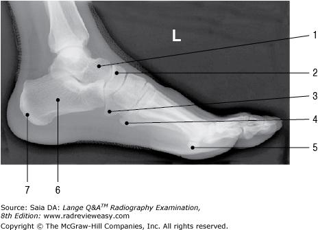
The bone labeled number 3 in Figure 7–15 is the
A talus
B cuboid
C navicular
D lateral cuneiform
B cuboid
-The bones of the foot include the 7 tarsal bones, 5 metatarsal bones, and 14 phalanges. The calcaneus (os calsis), or heel bone, is the largest tarsal (numbers 6 and 7). It serves as attachment for the Achilles tendon posteriorly, articulates anteriorly with the cuboid bone (number 3), presents three articular surfaces superiorly for its articulation with the talus (number 1), and has a prominent shelf on its anteromedial edge called the sustentaculum tali. The inferior surface of the talus (astragalus) articulates with the superior calcaneus to form the three-faceted subtalar joint. The talus also articulates anteriorly with the navicular (number 2). Articulating anteriorly with the navicular are the three cuneiform bones—medial/first, intermediate/second, and lateral/third. The navicular articulates laterally with the cuboid.
Skeletal conditions characterized by faulty bone calcification include
1.osteoarthritis.
2.osteomalacia.
3.rickets.
A 1 only
B 1 and 2 only
C 2 and 3 only
D 1, 2, and 3
C 2 and 3 only
-Rickets and osteomalacia are skeletal disorders characterized by abnormal calcification processes. In osteomalacia, bones become soft and are easily misshapen. Rickets affects the growing bones of children and is also characterized by soft, misshapened bones—as a result of calcium salts not being deposited in bone matrix. Osteoarthritis is a degeneration of articular cartilage; when these surfaces then attempt to articulate and move, bone friction and pain occur.
A patient suffering from orthopnea would experience the least discomfort in which body position?
A Fowler
B Trendelenburg
C Recumbent
D Erect
D Erect
-Orthopnea is a respiratory condition in which the patient has difficulty breathing (dyspnea) in any position other than erect. The patient is usually comfortable in the erect, standing, or seated position. The Trendelenburg position places the patient's head lower than the rest of the body, the Fowler position is a semierect position, and the recumbent position is lying down.
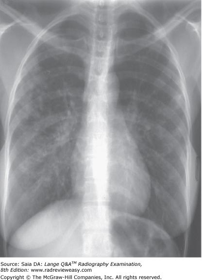
The PA chest image shown in Figure 4–13 exhibits which of the following qualities?
- Adequate penetration of the heart
- Long-scale contrast
- Adequate inspiration
A 1 only
B 1 and 2 only
C 2 and 3 only
D 1, 2, and 3
D 1, 2, and 3
-The PA projection of the chest shown in the figure demonstrates faint visualization of the thoracic vertebrae through the heart—characteristic of adequate penetration. Many shades of gray are seen, illustrating a long scale of contrast. The image is representative of having been exposed at the ideal milliampere-seconds and kilovoltage factors. Ten posterior ribs are seen; this illustrates adequate inspiration.
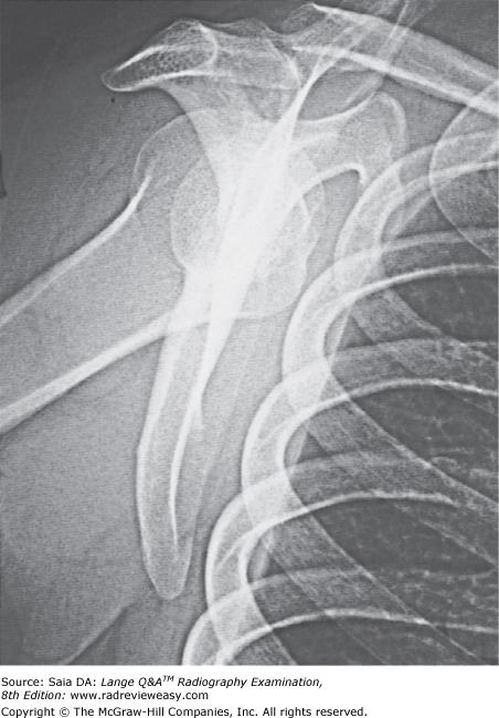
Which of the following statements is (are) true with respect to the radiograph shown in the Figure below?
- The acromion process is seen partially superimposed on the third rib.
- This projection is performed to evaluate the scapula.
- This projection is performed to evaluate the acromioclavicular articulation.
A 1 only
B 2 only
C 1 and 2 only
D 2 and 3 only
B 2 only
-The radiograph in the Figure illustrates a lateral projection of the scapula. The axillary and vertebral borders are superimposed. The acromion and coracoid process are visualized; the coracoid process is partially superimposed on the axillary portion of the third rib. A scapular Y projection is often performed to demonstrate shoulder dislocation, but the affected arm is left to rest at the patient's side; the arm in this radiograph is abducted somewhat to better view the body of the scapula.
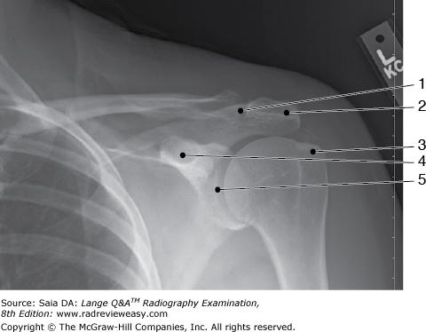
What projection was used to obtain the image seen in Figure 2–41?
A AP, internal rotation
B AP, external rotation
C AP, neutral position
D AP axial
B AP, external rotation
-An AP, external rotation, projection of the shoulder is pictured. The hand is supinated, and the arm is in the anatomical position. Therefore, the greater tubercle (number 3) is well visualized. The greater portion of the clavicle is seen, the acromioclavicular joint (number 1), the acromion process (number 2), the coracoid process (number 4), and the glenohumeral joint (number 5). The coronoid process is located on the ulna.
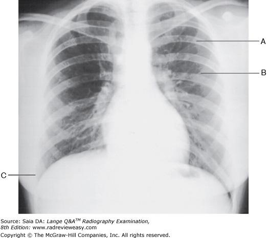
The letter B in Figure 2–13 indicates a
A left anterior rib
B right posterior rib
C left posterior rib
D right anterior rib
A left anterior rib
-A PA projection of the chest is shown. Adequate inspiration is demonstrated by visualization of 10 posterior ribs above the diaphragm. Rotation of the chest is demonstrated by asymmetrical sternoclavicular joints. The apices and costophrenic angles should be included on every chest radiograph. The letter A indicates a left posterior rib, B represents a left anterior rib, and C represents the right costophrenic angle.
When examining a patient whose elbow is in partial flexion,
A the AP projection requires two separate positions and exposures.
B the AP projection is made through the partially flexed elbow, resting on the olecranon process, CR perpendicular to IR.
C the AP projection is made through the partially flexed elbow, resting on the olecranon process, CR parallel to the humerus.
D the AP projection is eliminated from the routine.
A the AP projection requires two separate positions and exposures.
-When a patient's elbow needs to be examined in partial flexion, the lateral projection offers little difficulty, but the AP projection requires special attention. If the AP projection is made with a perpendicular CR and the olecranon process resting on the table-top, the articulating surfaces are obscured. With the elbow in partial flexion, two exposures are necessary to achieve an AP projection of the elbow joint articular surfaces. One is made with the forearm parallel to the IR (humerus elevated), which demonstrates the proximal forearm. The other is made with the humerus parallel to the IR (forearm elevated), which demonstrates the distal humerus. In both cases, the CR is perpendicular if the degree of flexion is not too great or angled slightly into the joint space with greater degrees of flexion.
Which of the following techniques would provide a posteroanterior (PA) projection of the gastroduodenal surfaces of a barium-filled high and transverse stomach?
A Place the patient in a 35- to 40-degree right anterior oblique (RAO) position.
B Place the patient in a lateral position.
C Angle the CR 35 to 45 degrees cephalad.
D` Angle the CR 35 to 45 degrees caudad.
C Angle the CR 35 to 45 degrees cephalad.
-In the PA position, portions of the barium-filled hypersthenic stomach superimpose on themselves. Thus, patients with a hypersthenic body habitus usually present a high transverse stomach with poorly defined curvatures. If the PA stomach is projected with a 35- to 45-degree cephalad CR, the stomach “opens up.” That is, the curvatures, the antral portion, and the duodenal bulb all appear as a sthenic habitus stomach would appear. A 35-to 40-degree RAO position is used to demonstrate many of these structures in the average, or sthenic, body habitus. A lateral position is used to demonstrate the anterior and posterior gastric surfaces and retrogastric space.
Figure A was made in which of the following positions?
A AP
B Medial oblique
C Lateral oblique
D Partial flexion
C Lateral oblique
-The radiograph is a lateral oblique (external rotation) projection of the elbow, removing the proximal radius from superimposition with the ulna and demonstrating its articulation with the ulna at the radial notch, that is, the proximal radioulnar articulation. An AP projection of the elbow would demonstrate partial overlap of the proximal radius and ulna. A medial oblique would demonstrate complete overlap of the proximal radius and ulna; this position is used to demonstrate the coronoid process in profile and the olecranon process within the olecranon fossa.
Which of the following positions is required to demonstrate small amounts of air in the peritoneal cavity?
A Lateral decubitus, affected side up
B Lateral decubitus, affected side down
C AP Trendelenburg
D AP supine
A Lateral decubitus, affected side up
-Air or fluid levels will be clearly delineated only if the CR is directed parallel to them. Therefore, to demonstrate air or fluid levels, the erect or decubitus position should be used. Small amounts of fluid within the peritoneal or pleural space are best demonstrated in the lateral decubitus position, affected side down. Small amounts of air within the peritoneal or pleural space are best demonstrated in the lateral decubitus position, affected side up.
Operative cholangiography may be performed to
1.visualize biliary stones or a neoplasm.
2.determine function of the hepatopancreatic ampulla.
3.examine the patency of the biliary tract.
A 1 and 2 only
B 1 and 3 only
C 2 and 3 only
D 1, 2, and 3
D 1, 2, and 3
-Operative cholangiography may be performed to visualize biliary stones or a neoplasm, determine the function of the hepatopancreatic ampulla, and examine the patency of the biliary tract. Any strictures or obstructions may be localized when contrast medium is introduced into the catheter and images are obtained. It is important that no air bubbles are introduced into the biliary tract because they can imitate radiolucent stones. The radiographer can coordinate the time of exposure with the anesthesiologist to obtain the radiographs during suspended respiration.
Which of the following examinations most likely would be performed to diagnose Wilm's tumor?
A BE
B Upper GI
C IVU
D Bone survey
C IVU
-Wilm's tumor is a rapidly developing tumor of the kidney(s). It is the most common childhood renal tumor, usually affecting only one kidney. Newer treatments are effective in controlling about 90% of these tumors. As the kidneys are affected, an IVU would be the most appropriate of the examinations listed. Other useful examinations would be CT scan and sonography.
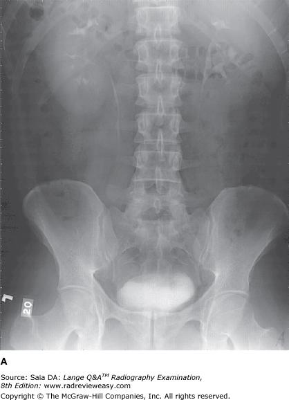
Which of the following statements referring to the images below is (are) correct?
- Image A was performed AP.
- Image B was performed AP.
- The AP image was obtained using ureteral compression.
A 1 only
B 2 only
C 1 and 3 only
D 2 and 3 only
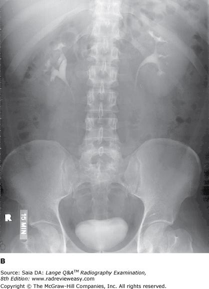
B 2 only
-Radiograph A was performed PA, and radiograph B performed AP, as evidenced by the bony pelvis anatomy. The PA projection (image A) shows the ilia more foreshortened, giving the pelvis a “closed” appearance, whereas in the AP projection the ilia and bladder area appears more “open.” There was an appropriate selection of exposure factors, for the required anatomic structures are well visualized—renal shadows, psoas muscle, lumbar transverse processes, and inferior margin of the liver. There is no evidence of ureteral compression, which would be seen as air-filled bladders, or some other type of compression device, placed over the distal ureters.

The figure below illustrates a sectional image of the abdomen. Which of the following is represented by the number 13?
A Gallbladder
B Portal vein
C Inferior vena cava
D Aorta
B Portal vein
-A cross-sectional image of the abdomen is shown in the figure. The large, homogeneous structure on the right, labeled 14, is the liver. The gallbladder is often seen darker on the medial border of the liver but is not visualized here. The left kidney is labeled 5; the right kidney is seen clearly on the opposite side labeled 11. The vertebra is seen in the center, and the psoas muscles are seen just posterior to the vertebra. Just anterior to the body of the vertebra is the circular aorta, labeled 7. The inferior vena cava(number 12) is seen to the right of the aorta. The circular structure just anterior to the inferior vena cava is the portal vein (number 13). Number 1 is the stomach, number 2 is the splenic/left colic flexure, number 3 is the pancreas, and number 4 is the spleen. Numbers 6 and 10 are portions of the left and right adrenal glands—not normally seen at this level. Number 8 is the celiac trunk; the common hepatic artery is seen branching to the right, and the splenic artery is seen branching to the left. Number 9 is a part of the diaphragmatic crura connecting the vertebrae and diaphragm.
Which of the following radiologic examinations requires preparation consisting of a low-residue diet, cathartics, and enemas?
A Upper GI series
B Small bowel series
C Barium enema (BE)
D Intravenous (IV) cystogram
C Barium enema (BE)
-To have high diagnostic quality, a barium enema (BE) examination requires rigorous and complete patient preparation. This usually consists of a modified low-residue diet for a few days before the examination, cathartics the day before, and cleansing enemas the morning of the examination. Instructions for a upper GI series, small bowel series, and IV cystogram are usually to be NPO after midnight.
Which of the following shoulder projections can be used to evaluate the lesser tubercle in profile?
A External rotation position
B Internal rotation position
C Neutral rotation position
D Inferosuperior axial position
B Internal rotation position
-The internal rotation position places the humeral epicondyles perpendicular to the IR, the humerus in a true lateral position, and the lesser tubercle in profile. The external rotation position places the humeral epicondyles parallel to the IR, the humerus in a true AP position, and the greater tubercle in profile. The neutral position is used often for the evaluation of calcium deposits in the shoulder joint.
Which of the following articulations participate in the formation of the elbow joint?
1. Between the humeral trochlea and the semilunar/trochlear notch
2. Between the capitulum and the radial head
3. The proximal radioulnar joint
A 1 only
B 1 and 2 only
C 2 and 3 only
D 1, 2, and 3
D 1, 2, and 3
-The distal humerus articulates with the radius and ulna to form a part of the elbow joint. The lateral aspect of the distal humerus presents a raised, smooth, rounded surface, the capitulum, that articulates with the superior surface of the radial head. The trochlea is on the medial aspect of the distal humerus and articulates with the semilunar notch of the ulna. All three articulations are enclosed in a common capsule to form the elbow joint proper.
During IV urography, the prone position generally is recommended to demonstrate
- the filling of the ureters
- the renal pelvis
- the superior calyces
A 1 only
B 1 and 2 only
C 1 and 3 only
D 1, 2, and 3
B 1 and 2 only
-The kidneys lie obliquely in the posterior portion of the trunk with their superior portion angled posteriorly and their inferior portion and ureters angled anteriorly. Therefore, to facilitate filling of the most anteriorly placed structures, the patient is examined in the prone position. Opacified urine then flows to the most dependent part of the kidney and ureter—the ureteropelvic region, inferior calyces, and ureters.
The carpal scaphoid can be demonstrated in which of the following projection(s) of the wrist?
- PA oblique
- PA with radial flexion
- PA with elbow elevated 20 degrees
A 1 only
B 1 and 2 only
C 1 and 3 only
D 1, 2, and 3
A 1 only
-The scaphoid can be difficult to image because its curved shape lends itself to foreshortening and self-superimposition. The lateral carpals, especially the scaphoid, are well demonstrated in the PA oblique projection. The ulnar flexion maneuver helps to overcome the scaphoid's self-superimposition. The scaphoid may also be demonstrated with less foreshortening with the wrist PA and elevated 20 degrees. The CR is directed perpendicular to the carpal scaphoid.
The medial carpals, especially the pisiform, are well demonstrated in the AP oblique projection with the radial flexion maneuver.
Which position of the shoulder demonstrates the lesser tubercle in profile medially?
A AP
B External rotation
C Internal rotation
D Neutral position
C Internal rotation
-The external rotation position is the true AP position and places the greater tubercle in profile laterally and places the lesser tubercle anteriorly. The internal rotation position demonstrates the lesser tubercle in profile medially and places the humerus in a true lateral position; the greater tubercle is seen superimposed on the humeral head. The epicondyles should be superimposed and perpendicular to the IR. The neutral position places the epicondyles about 45 degrees to the IR and places the greater tubercle anteriorly but still lateral to the lesser tubercle.
To demonstrate the pulmonary apices with the patient in the AP position, the
A central ray is directed 15° to 20° cephalad.
B central ray is directed 15° to 20° caudad.
C exposure is made on full exhalation.
D patient's shoulders are rolled forward.
A central ray is directed 15° to 20° cephalad.
-When the shoulders are relaxed, the clavicles are usually carried below the pulmonary apices. To examine the portions of the lungs lying behind the clavicles, the central ray is directed cephalad 15° to 20° to project the clavicles above the apices when the patient is examined in the AP position.
The term that refers to parts away from the source or beginning is
A cephalad
B proximal
C distal
D lateral
C distal
-There are many terms (with which the radiographer must be familiar) that are used to describe radiographic positioning techniques. Cephalad refers to that which is toward the head, and caudad refers to that which is toward the feet. Structures close to the source or beginning are said to be proximal, whereas those lying away from the source or origin are distal. Parts close to the midline are said to be medial, and those away from the midline are lateral.
Medial displacement of a tibial fracture would be best demonstrated in the
A AP projection
B lateral projection
C medial oblique projection
D lateral oblique projection
A AP projection
-A frontal projection (AP or PA) demonstrates the medial and lateral relationships of structures. A lateral projection demonstrates the anterior and posterior relationships of structures. Two views, at right angles to each other, generally are taken of most structures.
An aspirated foreign body is more likely to enter the lower respiratory tract via the
A left main stem bronchus.
B right main stem bronchus.
C bronchioles.
D alveoli.
B right main stem bronchus.
-The right and left main stem (primary) bronchi branch from the distal trachea; the right branch supplies air to the right lung, and the left branch supplies air to the left lung. The right main stem bronchus is shorter, wider, and more vertical than the left—making it the most likely route for aspirated foreign bodies to enter the right lung.
Endoscopic retrograde cholangiopancreatography (ERCP) usually involves
- cannulation of the hepatopancreatic ampulla
- introduction of contrast medium into the common bile duct
- introduction of barium directly into the duodenum
A 1 only
B 1 and 2 only
C 1 and 3 only
D 1, 2, and 3
B 1 and 2 only
-ERCP may be performed to investigate abnormalities of the biliary system or pancreas. The patient's throat is treated with a local anesthetic in preparation for the passage of the endoscope. The hepatopancreatic ampulla (of Vater) is located, and a cannula is passed through it so that contrast medium may be introduced into the common bile duct. Spot images of the common bile duct and pancreatic duct are taken frequently in the oblique position. Direct injection of barium mixture into the duodenum occurs during an enteroclysis procedure of the small bowel.
Which of the following is (are) valid evaluation criteria for a lateral projection of the forearm?
- The radius and the ulna should be superimposed distally.
- The coronoid process and the radial head should be partially superimposed.
- The humeral epicondyles should be superimposed.
A 1 only
B 1 and 2 only
C 2 and 3 only
D 1, 2, and 3
D 1, 2, and 3
-To accurately position a lateral forearm, the elbow must form a 90-degree angle with the humeral epicondyles superimposed. The radius and ulna are superimposed distally. Proximally, the coronoid process and radial head are partially superimposed. Failure of the elbow to form a 90-degree angle or the hand to be lateral results in a less than satisfactory lateral projection of the forearm.

Which of the following positions will move the fundus of the gallbladder shown in Figure 7–6 away from the superimposed transverse process?
A RAO
B LAO
C LPO
D Left lateral decubitus
B LAO
-The image shown is an erect PA projection of a patient of hyposthenic body habitus. Note the low position of the gallbladder. It is a result of body habitus and position (viscera assume a lower position in the erect position). The gallbladder may be moved away from the spine by using the LAO position. The right lateral decubitus position also will move the gallbladder away from the spine.
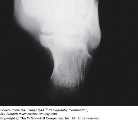
The radiograph shown in Figure 7–12 can be produced with the
- long axis of the plantar surface perpendicular to the IR
- CR 40 degrees cephalad to the base of the third metatarsal
- CR 20 degrees cephalad to the talotibial joint
A 1 only
B 2 only
C 1 and 2 only
D 1 and 3 only
C 1 and 2 only
-The radiograph illustrates a plantodorsal projection of the calcaneus. The patient usually is positioned with the leg extended and the long axis of the plantar surface perpendicular to the tabletop/IR. The CR is directed 40 degrees cephalad to the base of the third metatarsal. Structures that should be visualized include the sustentaculum tali, trochlear process, and calcaneal tuberosity.
The pyloric canal and duodenal bulb are best demonstrated during an upper GI series in which of the following positions?
A RAO
B Left lateral
C Recumbent PA
D Recumbent AP
A RAO
-The RAO position affords a good view of the pyloric canal and duodenal bulb. It is also a good position for the barium-filled esophagus, projecting it between the vertebrae and the heart. The left lateral projection of the stomach demonstrates the left retrogastric space, the recumbent PA projection is used as a general survey of the gastric surfaces, and the recumbent AP projection with slight left oblique affords a double contrast of the pylorus and duodenum.
The sternoclavicular joints will be best demonstrated in which of the following positions?
A Apical lordotic
B Anterior oblique
C Lateral
D Weight-bearing
B Anterior oblique
-The (diarthrotic) sternoclavicular joints are formed by the medial (sternal) extremities of the clavicles and the clavicular notches of the manubrium (of the sternum). They can be demonstrated in the LAO and RAO positions. The LAO projection demonstrates the left sternoclavicular joint, whereas the RAO projection demonstrates the joint on the right. The patient is obliqued about 15 degrees with the side of interest adjacent to the IR.
A modified axiolateral inferosuperior projection of the femoral neck is particularly useful
1.when the "cross-table" axiolateral is contraindicated.
2.for patients with bilateral hip fractures.
3.for patients with limited movement of the unaffected leg.
A 1 only
B 1 and 2 only
C 1 and 3 only
D 1, 2, and 3
D 1, 2, and 3
-Typically, traumatic injury to the hip requires a cross-table (axiolateral) lateral projection. Occasionally, this projection may be contraindicated, for example, a patient with suspected bilateral hip fractures, or one who is unable to move the unaffected hip out of the way as required by the axiolateral. In these instances, the axiolateral inferosuperior trauma projection (Clements–Nakayama method) can be employed. The patient is recumbent with lateral surface of affected side close to table/stretcher edge. The CR is directed almost horizontally to the affected femoral neck (inferosuperior), with a 15° posterior angulation. Correct placement and angulation of the grid cassette is essential to avoid grid cutoff.

Which of the following statements regarding the radiograph in Figure A is (are) true?
1.The tibial eminences are well visualized.
2.The intercondyloid fossa is demonstrated between the femoral condyles.
3.The femorotibial articulation is well demonstrated.
A 1 only
B 1 and 2 only
C 1 and 3 only
D 2 and 3 only
C 1 and 3 only
-The pictured radiograph is an AP projection of the knee with the knee extended. The tibial intercondylar eminences are well demonstrated on the tibial plateau, and the femorotibial joint is well visualized. The intercondyloid fossa is not demonstrated here. A "tunnel" view of the knee is required to demonstrate the intercondyloid fossa.
An esophagram would most likely be requested for patients with which of the following esophageal disorders/symptoms?
- Varices
- Achalasia
- Dysphasia
A 1 only
B 1 and 2 only
C 1 and 3 only
D 1, 2, and 3
B 1 and 2 only
-Dilated, twisted veins, or varices, of the esophagus are frequently associated with obstructive liver disease or cirrhosis of the liver. These esophageal veins enlarge and can rupture, causing serious hemorrhage. Achalasia is dilation of the esophagus as a result of the cardiac sphincter's failure to relax and allow food to pass into the stomach. Dysphasia is a speech impairment resulting from a brain lesion; it is unrelated to the esophagus. Dysphagia refers to difficulty swallowing and is the most common esophageal complaint. Hiatal hernia is another common esophageal problem; it is characterized by protrusion of a portion of the stomach through the cardiac sphincter. It is a common condition, and many individuals with the condition are asymptomatic. Each of these conditions of the esophagus may be evaluated with an esophagogram. Positions usually include the posteroanterior, right anterior oblique, and right lateral positions.

Which of the following correctly identifies the head of the ulna in the illustration in Figure 6–22?
A Number 3
B Number 4
C Number 5
D Number 9
D Number 9
-An anterior view of the forearm is shown. The proximal anterior surface of the ulna (number 8) presents a rather large pointed process at the anterior margin of the semilunar (trochlear) notch (number 5) called the coronoid process (number 6). The olecranon process is identified as number 4, and the radial notch of the ulna is number 7. Distally, the ulnar head is number 9, and the styloid process is labeled 10. The radius (number 12) is the lateral bone of the forearm. The radial head is number 3, the radial neck is number 2, and the radial tuberosity is number 1. Distally, the radial styloid process is labeled 11.
Which of the following is (are) located on the proximal aspect of the humerus?
- Intertubercular groove
- Capitulum
- Coronoid fossa
A 1 only
B 1 and 2 only
C 1 and 3 only
D 1, 2, and 3
A 1 only
-The intertubercular (bicipital) groove is located on the proximal humerus, distal to the head, between the greater and lesser tubercles. The distal humerus articulates with the radius and ulna to form the elbow joint. The lateral aspect of the distal humerus presents a raised, smooth, rounded surface, the capitulum, which articulates with the superior surface of the radial head. The trochlea is on the medial aspect of the distal humerus and articulates with the semilunar notch of the ulna. Just proximal to the capitulum and the trochlea are the lateral and medial epicondyles; the medial is more prominent and palpable. The coronoid fossa is found on the anterior distal humerus and functions to accommodate the coronoid process with the elbow in flexion.
Which of the following articulations participate(s) in formation of the ankle mortise?
- Talotibial
- Talocalcaneal
- Talofibular
A 1 only
B 1 and 3 only
C 2 and 3 only
D 3 only
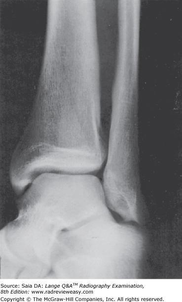
B 1 and 3 only
-The ankle mortise, or ankle joint, is formed by the articulation of the tibia, fibula, and talus (Figure 2–45). Two articulations form the ankle mortise: the talotibial and talofibular articulations. The calcaneus is not associated with formation of the ankle mortise.
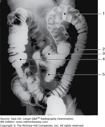
The structure indicated by the number 5 in Figure 6–11 is the
A sigmoid.
B ileum.
C cecum.
D ascending colon.
B ileum.
-The figure shown is a double-contrast BE, oblique position. Since the left colic/splenic flexure (number 1) is “open,” this is either a RPO or LAO position. Also demonstrated are the descending colon (number 2), and transverse colon (number 3). Barium has refluxed into the ileum (number 5).
Which of the following articulate(s) with the bases of the metatarsals?
- The heads of the first row of phalanges
- The cuboid
- The cuneiforms
A 1 only
B 1 and 2 only
C 2 and 3 only
D 1, 2, and 3
C 2 and 3 only
-The foot is composed of the 7 tarsal bones, 5 metatarsals, and 14 phalanges. The metatarsals and phalanges are miniature long bones; each has a shaft, base (proximal), and head (distal). The bases of the first to third metatarsals articulate with the three cuneiforms. The bases of the fourth and fifth metatarsals articulate with the cuboid. The heads of the metatarsals articulate with the bases of the first row of phalanges.
How can OID be reduced for a PA projection of the wrist?
A Extend the fingers.
B Flex the metacarpophalangeal joints.
C Extend the forearm.
D Oblique the metacarpals 45 degrees.
B Flex the metacarpophalangeal joints.
-When the hand is pronated and the fingers are extended for a PA projection of the wrist, the wrist arches, and an OID is introduced between the wrist and the cassette. To reduce this OID, the metacarpophalangeal joints should be flexed slightly. This maneuver will bring the anterior surface of the wrist into contact with the cassette.
Examples of synovial pivot articulations include the
- atlantoaxial joint
- radioulnar joint
- temporomandibular joint
A 1 only
B 1 and 2 only
C 2 and 3 only
D 1, 2, and 3 only
B 1 and 2 only
-Synovial pivot joints are diarthrotic, that is, freely movable. Pivot joints permit rotation motion. Examples include the proximal radioulnar joint that permits supination and pronation of the hand. The atlantoaxial joint is the articulation between C1 and C2 and permits rotation of the head. The temporomandibular joint is diarthrotic, having both hinge and planar movements.
Which of the following are mediastinal structures?
- Heart
- Trachea
- Esophagus
A 1 only
B 1 and 2 only
C 2 and 3 only
D 1, 2, and 3
D 1, 2, and 3
-The thoracic cavity contains the lungs and heart and is separated from the abdominal cavity by the diaphragm. The mediastinum is the space between the lungs which contains the heart and great vessels, trachea, esophagus, thymus, lymph nodes, and connective tissue.
Below-diaphragm ribs are better demonstrated when
A respiration is suspended at the end of full inhalation.
B exposed using shallow breathing technique.
C the patient is in the recumbent position.
D the patient is in the AP erect position.
C the patient is in the recumbent position.
-The ribs below the diaphragm are best demonstrated with the diaphragm elevated. This is accomplished by placing the patient in a recumbent position and by taking the exposure at the end of exhalation. Conversely, the ribs above the diaphragm are best demonstrated with the diaphragm depressed. Placing the patient in the erect position and taking the exposure at the end of deep inspiration accomplishes this.
Which of the following structures will be filled with barium in the AP recumbent position of a sthenic patient during an upper GI examination?
A Duodenal bulb
B Descending duodenum
C Pyloric vestibule
D Gastric fundus
D Gastric fundus
-The stomach is normally angled with the fundus lying posteriorly and the body, pylorus, and duodenum inferior to the fundus and angling anteriorly. Therefore, when the patient ingests barium and lies AP recumbent, the heavy barium gravitates easily to the fundus and fills it. With the patient PA recumbent, barium gravitates inferiorly to the body, pylorus, and duodenum, displacing air into the fundus.
In the 45-degree medial oblique projection of the ankle, the
- talotibial joint is visualized
- tibiofibular joint is visualized
- plantar surface should be vertical
A 1 only
B 2 only
C 2 and 3 only
D 1, 2, and 3
C 2 and 3 only
-The medial oblique projection of the ankle can be performed either as a 15- to 20-degree oblique or as a 45-degree oblique. The 15- to 20-degree oblique projection demonstrates the ankle mortise, that is, the articulations between the talus, tibia, and fibula. The 45-degree oblique opens the distal tibiofibular joint. In all three cases, although the MSP can change, the plantar surface must be vertical.
Widening of the intercostal spaces is characteristic of which of the following conditions?
A Pneumothorax
B Emphysema
C Pleural effusion
D Pneumonia
B Emphysema
-Chest radiographs demonstrating emphysema will show the characteristic irreversible trapping of air that increases gradually and overexpands the lungs. This produces the characteristic “flattening” of the hemidiaphragms and widening of the intercostal spaces. The increased air content of the lungs requires a compensating decrease in technical factors. Pneumonia is inflammation of the lungs, usually caused by bacteria, virus, or chemical irritant. Pneumothorax is a collection of air or gas in the pleural cavity (outside the lungs), with an accompanying collapse of the lung. Pleural effusion is excessive fluid between the parietal and visceral layers of pleura.
Inspiration and expiration projections of the chest are performed to demonstrate
- partial or complete collapse of pulmonary lobe(s)
- air in the pleural cavity
- foreign body
A 1 only
B 1 and 2 only
C 1 and 3 only
D 1, 2, and 3
D 1, 2, and 3
-The phase of respiration is exceedingly important in thoracic radiography because lung expansion and the position of the diaphragm strongly influence the appearance of the finished radiograph. Inspiration and expiration radiographs of the chest are taken to demonstrate air in the pleural cavity (pneumothorax), to demonstrate atelectasis (partial or complete collapse of one or more pulmonary lobes) or the degree of diaphragm excursion, or to detect the presence of a foreign body. The expiration image will require a somewhat greater exposure (6–8 kV more) to compensate for the diminished quantity of air in the lungs.
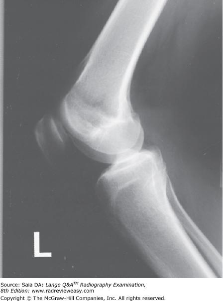
To better visualize the knee-joint space in the radiograph in Figure 2–31, the radiographer should
A flex the knee more acutely
B flex the knee less acutely
C angle the CR 5 to 7 degrees cephalad
D angle the CR 5 to 7 degrees caudad
C angle the CR 5 to 7 degrees cephalad
-In the lateral projection of the knee, the joint space is obscured by the magnified medial femoral condyle unless the CR is angled 5 to 7 degrees cephalad. The degree of flexion of the knee is important when evaluating the knee for possible transverse patellar fracture. In such a case, the knee should not be flexed more than 10 degrees. The knee normally should be flexed 20 to 30 degrees in the lateral position.
What is the position of the stomach in a hypersthenic patient?
A High and vertical
B High and horizontal
C Low and vertical
D Low and horizontal
B High and horizontal
-The position, shape, and motility of various organs can differ greatly from one body habitus to another. The hypersthenic individual is large and heavy; the lungs and heart are high, the stomach is high and transverse, the gallbladder is high and lateral, and the colon is high and peripheral. In contrast, the other habitus extreme is the asthenic individual. This patient is slender and light and has a long and narrow thorax, a low and long stomach, a low and medial gallbladder, and a low medial and redundant colon. The radiographer must consider these characteristic differences when radiographing individuals of various body types.
Which of the following structures is (are) located in the right upper quadrant (RUQ)?
- Spleen
- Gallbladder
- Hepatic flexure
A 1 only
B 1 and 2 only
C 2 and 3 only
D 1, 2, and 3
C 2 and 3 only
-The abdomen can be divided into four quadrants or nine regions. The liver, gallbladder, and hepatic/right colic flexure are all located in the RUQ. The stomach and spleen are both normally located in the LUQ.
The greater tubercle should be visualized in profile in which of the following?
A AP shoulder, external rotation
B AP shoulder, internal rotation
C AP elbow
D Lateral elbow
A AP shoulder, external rotation
-The greater and lesser tubercles are prominences on the proximal humerus, separated by the bicipital groove. The AP projection of the humerus in external rotation demonstrates the greater tubercle in profile. With the arm placed in internal rotation, the humerus is placed in a true lateral position and the lesser tubercle is demonstrated.
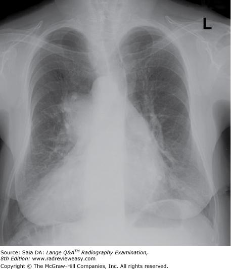
The figure below demonstrates which of the following conditions?
A right upper lobe atelectasis
B left upper lobe atelectasis
C pneumothorax
D dextrocardia
D dextrocardia
-The figure illustrates a PA projection of the chest and the side marker correctly placed. The heart is seen on the right side—this is termed dextrocardia. Atelectasis (partial or complete collapse of lung) would be demonstrated as increased tissue density in the affected area. A classic pneumothorax (air within the thoracic cavity) would demonstrate an absence of lung markings in the affected area and flattening of the hemidiaphragm on the affected side. A small pneumothorax can be easily missed on a chest image demonstrating excessive receptor exposure.
Which of the following conditions would require an increase in exposure factors?
1.Congestive heart failure
2.Pleural effusion
3.Emphysema
A 1 only
B 1 and 2 only
C 1 and 3 only
D 1, 2, and 3
B 1 and 2 only
-Emphysema is abnormal distention of the alveoli (or tissue spaces) with air. The presence of abnormal amounts of air makes it necessary to decrease from normal exposure factors. Congestive heart failure and pleural effusion involve abnormal amounts of fluid in the chest and thus require an increase in exposure factors
Which of the following is most likely to be the correct routine for a radiographic examination of the forearm?
A PA and medial oblique
B AP and lateral oblique
C PA and lateral
D AP and lateral
D AP and lateral
-To demonstrate the radius and ulna free of superimposition, the forearm must be radiographed in the AP position, with the hand supinated. Pronation of the hand causes overlapping of the proximal radius and ulna. Two views, at right angles to each other, are generally required for each examination. Therefore, AP and lateral is the usual routine for an examination of the forearm.
When examining a patient whose elbow is in partial flexion, how should an AP projection be obtained?
- With humerus parallel to IR, CR perpendicular
- With forearm parallel to IR, CR perpendicular
- Through the partially flexed elbow, resting on the olecranon process, CR perpendicular
A 1 only
B 1 and 2 only
C 2 and 3 only
D 1, 2, and 3
B 1 and 2 only
-When a patient's elbow needs to be examined in partial flexion, the lateral projection offers little difficulty, but the AP projection requires special attention. If the AP radiograph is made with a perpendicular CR and the olecranon process resting on the tabletop, the articulating surfaces are obscured. With the elbow in partial flexion, two exposures are necessary. One is made with the forearm parallel to the IR (humerus elevated), which demonstrates the proximal forearm. The other is made with the humerus parallel to the IR (forearm elevated), which demonstrates the distal humerus. In both cases, the CR is perpendicular if the degree of flexion is not too great or is angled slightly into the joint space with greater degrees of flexion.
Which of the following projections will best demonstrate the carpal scaphoid?
A Lateral wrist
B Ulnar deviation
C Radial deviation
D Carpal tunnel
B Ulnar deviation
-The carpal scaphoid is somewhat curved and consequently foreshortened radiographically in the PA projection/position. To better separate it from the adjacent carpals, the ulnar deviation maneuver is frequently employed. In addition to correcting foreshortening of the scaphoid, ulnar deviation opens the interspaces between adjacent lateral carpals. Radial deviation is used to better demonstrate medial carpals.
Which of the following examinations require(s) restriction of a patient's diet?
- Barium enema
- Pyelogram
- Metastatic survey
A 1 only
B 1 and 2 only
C 1 and 3 only
D 2 and 3 only
B 1 and 2 only
-A patient who is having a BE generally is required to have a low-residue diet for 1 or 2 days, followed by cathartics and cleansing enemas prior to the examination. Any retained fecal material can simulate or obscure pathology. A patient who is scheduled for a pyelogram must have the preceding meal withheld to avoid the possibility of aspirating vomitus in case of an allergic reaction. A metastatic survey does not require the use of contrast media, and no patient preparation is necessary.
Which of the following equipment is mandatory for performance of a myelogram?
A Cine camera
B 105-mm spot film
C Tilting x-ray table
D Tomography
C Tilting x-ray table
-Myelography requires that contrast medium be instilled into the lumbar subarachnoid space and distributed via gravity to various levels of the subarachnoid space. This gravitational distribution is accomplished through the use of an x-ray table that is capable of angling or tilting during the procedure.
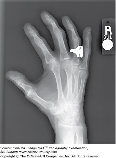
Which of the following statements regarding Figure 2–10 is (are) true?
- Correct degree of rotation is present.
- Midphalanges are foreshortened.
- Fingers are parallel to the IR.
A 1 only
B 1 and 2 only
C 2 and 3 only
D 1, 2, and 3
B 1 and 2 only
-A PA oblique projection of the hand is shown. The correct degree of obliquity (45 degrees) is evidenced by no overlap of midshaft third, fourth, and fifth metacarpals and minimal overlap of their heads. The interphalangeal joint spaces are not visualized because the fingers are not elevated to be parallel to the IR, hence, they are foreshortened.
In which of the following conditions is a double-contrast BE essential for demonstration of the condition?
- Polyps
- Colitis
- Diverticulosis
A 1 only
B 1 and 2 only
C 1 and 3 only
D 1, 2, and 3
B 1 and 2 only
-Double-contrast studies of the large bowel are particularly useful for demonstration of the bowel walland anything projecting into it, for example, polyps. Polyps are projections of the bowel wall mucous membrane into the bowel lumen. Colitis is inflammation of the large bowel, often associated with ulcerations of the mucosal wall. A single-contrast study most likely would obliterate these mucosal conditions, but coating of the bowel mucosa with barium and subsequent filling of the bowel with air (double contrast) provide optimal delineation. Single-contrast studies will demonstrate projections/outpouchings from the intestinal wall such as diverticulitis.
Which of the following is (are) associated with a Colles' fracture?
- Transverse fracture of the radial head
- Chip fracture of the ulnar styloid
- Posterior or backward displacement
A 1 only
B 1 and 3 only
C 2 and 3 only
D 1, 2, and 3
C 2 and 3 only
-A Colles fracture usually is caused by a fall onto an outstretched (extended) hand to “brake” a fall. The wrist then suffers an impacted transverse fracture of the distal inch of the radius with an accompanying chip fracture of the ulnar styloid process. Because of the hand position at the time of the fall, the fracture usually is displaced backward approximately 30 degrees. The proximal radius, the radial head, is not involved in this type fracture.
The main forms of COPD include
- chronic bronchitis.
- pulmonary emphysema.
- asthma.
A 1 only
B 1 and 2 only
C 2 and 3 only
D 1, 2, and 3
B 1 and 2 only
-Chronic obstructive pulmonary disease (COPD) is the abbreviation for chronic obstructive pulmonary disease; it refers to a group of disorders. COPD has two main forms, chronic bronchitis and emphysema. Many experts believe that asthma should be included, others do not. COPD is irreversible and decreases the ability of the lungs to perform their ventilation functions. There is often less than half the normal expected maximal breathing capacity.
A lesion with a stalk projecting from the intestinal mucosa into the lumen is a(n)
A fistula
B polyp
C diverticulum
D abscess
B polyp
-A polyp is a tumor with a pedicle (stalk) that is found commonly in vascular organs projecting inward from its mucosal wall. Polyps usually are removed surgically because, although usually benign, they can become malignant. A diverticulum is an outpouching from the wall of an organ, such as the colon. A fistula is an abnormal tube-like passageway between organs or between an organ and the surface. An abscess is a localized collection of pus as a result of inflammation.
Which of the following positions will most effectively move the gallbladder away from the vertebrae in an asthenic patient?
A LAO
B RAO
C LPO
D Erect
A LAO
-The position of the gallbladder varies with the body habitus of the patient. Hypersthenic patients are more likely to have their gallbladder located high and lateral. The asthenic patient's gallbladder is most likely to occupy a low and medial position, occasionally superimposed on the vertebrae or iliac fossa. The LAO position is used most often to move the gallbladder away from the spine. The erect position would make the gallbladder move even more inferior and medial.
A patient is usually required to drink barium sulfate suspension to demonstrate which of the following structures?
1.Esophagus
2.Pylorus
3.Ilium
A 1 only
B 1 and 2 only
C 2 and 3 only
D 1, 2, and 3
B 1 and 2 only
-Oral administration of barium sulfate is used to demonstrate the upper digestive tract: the esophagus; the fundus, body, and pylorus of the stomach; and the small bowel, consisting of duodenum, jejunum, and ileum (i.e. small bowel..not ilium). Consistent care must be taken to read and record patient information accurately and correctly. The large bowel is usually demonstrated via rectal administration of barium.
Which of the following articulates with the base of the fifth metatarsal?
A First cuneiform
B Third cuneiform
C Navicular
D Cuboid
D Cuboid
-The bones of the foot include the seven tarsal bones, five metatarsal bones, and 14 phalanges. The calcaneus (os calsis) serves as the attachment for the Achilles tendon and articulates anteriorly with thecuboid bone.
Articulating anteriorly with the navicular are the three cuneiform bones: medial/first, intermediate/second, and lateral/third. The navicular articulates laterally with the cuboid. The bases of the fourth and fifth metatarsals articulate with the cuboid. The fifth (most lateral) metatarsal projects laterally and presents a large tuberosity at its base making it very susceptible to fracture.
All the following positions are likely to be employed for both single- and double-contrast examinations of the large bowel except
A lateral rectum.
B AP axial rectosigmoid.
C right and left lateral decubitus abdomen.
D RAO and LAO abdomen.
C right and left lateral decubitus abdomen.
-Radiographic examinations of the large bowel generally include the AP or PA axial position to “open” the S-shaped sigmoid colon, the lateral position especially for the rectum, and the LAO and RAO (or LPO and RPO) positions to “open” the colic flexures. The left and right decubitus positions usually are employed only in double-contrast barium enemas to better demonstrate double contrast of the medial and lateral walls of the ascending and descending colon.
To obtain an exact axial projection of the clavicle, place the patient
A supine and angle the central ray 30° caudally.
B prone and angle the central ray 30° cephalad.
C supine and angle the central ray 15° cephalad.
D in a lordotic position and direct the central ray at right angles to the coronal plane of the clavicle.
D in a lordotic position and direct the central ray at right angles to the coronal plane of the clavicle.
-The exact axial projection is performed by placing the patient in a lordotic position, leaning against the vertical grid device. This places the clavicle at right angles, or nearly so, to the plane of the IR. The central ray is directed to enter the inferior border of the clavicle, at right angles to its coronal plane. Other axial projections may include a prone position with a 25° to 30° caudal angle. However, none of these produce an exact axial projection of the clavicle.
Following the ingestion of a fatty meal, what hormone is secreted by the duodenal mucosa to stimulate contraction of the gallbladder?
A Insulin
B Cholecystokinin
C Adrenocorticotropic hormone
D Gastrin
B Cholecystokinin
-About 30 minutes after the ingestion of fatty foods, cholecystokinin is released from the duodenal mucosa and absorbed into the bloodstream. As a result, the gallbladder is stimulated to contract, releasing bile into the intestine
Cells concerned with the formation and repair of bone are
A osteoblasts.
B osteoclasts.
C osteomas.
D osteons.
A osteoblasts.
-Osteoblasts are cells of mesodermal origin that are concerned with formation and repair of bone.Osteoclasts are cells concerned with the breakdown and resorption of old or dead bone. An osteoma is a benign bony tumor. An osteon is the microscopic unit of compact bone, consisting of a haversian canal and its surrounding lamellae.
Which of the following will be demonstrated best in the 45-degree right anterior oblique (RAO) position?
A Right axillary ribs
B Left axillary ribs
C Sternum in the heart shadow
D Left scapular Y
B Left axillary ribs
-The axillary portions of ribs are demonstrated in a 45-degree oblique position. In order to place the axillary portions parallel to the image receptor (IR), the affected side is away from the IR in the PA oblique(RAO and LAO) positions and toward the IR in the AP oblique (RPO and LPO) positions. Radiography of the sternum, in the slight RAO position, requires greater obliquity for thinner patients and lesser obliquity for thicker patients. The scapular Y position of the shoulder is performed to demonstrate dislocation and requires a rotation of 45–60 degrees, with the affected side closest to the IR.
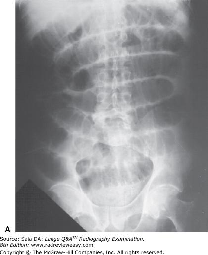
Which of the radiographs shown in Figure 4–5 most likely required the greater exposure?
A Image A
B Image B
C No difference in exposure was required
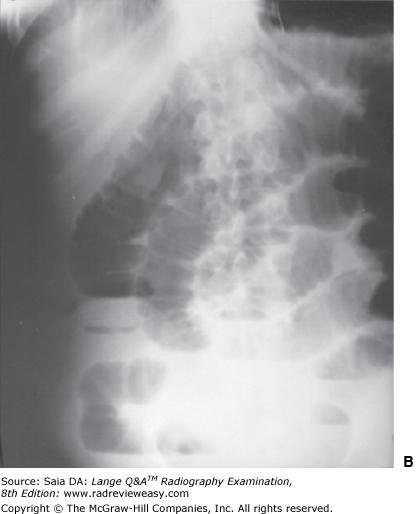
B Image B
-Of the two radiographs illustrated, image A was made recumbent, and image B was made in the erect position; this may be discerned by the presence of clearly defined air–fluid levels in the lower abdomen. Abdominal viscera move to a lower position in the erect position, making the abdomen “thicker” and requiring an increase in exposure (usually the equivalent of about 10 kV).
When the erect position is requested as part of an IVU, it is used to demonstrate
A the adrenal glands.
B the renal surfaces.
C kidney mobility.
D the bladder neck.
C kidney mobility.
-The erect position in IV urography may be part of the departmental routine, but more often than not it is requested as a supplemental view to rule out nephroptosis. With the patient erect, the kidneys normally change position, dropping no more than 2 inches. More marked dropping of the kidney is termed nephroptosis, a condition that is actually due to loss of the surrounding perinephric fat.
All of the following statements regarding the inferosuperior axial (nontrauma, Lawrence method) projection of the shoulder are true, except
A the coracoid process and lesser tubercle are seen in profile.
B the arm is abducted about 90° from the body.
C the arm should be in internal rotation.
D the CR is directed medially 25° to 30° through the axilla.
C the arm should be in internal rotation.
-The inferosuperior axial (nontrauma, Lawrence method) projection of the shoulder demonstrates the glenohumeral joint and adjacent structures. The patient is supine with arm abducted 90°, and in external rotation. The (horizontal) CR is directed medially 25° to 30° through the axilla. The coracoid process and lesser tubercle are seen in profile.
Which of the following indicates the scapular costal surface seen in the figure below?
A D
B H
C K
D M
C K
-The radiograph illustrates an AP projection of the scapula; abduction of the arm moves the scapula away from the rib cage, revealing a greater portion of the scapula than would be visualized with the arm at the side. A number of bony structures are identified: the acromion process (A), the humeral head (B), glenoid fossa (C), scapular spine (D), clavicle (E), supraspinatus fossa (F), acromioclavicular joint (G), scapular notch (H), coracoid process (I), inferior angle/apex (j), body/costal surface (K), lateral/axillary border (L), axillary part upper rib (M).
Dorsal decubitus projections of the chest are used to evaluate small amounts of
- fluid in the posterior chest
- air in the posterior chest
- fluid in the anterior chest
A 1 only
B 1 and 2 only
C 2 and 3 only
D 1, 2, and 3
A 1 only
-The dorsal decubitus position is obtained with the patient supine and the x-ray beam directed horizontally. The finished image looks similar to a lateral projection of the chest. However, small amounts of fluid will gravitate to the posterior chest, and small amounts of air will rise to the anterior chest. The ventral decubitus position is obtained with the patient prone and the x-ray beam directed horizontally. The finished image can demonstrate small amounts of fluid anteriorly and small amounts of air posteriorly.
Which of the following projections of the ankle would best demonstrate the mortise?
A Medial oblique 15 to 20 degrees
B Lateral oblique 15 to 20 degrees
C Medial oblique 45 degrees
D Lateral oblique 45 degrees
A Medial oblique 15 to 20 degrees
-The 15-degree medial oblique projection is used to demonstrate the ankle mortise (joint). Although the joint is well demonstrated in the 15-degree medial oblique projection, there is some superimposition of the distal tibia and fibula, and greater obliquity is required to separate the bones. To best demonstrate the distal tibiofibular articulation, a 45-degree medial oblique projection of the ankle is required.
In which of the following projections is the talofibular joint best demonstrated?
A AP
B Lateral oblique
C Medial oblique
D Lateral
C Medial oblique
-The AP projection demonstrates superimposition of the distal fibula on the talus; the joint space is not well seen. The 15- to 20-degree medial oblique position shows the entire mortise joint; the talofibular joint is well visualized, as well as the talotibial joint. There is considerable superimposition of the talus and fibula in the lateral and lateral oblique projections.

Which of the following is represented by the number 3 in the figure below?
A Inferior vena cava
B Aorta
C Gallbladder
D Psoas muscle
B Aorta
-A cross-sectional image of the abdomen is shown in Figure 2–36. The large structure on the right, labeled number 1, is the liver. The gallbladder is seen somewhat darker on the medial border of the liver. The left kidney is labeled number 4; the right kidney is seen clearly on the other side. The vertebra is labeled number 5, and the psoas muscles are seen just posterior to the vertebra. Just anterior to the body of the vertebra is the circular aorta, labeled number 3 (some calcification can be seen brighter). The somewhat flattened inferior vena cava (number 2) is seen to the left of and slightly anterior to the aorta.
In which type of fracture are the fractured ends of bone forced through the skin?
A Closed
B Compound
C Compression
D Depressed
B Compound
-The type of fracture in which the fractured/splintered ends of bone are forced through the skin is a compound fracture. In a closed fracture, no bone protrudes through the skin. Compression fractures are seen in stressed areas, such as the vertebrae. A depressed fracture would not protrude but rather would be pushed in.
For an AP projection of the knee on a patient whose measurement from ASIS to tabletop is 21 cm, which CR direction will best demonstrate the knee joint?
A 5 degrees caudad
B 10 degrees caudad
C 5 degrees cephalad
D 0 degrees (perpendicular)
D 0 degrees (perpendicular)
-The knee is formed by the proximal tibia, the patella, and the distal femur, which articulate to form the femorotibial and femoropatellar joints. Body habitus will change the relationship of the knee-joint space with the tabletop/IR considerably. The CR should be directed to ½ inch below patellar apex (knee joint). The direction of CR depends on distance between the ASIS and tabletop/IR. When this distance is up to 19 cm (thin pelvis), the CR should be directed 3 to 5 degrees caudad; when the distance is between 19 to 24 cm, the CR is directed vertically/perpendicular (0 degrees); when the distance is greater than 24 cm (thick pelvis), the CR is directed 3 to 5 degrees cephalad.
For the average patient, the CR for a lateral projection of a barium-filled stomach should enter
A midway between the midcoronal line and the anterior abdominal surface
B midway between the vertebral column and the lateral border of the abdomen
C at the midcoronal line at the level of the iliac crest
D perpendicular to the level of L2
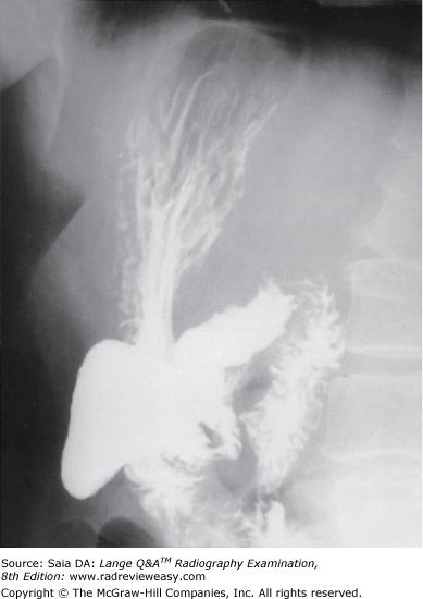
A midway between the midcoronal line and the anterior abdominal surface
-Lateral projections of the barium-filled stomach (Figure 2–61) may be performed recumbent or upright for demonstration of the retrogastric space. With the patient in the (usually right) lateral position, the CR is directed to a point midway between the midcoronal line and the anterior surface of the abdomen at the level of L1. When the patient is in the LPO or RAO position, the CR should be directed midway between the vertebral column and the lateral border of the abdomen. For the PA projection, the CR is directed perpendicular to the IR at the level of L2.
Which of the following is (are) valid criteria for a lateral projection of the forearm?
1.The radius and ulna should be superimposed proximally and distally.
2.The coronoid process and radial head should be superimposed.
3.The radial tuberosity should face anteriorly.
A 1 only
B 1 and 2 only
C 2 and 3 only
D 1, 2, and 3
C 2 and 3 only
-To accurately position a lateral forearm, the elbow must form a 90° angle with the humeral epicondyles superimposed. The radius and ulna are superimposed only distally. Proximally, the coronoid process and radial head are superimposed, and the radial head faces anteriorly. Failure of the elbow to form a 90° angle or the hand to be lateral results in a less than satisfactory lateral projection of the forearm.
Which of the following projections require(s) that the humeral epicondyles be perpendicular to the IR?
- AP humerus
- Lateral forearm
- Internal rotation shoulder
A 1 only
B 1 and 2 only
C 2 and 3 only
D 1, 2, and 3
C 2 and 3 only
-When the arm is placed in the AP position, the epicondyles are parallel to the plane of the IR, and the shoulder is placed in external rotation. In this position, an AP projection of the humerus, elbow, and forearm can be obtained; it places the greater tubercle of the humerus in profile. For the lateral projection of the humerus and the internal rotation projection of the shoulder, the arm is internally rotated, elbow somewhat flexed, with the back of the hand against the thigh, and the epicondyles are superimposed and perpendicular to the IR. The lateral projections of the humerus, elbow, and forearm all require that the epicondyles be perpendicular to the plane of the cassette.
The stomach of an asthenic patient is most likely to be located
A high, transverse, and lateral.
B low, transverse, and lateral.
C high, vertical, and toward the midline.
D low, vertical, and toward the midline.
D low, vertical, and toward the midline.
-The four body types (from largest to smallest) are hypersthenic, sthenic, hyposthenic, and asthenic. The abdominal viscera of the asthenic person are generally located quite low, vertical, and toward the midline. The opposite is true of the hypersthenic individual: Organs are located high, transverse, and lateral.
Which of the following fracture classifications describes a small bony fragment pulled from a bony process?
A Avulsion fracture
B Torus fracture
C Comminuted fracture
D Compound fracture
A Avulsion fracture
-An avulsion fracture is a small bony fragment pulled from a bony process as a result of a forceful pull of the attached ligament or tendon. A comminuted fracture is one in which the bone is broken or splintered into pieces. A torus fracture is a greenstick fracture with one cortex buckled and the other intact. A compound fracture is an open fracture in which the fractured ends have perforated the skin.
Free air in the abdominal cavity is demonstrated in which of the following?
1.Lateral recumbent abdomen
2.Erect AP abdomen
3.Left lateral decubitus abdomen
A 1 only
B 2 only
C 1 and 2 only
D 2 and 3 only
D 2 and 3 only
-When air-fluid levels are to be demonstrated, it is important to direct the central ray horizontally. If the central ray is angled or directed vertically, the air or fluid level will be distorted or entirely obliterated. Free air in the abdominal cavity is best visualized when the patient is left lateral decubitus or erect AP. The decubitus allows the air to accumulate around the homogeneous liver.
Which of the following radiologic procedures requires that a contrast medium be injected into the renal pelvis via a catheter placed within the ureter?
A Nephrotomography
B Retrograde urography
C Cystourethrography
D IVU
B Retrograde urography
-Retrograde urography requires ureteral catheterization so that a contrast medium can be introduced directly into the pelvicalyceal system. This procedure provides excellent opacification and structural information but does not demonstrate the function of these structures. IV studies such as the IVU demonstrate function. Cystourethrography is an examination of the bladder and urethra, frequently performed during voiding. Nephrotomography is performed after IV administration of a contrast agent; it may be used to evaluate small intrarenal lesions and renal hypertension.
In the lateral projection of the knee, the central ray is angled 5° cephalad to prevent superimposition of which of the following structures on the joint space?
A Lateral femoral condyle
B Medial femoral condyle
C Patella
D Tibial eminence
B Medial femoral condyle
-For the lateral projection of the knee, the patient is turned onto the affected side. This places the lateral femoral condyle closest to the IR and the medial femoral condyle remote from the IR. Consequently, there is significant magnification of the medial femoral condyle and, unless the central ray is angled slightly cephalad, subsequent obliteration of the joint space.
What instructions might a patient be given following an upper GI examination?
1.Drink plenty of fluids.
2.Take a mild laxative.
3.Increase dietary fiber.
A 1 only
B 1 and 2 only
C 2 and 3 only
D 1, 2, and 3
D 1, 2, and 3
-Barium can dry and harden in the large bowel, causing symptoms ranging from mild constipation to bowel obstruction. It is therefore essential that the radiographer provide clear instructions, especially to outpatients, for follow-up care, along with the rationale for this care. To avoid the possibility of fecal impaction, patients should drink plenty of fluids for the next few days, increase their dietary fiber, and take a mild laxative such as milk of magnesia.
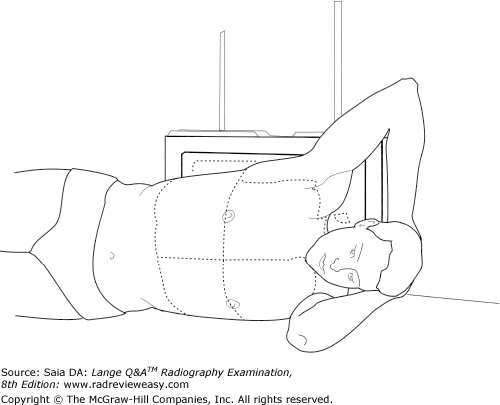
All the following statements regarding the position shown in Figure 2–17 are true except
A a left pleural effusion could be demonstrated.
B a right pneumothorax could be demonstrated.
C a left lateral decubitus position is illustrated.
D the CR is directed vertically to the level of T7.
D the CR is directed vertically to the level of T7.
-The illustration shows the patient positioned on his left side with the cassette behind his back. This is a left lateral decubitus position. The x-ray beam is directed horizontally in decubitus positions to demonstrate air–fluid levels. Air or fluid levels will be clearly delineated only if the CR is directed parallel to them. Fluid levels will be best detected on the down side (in this case, left); air levels will be best detected on the up side (in this case, right). If the patient were lying on the right side, it would be a right lateral decubitus position. If the patient were lying on his or her back with a horizontal x-ray beam, it would be a dorsal decubitus position. Lying prone with a horizontal x-ray beam is termed a ventral decubitus position.
Which of the following positions can be used to demonstrate the axillary ribs of the right thorax?
- RAO
- LAO
- RPO
A 1 only
B 1 and 2 only
C 2 and 3 only
D 1, 2, and 3
C 2 and 3 only
-The axillary portion of the ribs is best demonstrated in a 45-degree oblique position. The axillary ribs are demonstrated in the AP oblique projection with the affected side adjacent to the IR and in the PA oblique projection with the affected side away from the IR. Therefore, the right axillary ribs would be demonstrated in the RPO (AP oblique with affected side adjacent to the IR) and LAO (PA oblique with affected side away from the IR) positions.

The structure labeled number 2 in Figure 6–3, image B is the
A caudate lobe of liver
B duodenum
C colon
D gallbladder
D gallbladder
-Image B is an axial CT section of the abdomen with contrast. Structures demonstrated include the barium-filled stomach (number 1), the gallbladder (number 2), duodenum (number 3), pancreas (number 4), IVC (number 5), left adrenal gland (number 6), aorta (number 7), spleen (number 8), left kidney (number 9), right kidney (number 10), and liver (number 11).
Which of the following projections will best demonstrate acromioclavicular separation?
A AP recumbent, affected shoulder
B AP recumbent, both shoulders
C AP erect, affected shoulder
D AP erect, both shoulders
D AP erect, both shoulders
-Acromioclavicular (AC) joints usually are examined when separation or dislocation is suspected. They must be examined in the erect position because in the recumbent position a separation appears to reduce itself. Both AC joints are examined simultaneously for comparison because separations may be minimal.
For the AP projection of the scapula, the
- patient's arm is abducted at right angles to the body.
- patient's elbow is flexed.
- exposure is made during quiet breathing.
A 1 and 2 only
B 1 and 3 only
C 3 only
D 1, 2, and 3
D 1, 2, and 3
-With the patient in the AP position, the scapula and upper thorax are normally superimposed. With the arm abducted, the elbow flexed, and the hand usually supinated, much of the scapula is drawn away from the ribs. The patient should not be rotated toward the affected side because this causes superimposition of ribs on the scapula. The exposure is made during quiet breathing to obliterate pulmonary vascular markings.
Deoxygenated blood from the head and thorax is returned to the heart by the
A pulmonary artery
B pulmonary veins
C superior vena cava
D thoracic aorta
C superior vena cava
-Deoxygenated (venous) blood from the upper body (i.e., head, neck, thorax, and upper extremities) empties into the superior vena cava. Deoxygenated (venous) blood from the lower body (i.e., abdomen, pelvis, and lower extremities) empties into the inferior vena cava. The superior and inferior venae cavae empty into the right atrium. The coronary sinus, which returns venous blood from the heart, also empties into the right atrium. Deoxygenated blood passes from the right atrium through the tricuspid valve into the right ventricle. From the right ventricle, blood is pumped (during ventricular systole) through the pulmonary semilunar valve into the pulmonary artery—the only artery that carries deoxygenated blood. From the pulmonary artery, blood travels to the lungs, picks up oxygen, and is carried by the four pulmonary veins (the only veins carrying oxygenated blood) to the left atrium. The oxygenated blood passes through the mitral (or bicuspid) valve during atrial systole and into the left ventricle. During ventricular systole, oxygenated blood from the left ventricle passes through the aortic semilunar valve into the aorta and into the systemic circulation.
Which of the following structures will usually contain air, in the PA recumbent position on a sthenic patient, during a double-contrast upper GI (UGI) examination?
A Duodenal bulb
B Descending duodenum
C Pyloric vestibule
D Gastric fundus
D Gastric fundus
-The stomach is normally angled with the fundus lying posteriorly and the body, pylorus, and duodenum inferior to the fundus and angling anteriorly. Therefore, when the patient ingests barium and lies AP recumbent, the heavy barium gravitates easily to the fundus and fills it. With the patient PA recumbent, barium gravitates inferiorly to the body, pylorus, and duodenum, displacing air into the fundus.
Which of the following positions is most likely to offer the best visualization of the pulmonary apices?
A Lateral decubitus
B Dorsal decubitus
C Erect lateral
D AP axial lordotic
D AP axial lordotic
-The pulmonary apices are often at least partially obscured by the clavicles. To visualize the entire lung apex and any suspicious areas, the clavicles must be “removed.” This can be accomplished with the AP axial lordotic position. Through the arching of the patient's back and the cephalad angulation, the clavicles are projected upward and out of the pulmonary apices. Decubitus positions are used primarily to see air–fluid levels. Lateral and dorsal decubitus positions show fluid in the side that is down and air in the side that is up.
The ridge that marks the bifurcation of the trachea into the right and left primary bronchi is the
A root.
B hilus.
C carina.
D epiglottis.
C carina.
-The carina is an internal ridge located at the bifurcation of the trachea into right and left primary, or mainstem, bronchi. The epiglottis is a flap of elastic cartilage that functions to prevent fluids and solids from entering the respiratory tract during swallowing. The root of the lung attaches the lung, via dense connective tissue, to the mediastinum. The root of the left lung is at the level of T6, and the root of the right is at T5. The hilus (hilum) is the slitlike opening on the medial aspect of the lung through which arteries, veins, lymphatics, and so forth, enter and exit.
All the following are posterior structures except
A the linea aspera.
B the intertrochanteric line.
C the popliteal surface.
D the intercondyloid fossa.
B the intertrochanteric line.
-The femur is the longest and strongest bone in the body. The femoral shaft is bowed slightly anteriorly. The proximal end of the femur consists of a head, which is received by the acetabulum of the pelvis. The femoral head has a small notch, the fovea capitis femoris, for ligament attachment. The femoral neck, which joins the head and shaft, angles upward approximately 120 degrees and forward (in anteversion) approximately 15 degrees. The greater (lateral) and lesser (medial) trochanters are large processes on the posterior proximal femur. The greater trochanter is a prominent positioning landmark that lies in the same transverse plane as the public symphysis and coccyx. Theintertrochanteric crest runs obliquely between the trochanters; the intertrochanteric line runs anteriorly parallel to the crest. The femoral shaft presents a long, narrow ridge posteriorly called the linea aspera.
Its distal anterior portion presents the patellar surface—a triangular depression over which the patella glides during flexion. The distal posterior surface presents the popliteal surface—a depression that houses the popliteal artery. The medial and lateral femoral condyles are very prominent posterior structures, and between them is the deep intercondyloid fossa. Just above the condyles are the medial and lateral femoral epicondyles.
Which of the following conditions is limited specifically to the tibial tuberosity?
A Ewing sarcoma
B Osgood–Schlatter disease
C Gout
D Exostosis
B Osgood–Schlatter disease
-Osgood-Schlatter disease is most common in adolescent boys, involving osteochondritis of the tibial tuberosity epiphysis. The large patellar tendon actually will pull the tibial tuberosity away from the tibia. Immobilization generally will resolve the issue. Ewing sarcoma is a malignant bone tumor most common in young children. It attacks long bones and presents a characteristic “onion peel” appearance. Gout is a type of arthritis that most commonly attacks the knee and first metatarsophalangeal joint, although other joints also can be involved. High levels of uric acid in the blood are deposited in the joint.Exostosis is a bony growth arising from the surface of a bone and growing away from the joint. It is a benign and sometimes painful condition.
All the following can be associated with the distal ulna except
A head.
B radioulnar joint.
C styloid process.
D trochlear notch.
D trochlear notch.
-The distal ulna presents a head and styloid process and articulates with the distal radius to form the distal radioulnar joint. The ulna is slender distally but enlarges proximally and becomes the larger of the two bones of the forearm. At its proximal end, the ulna presents the olecranon process (posteriorly) and coronoid process (anteriorly) that are joined by a large articular cavity, the semilunar, or trochlear notch. The coronoid process fits into the humeral coronoid fossa during flexion, and the olecranon process fits into the humeral olecranon fossa during extension. Just distal and lateral to the semilunar/trochlear notch is the radial notch, which provides articulation for the radial head to form the proximal radioulnar articulation. The ulna is the principal bone of the elbow joint, and the radius is the principal bone of the wrist joint.
The instrument that is used frequently in quality-control programs to measure varying degrees of x-ray exposure is the
A aluminum step wedge.
B spinning top.
C densitometer.
D sensitometer
C densitometer.
-Every radiographic image is composed of a number of different densities. These densities may be measured and given a numeric value with a device called a densitometer. A sensitometer is another device used in QA programs; it is used to give a precise exposure to a film emulsion. An aluminum step wedge (penetrometer) may be used to show the effect of kilovoltage on contrast. A spinning top is used to test the accuracy of the x-ray machine's timer or rectifiers.
In which position of the shoulder is the lesser tubercle demonstrated in profile on the medial aspect of the humeral head?
A AP
B External rotation
C Internal rotation
D Neutral position
C Internal rotation
-The external rotation position is the true AP position and places the greater tubercle in profile laterally and places the lesser tubercle anteriorly. The internal rotation position demonstrates the lesser tubercle in profile medially and places the humerus in a true lateral position. The epicondyles should be superimposed and perpendicular to the IR. The neutral position places the epicondyles about 45 degrees to the IR and the greater tubercle is partially superimposed on the humeral head.
Which of the following positions is obtained with the patient lying supine on the radiographic table with the CR directed horizontally to the iliac crest?
A Left lateral decubitus position
B Right lateral decubitus position
C Ventral decubitus position
D Dorsal decubitus position
D Dorsal decubitus position
-A decubitus projection is obtained using a horizontal x-ray beam. The type of decubitus projection is dependent on the patient's recumbent position. When the patient is lying AP recumbent (i.e., supine), the patient is said to be in the dorsal decubitus position. When the patient is lying prone, he or she is in the ventral decubitus position. If the patient is lying in the left or right lateral recumbent position with the x-ray beam directed horizontally, the patient is said to be in the left or right lateral decubitus position, respectively.