Describe the actions of prime movers (agonists), antagonists, synergists, and fixators.
1. prime movers (agonists) - muscles that have the major responsibility for producing a specific movement
example - biceps brachii is prime mover of forearm flexion
2. antagonists - muscles that oppose or reverse a particular movement
example - triceps brachii extends the forearm
3. synergists - assist prime movers add force to a movement
reduce undesirable or unnecessary movement
example - when a muscle crosses two or more joints,its contraction affects all the spanned joints unless other musclesact as joint stabilizers
4. fixators - synergists that immobilize a bone or muscle’s origin
give prime mover a stable base on which to act
example - scapula is held to the axial skeleton only by muscles; fixator muscles immobilize the scapula
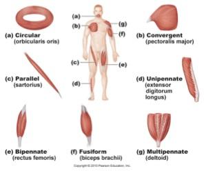
Describe different patterns of fascicle arrangement and name an example of each (Figure 10.2, page 322).
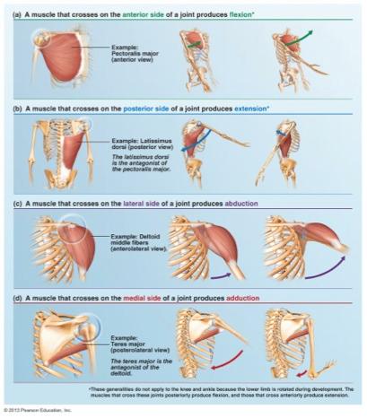
Infer the action of a muscle given the position of the muscle relative to the joint it crosses (Figure 10.1, page 321).
Correlate fascicle arrangement with a muscle’s range of motion and power.
range of motion - the longer & more nearly parallel the muscles are to a muscle’s long axis, the more the muscle can shorten
parallel fascicles - shorten the most, but not very powerful
muscle power - depends on total number of muscle fibers in the muscle
↑ number of muscle fibers = ↑ muscle power
Define the terms lever, fulcrum, effort, and load.
lever - a rigid bar that moves on a fixed point called a fulcrum
fulcrum - support or point on which a lever pivots
effort - force applied to a lever
load - object moved by effort; resistance
Explain mechanical advantage and mechanical disadvantage. Differentiate between a power lever and a speed lever.
power lever - lever that operates at a mechanical advantage
speed lever - allows a load to be moved rapidly over a large distance with a wide range of motion
State the arrangement of load, fulcrum, and effort in first-class, second-class, and third-class levers (Figure 10.4, page 325). Describe an example of each in the body.
...
List the general characteristics of the muscles of facial expression.
control facial expressions
lie in scalp & face, just deep to the skin
thin and variable in shape & strength
insert into skin or other muscles, not bone; adjacent muscles often fused
important in nonverbal communication
all innervated by cranial nerve VII (facial nerve)
Describe the epicranius. List its three main parts and describe the function of each.
Epicranius (occipitofrontalis) (epi = over, cran = skull) - bipartite (two part) muscle with a wide aponeurosis connecting the two muscle parts
frontal belly (front = forehead) - raises eyebrow
occipital belly (occipito = base of skull) - pulls scalp posteriorly
epicranial aponeurosis - connective tissue sheet connecting the frontal belly & occipital belly
Explain the functions of each of the 11 muscles of the face.
Corrugator supercilii (corrugo = wrinkle; supercilium = eyebrow) - draws eyebrows together
Orbicularis oculi (orb = circular; ocul = eye) - closes eye
Levator labii superioris (leva = raise; labi =lip; superior = above) - opens lips
Zygomaticus (zygomatic = cheekbone) - raises lateral corners of mouth upwards (smiling muscle)
Buccinator (bucc = cheek) - compresses cheek
Risorius (risor = laughter) - draws corner of lip laterally (synergist of zygomaticus)
Orbicularis oris (or = mouth) - closes lips
Mentalis (ment = chin) - wrinkles chin
Depressor labii inferioris (depressor = depresses; infer = below) - draws lower lip inferiorly
Depressor anguli oris (angul = angle, corner; or = mouth) - draws corners of lip downwards & laterally
Platysma (platy = broad, flat) - tenses skin of neck
List the general characteristics of the muscles of mastication.
Characteristics
mastication - chewing
buccinator muscle - aids muscles of mastication by compressing the cheek and keeping food between the grinding surfaces of the teeth during chewing
all are innervated by cranial nerve V (trigeminal nerve)
Name the cranial nerve that innervates the following groups of muscles: muscles of facial expression, muscles of mastication, and extrinsic tongue muscles.
...
Describe the functions of each of the 4 muscles of mastication
Temporalis (tempora = time) - a prime mover of jaw closure; elevates & retracts mandible
Masseter (maseter = chewer) - another prime mover of jaw closure; elevates mandible
Lateral pterygoid (lateral = away from median plane; pterygoid = winglike) - forward sliding & side-to-side grinding movements of lower teeth
Medial pterygoid (medial = toward median plane) - promotes side-to-side grinding movements
Differentiate between intrinsic and extrinsic tongue muscles.
intrinsic tongue muscles - learn later in A&P II
made up of muscle fibers that curl, squeeze, and fold during speaking & chewing
change the shape of tongue & make it nimble
do not really move the tongue
extrinsic tongue muscles - learn now
anchor & move the tongue
all are innervated by cranial nerve XII (hypoglossal
Describe the functions of each of the 3 muscles promoting tongue movement.
Genioglossus (geni = chin; glossus = tongue) - protracts tongue
Styloglossus (stylo = pertaining to styloid process) - retracts & elevates tongue
Hyoglossus (hyo = pertaining to hyoid bone) - depres
Differentiate between suprahyoid and infrahyoid muscles.
suprahyoid muscles - above (superior to) the hyoid bone
infrahyoid muscles - below (inferior to) the hyoid bone
Describe the functions of each of the 4 suprahyoid muscles and 5 infrahyoid muscles.
Digastric (di = two; gaster = belly) - anterior & posterior bellies united by a central tendon connected to hyoid bone; open mouth and depress mandible
Mylohyoid (myle = molar) - elevates hyoid bone & floor of mouth
Stylohyoid - elevates & retracts hyoid bone
Geniohyoid ( geni = chin) - pulls hyoid bone superiorly & anteriorly
Thyrohyoid (thyro = thyroid cartilage) - superior continuation of sternohyoid muscle; depresses hyoid; elevates larynx if hyoid fixed
Sternothyroid (sterno = sternum) - pulls larynx & hyoid bone inferiorly
Omohyoid (omo = shoulder) - superior & inferior bellies originate at scapula; depresses & retracts hyoid
Sternohyoid - pulls larynx & hyoid bone inferiorly
Pharyngeal constrictor muscles - superior, middle, & inferior muscles; constricts pharynx when swallowing
Describe the muscular actions that occur during swallowing.
1.suprahyoid muscles pull hyoid bone upward & forward, widening the pharynx (muscular tube from region posterior to nasal cavities to esophagus); larynx (voice box) is also pulled upward which covers it with the epiglottis (elastic cartilage at back of throat which prevents inhalation of food into the lungs)
2. small muscles elevate the soft palate (soft portion in the roof of the mouth) to close off the nasal passages; prevents food from entering the nasal cavity
3. pharyngeal constrictor muscles propel food into the esophagus
4. infrahyoid muscles return hyoid bone and larynx to their more inferior positions as swallowing ends
Describe the functions of each of the anterolateral neck muscles.
Sternocleidomastoid (sterno = sternum, cleido = clavicle, mastoid = mastoid process) - flexes & laterally rotates head
suprahyoid & infrahyoid muscles are synergists to head flexion
Scalene muscles (scalene = uneven) - anterior, middle, posterior; elevate first two ribs to aid in inspiration
Describe the functions of each of the intrinsic muscles of the back.
Splenius (splenion = bandage) - extends or hyperextends head
Erector spinae- prime mover of back extension; consists of three columns on each side of vertebral column
Iliocostalis (ilio = ileum; cost = rib) - lateral
Longissimus (longissimus = longest) - intermediate
Spinalis (spin = vertebral column, spine) - medial
Semispinalis (semi = half) - composite muscle (capitis, cervicis, thoracis); extends vertebral column & head and rotates them to opposite side
Quadratus lumborum (quad = four-sided; lumb = lumbar region) - flexes vertebral column laterally when muscles act separately on each side
Contrast the functions of the external intercostals and internal intercostals. Explain how the intercostal muscles aid in breathing
...
Describe the diaphragm and list its 3 openings. Explain how its contraction and relaxation cause inspiration and expiration.
prime mover of inspiration
broad, dome-shaped structure
flattens on contraction
increases thorax area when contracted
large central tendon
openings for aorta, inferior vena cava, & esophagus
innervated by phrenic nerves
Describe the direction of muscle fibers in the 4 muscles of the abdominal wall. Explain how this arrangement contributes to abdominal wall strength. Describe the linea alba.
1.External oblique (oblique = running at an angle) - largest, most superficial of the 3 lateral muscles; flexes vertebral column & compresses abdominal wall
2. Internal oblique - most fibers run upward & medially; same action as external oblique
3. Transversus abdominis (transverse = running straight across; abdom = abdomen) - deepest, horizontal fibers; compresses abdominal contents
4. Rectus abdominis (rectus = straight) - medial superficial; “six pack”/“washboard” abs; flexes & rotates lumbar regiono f vertebral column
linea alba (“white line”) - fused rectus abdomen is aponeuroses
Describe the 2 muscles of the pelvic diaphragm. Describe their functions.
1. Levator ani (ani = anus) - broad, thin tripartite muscle (2 parts visible in diagram); supports pelvic organs
2. Coccygeus (coccy = coccyx) - supports pelvic organs
close inferior outlet of the bony pelvis
support pelvic organs
elevate pelvic floor to help release feces
resist increased intra-abdominal pressure to prevent expulsion of contents of bladder, rectum, & uterus
26. Define the term perineum. Name the 2 sphincters and describe their functions.
Perineum - body region inferior to the pelvic diaphragm; stretches between the two sides of the pubic arch
1. External urethral sphincter (sphin = squeeze) - urination
2. External anal sphincter - defecation
27. Explain the functions of each of the 3 muscles of the anterior thorax and 3 muscles of the posterior thorax
1. Subclavius (sub = under, beneath; clav = clavicle) - helps stabilize & depress pectoral girdle
2. Pectoralis minor (pectus = chest, breast; minor = lesser) - with ribs fixed, pulls scapula forward & downward
3. Serratus anterior (serratus = saw) - rotates scapula
1. Trapezius (trapezion = irregular four-sided figure) - prime mover of shoulder elevation
2. Rhomboids (rhomboid = diamond shaped) - minor and major; stabilize scapula
3. Levator scapulae - another prime mover of shoulder elevation
28. Describe the functions of each of the 9 muscles crossing the shoulder joint.
Deltoid (delta = triangular) - prime mover of arm abduction
Pectoralis major (pectus = breast, chest; major = larger) - prime mover of arm flexion; adducts arm
Latissimus dorsi (latissimus = widest; dorsi = back) - prime mover of arm extension; adducts arm
Rotator cuff muscles (4) - reinforce shoulder capsule; act as synergists & fixators for arm movements
Subscapularis (sub = under; scapular = scapula)
Supraspinatus (supra = above, over; spin = spine)
Infraspinatus (infra = below)
Teres minor (teres = round; minor = lesser)
Coracobrachialis (coraco = coracoid; brachi = arm) - synergist; doesn’t contribute to shoulder joint reinforcement; flexes and adducts humerus
Teres major (teres = round; major = greater) - extends and adducts humerus
29. List 4 rotator cuff muscles
Subscapularis (sub = under; scapular = scapula)
Supraspinatus (supra = above, over; spin = spine)
Infraspinatus (infra = below)
Teres minor (teres = round; minor = lesser)
30. Define the term fascia.
layers of connective tissue that cover and separate muscles
31. Upper arm: explain the general functions of muscles in the anterior and posterior compartments; list which muscles are located in each compartment
Posterior muscles - forearm extension
Triceps brachii (triceps = three heads; brachi = arm) - prime mover of forearm extension
Anconeus (acon = elbow) - weak synergist of triceps brachii
Anterior muscles - forearm flexion
Biceps brachii (biceps = two heads) - flexes forearm & supinates forearm
Brachialis - deep to biceps; strongest forearm flexor
Brachioradialis ( radi = radius, ray) - synergist in forearm flexion; helps stabilize elbow
32. Forearm: explain the general functions of muscles in the anterior and posterior compartments; list which muscles are located in each compartment; name which muscles pronate or supinate the forearm
Posterior compartment - extends wrist & fingers
Anterior compartment - flexes wrist & fingers
Pronator quadratus (pronation + turning palm posteriorly, or down; quad = square, four-sided) - pronates forearm
Pronator teres (teres = round) - pronates forearm
Supinator (supination = turning palm anteriorly or upward) - synergist with biceps brachii in supinating forearm
33. Describe the actions of the intrinsic muscles of the hand.
Intrinsic hand muscles - small muscles lying entirely in the hand
all found in the palm; no muscles on hand’s dorsal side
move metacarpals & fingers
control precise movements (e.g., threading a needle)
main finger abductors & adductors
produce opposition of thumb
34. Thigh: explain the general functions of muscles in the anterior, posterior, and medial compartments; identify which muscles are thigh flexors and which are thigh extenders
...
35. List 3 hamstring muscles.
Biceps femoris (biceps = two heads)
Semitendinosus (semi = half; tendinosus = tendon)
Semimembranosus (semi = half; membranosus = membrane)
36. Explain the functions of the 3 gluteal muscles.
...
37. Explain the importance of thigh abduction and thigh adduction.
thigh abduction & adduction are important to balance; those movements shift the body’s weight over the limb that is on the ground
“pulled groin” - strain or stretching of this muscle group
38. Describe the structure and function of deep leg fascia
Deep leg fascia - is continuous with fascia lata of the thigh
prevents excessive swelling of muscles during exercise
aids return of venous blood to the heart
39. Leg: explain the general functions of the anterior, lateral, and posterior compartments; list which muscles are located in each compartment
Anterior compartment - dorsiflexes foot & extends toes
Lateral compartment - plantar flexes & everts foot
Posterior compartment - plantar flexes foot & flexes toes
40. Describe the functions of the intrinsic foot muscles.
help flex, extend, abduct, & adduct toes
support foot arches
one muscle on superior aspect of foot; several muscles on plantar aspect (sole)