Morphogenesis
the formation and differentiation of tissues and organs of an animal
Prenatal Development: Overview
The structure of the human body is formed through a sequence of precisely timed events that begin with fertilization and end 38 weeks later with parturition or birth.
* End stage is called Parturiation (birth of baby)
Prenatal Development
38 weeks (gestation) are divided into
2 main stages:
- Embryonic Period: first 8 weeks
- Fetal Period: 9th week until birth 38th week
Embryonic Period ( 1st 8 weeks) : Overview
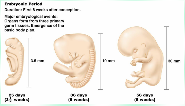
* First 8 weeks
* Initiated by fertilization and at the end of this stage the rudimentary organs and systems have formed with distinct human characteristics. Such as:
- Head is larger than body
- 1st brain waves occur in brain stem
- All major organ systems are present in rudimentary form
*Embryonic Period is the Development of the Basic Body Plan
Cells are programmed genetically to specialize (or differentiate) during this stage. Structural changes by weeks:
Fetal Period ( 9th week until 38th week)
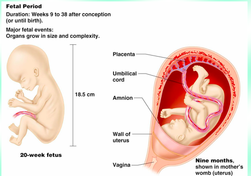
9th week to the 38 week after conception (or until birth); tremendous body growth with tissue differentiation and organ maturation.The period of growth and specialization of body structures.
The end of this stage is called parturition (birth of baby).
Development of The Basic Body Plan
Embryonic Period is the Development of the Basic Body Plan:
- Skin
- Body Outer Wall
- Body cavity and Inner Tube
- Kidneys and gonads
- Limbs
Embryonic Period: 1st 3 weeks
includes:
- #1 Fertilization ( occurs within the Uterine Tube)
- #2 Mitotic Divisions: aka Cleavage: mitosis- "cells divide and replicate"
- #3 Implantation ( occurs within the Uterine Wall)
- #4 Formation of 3 Germ Layers
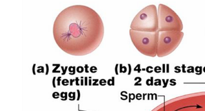
#1 Fertilization
Embryonic Period: 1st 3 weeks
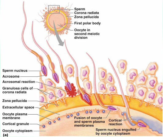
Fertizliation: the precursor to prenatal development. The coming together of 2 sets of chromosomes. NOTE: Mom-female has 23 chromosomes, Dad- male has 23 chromosomes -> End with 46 chromosomes. Number of chromosomes in the ovum (23) + sperm (23) produce a Fertilized egg or Zygote with a number of 46 chromosomes.
CONSISTS OF:
- Copulation
- Secondary Oocyte
- Acrosome
- Acrosomal Reaction
- Cortical Reaction
Define Copulation
#1
Fertilization
Embryonic Period: 1st 3 weeks
a male will ejaculate 100-500 million sperm into the vagina. Only approx. 100 will survive to fertilize the female egg.
Define Secondary Oocyte
#1
Fertilization
Embryonic Period: 1st 3 weeks
a mature female egg that remains in metaphase in the Meiotic II cell division.
Zona pellucida – a layer of protein and polysaccharides
Corona radiate – a layer of granulose cells capped by organelles
Define Acrosome
#1
Fertilization
Embryonic Period: 1st 3 weeks
cap on sperm head-contains protein digesting enzyme
Define Acrosomal Reaction
#1
Fertilization
Embryonic Period: 1st 3 weeks
the breakdown of the zona pellucida by digestive enzyme released from the acrosome of the sperm
Define Cortical Reaction
#1
Fertilization
Embryonic Period: 1st 3 weeks
the fusing of the male/female cell membranes and the release of the male nucleus in the female cytoplasm cause secretion of enzymes by oocyte to destroy sperm receptors in the zona pellucida.
The ovum (egg) completes the 2nd meiotic division.
Number of chromosomes in the ovum (23) + sperm (23) produce a Fertilized egg or Zygote with a number of 46 chromosomes
Prenatal Development now begins (gestation)
#2 Monotonic Divisions
Embryonic Period: 1st 3 weeks
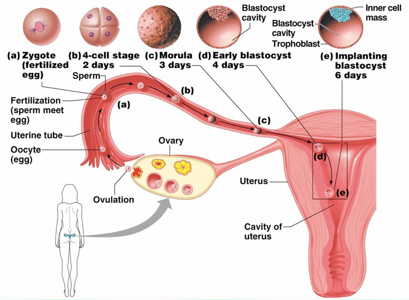
Monotonic Divisions : (f ormation of the Blastocyst )
During transportation through uterine tube to uterus
CONSISTS OF:
- Cleavage process: miotic division, cells get smaller
- Morula: ball of 16 or more cells. same size as zygote but not nutrients for growth
- Blastocysts : a hollow structure with 2 distinct cells
Define Cleavage Process
#2 Monotonic
Divisions
Embryonic Period: 1st 3 weeks
mitotic division, cells get smaller (Blastomeres)
Define: Morula
#2
Monotonic
Divisions
Embryonic Period: 1st 3 weeks
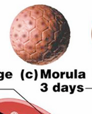
a ball of sixteen or more cells. It is the same size as a zygote and has no nutrients for growth.
note: Zygote= Fertlized Egg; Oocyte= Egg-that has not been fertilized yet; (it travels up the plebeian tube to become fertilized)
note: Morula turn into Blastocysts
Define: Blastocysts
#2 Monotonic
Divisions
Embryonic Period: 1st 3 weeks
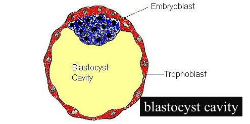
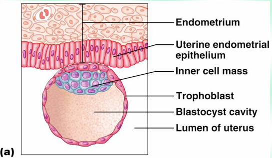
a hallow structure with two distinct cells.
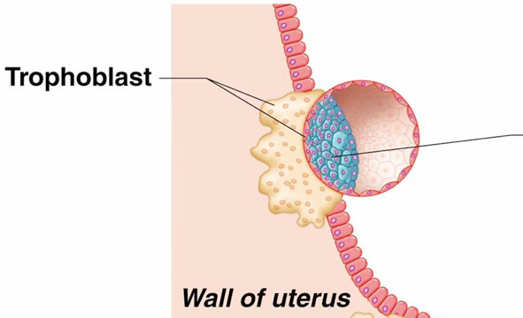
- Trophoblast- a single outer layer, forming the wall-> will become future placenta and membranes
- Inner cell mass (Embryoblast)- inner aggregation of cells-> will become future Embyro
- Blastocyst cavity- fluid-filled center
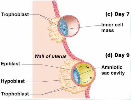
#3 Implantation
Embryonic Period: 1st 3 weeks

5-7 days and blastocyst attaches to the posterior wall of the uterus. Blastocyst implants itself and turns into a ->Trophblast-> Cytotrophoblast-> Syncytiotrophoblasts
CONSISTS OF:
- Trophoblast
- Cytotrophoblast
- Syncytiotrophoblast
Define: Tropoblast
#3
Implantation
Embryonic Period: 1st 3 weeks
secretes proteolytic enzymes that digest a portion of the endometrium (inner lining) of the uterine wall.
Define Cytotrophoblast
#3 Implantation
Embryonic Period: 1st 3 weeks

specialized trophoblast cells next to the wall.
Define: Syncytiotrophoblasts
# 3
Implantation
Embryonic Period: 1st 3 weeks
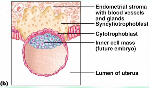
specialized cells of the Cytotrophoblast that multiple and fuse, extend finger-like projects into uterine wall to connect nutrient supply from Mother to Baby. Lacunae develop within and fill with maternal blood. Secretes substances to regulate pregnancy, such as Chorionic gonadotropin hormones (hCG). ( hint: pregnancy tests can detect hCG hormone)
#4: Formation of 3 Germ layers - Define 3 Germ Layers
Embryonic Period: 1st 3 weeks
all body tissues are derived from the 3 germ layers.
- Ectodermal layer- develop into the nervous system, epidermis, and portions of sensory organs.
- Mesodermal layer- develop into skeleton, muscles, blood, reproductive organs, dermis of skin and connective
- Endodermal layer - develop into lining of digestive tract, digestive organs, respiratory tract and lungs, urinary bladder and
#4: Formation of 3 Germ layers
Embryonic Period: 1st 3 weeks
CONSISTS OF:
- Innercell mass ( embryoblasts) splits into two cells
- Bilaminar Embroyic Disc
- Gastrulation- Trilaminar Dic
Define what happens to the : Innercell mass ( Embryoblasts)
#4:
Formation of 3 Germ layers
Embryonic Period: 1st 3 weeks

Inner cell mass (Embryoblast) differentiates into two sheets of cells:
- Epiblast – forms the amniotic sac w/ fluid
- Hypoblast – forms the yolk sac w/fluid
Define: Bilaminar Embryonic Disc
#4: Formation of 3 Germ
layers
Embryonic Period: 1st 3 weeks
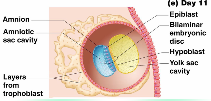
flattened mass of epiblast and hypoblast cells in contact.
basically the mass that connects the Epiblast and the Hypoblast ( yolk sac together)
Define: Gastrulation- Trilaminar Disc
#4: Formation of 3 Germ
layers
Embryonic Period: 1st 3 weeks
(beginning of 3rd wk) development of a Trilaminar disc from the epiblast cells into the three Primary Germ Layers.
note: beginning of 3rd week is when the three Primary Germ Layers start forming
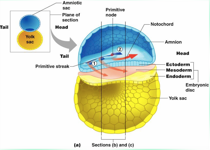
Define: Primitive Streak (last stage of Gastrulation)
#4:
Formation of 3 Germ layers
Embryonic Period:
1st 3 weeks
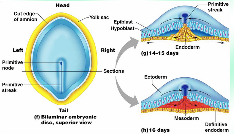
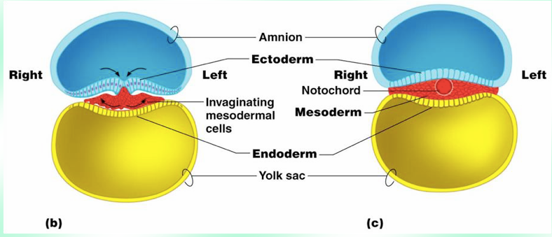
Primitive Steak: a band develops along the dorsal midline of the embryonic disc or epiblast.
- epiblast migrate through streak replace hypoblast to become Endoderm ( the top layer hint- " Eco system")
- second migration of epiblast form a middle layer to become Mesoderm ( the middle layer hint- meso- middle)
- remaining top layer of epiblast become Extoderm- ( bottom layer near yolk sac)
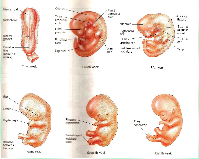
What Happens During the 3rd Week of the Embryonic Period?
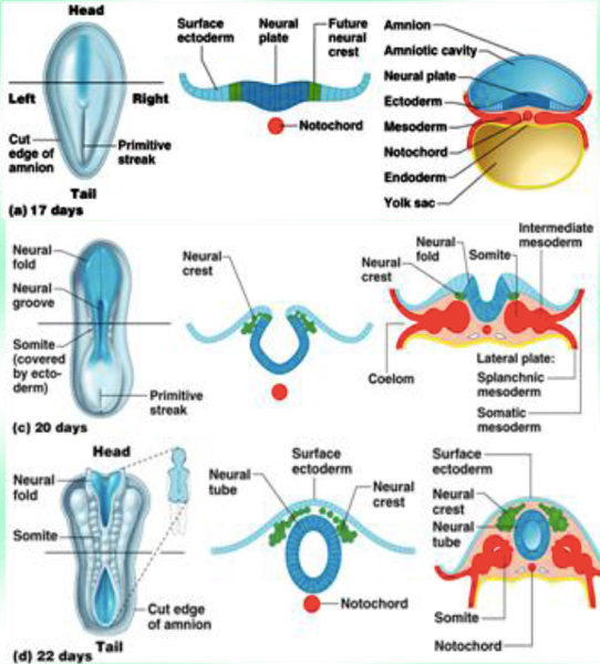
- Primitive node ( stemming from Primitive Streak) - Gives rise to head and notochord
- Notochord – from mesoderm, forms a midline rod that divides body rt/lft; provides support to the embryo; basis for the vertebral column.
- Neurulation - the ectoderm begins forming the spinal cord and brain. (neural folds, neural groove and neural tube)
-
Mesoderm- divides into 3 regions
- Somites – segmentation
- Intermediate mesoderm – kidneys/gonads
- Lateral plate – eventually develops Coelom

What Happens During 4th Week?
The Body begins to take shape during the 4th week of the Embryonic period.
- Folding begins – sides, front and back fold in.
- Ectoderm - becomes Central N. S. and outer skin
- Somites - 1) Sclerotomes – vertebrae and ribs ( think of-scleroous- bones 2) Myotomes - muscle 3) Dermatomes – skin (dermis)
What Begins to Fom During the End of the 4th Week?
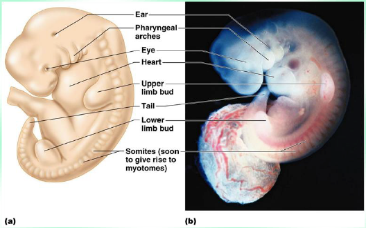
a) Body Stalk- involved w/formation of the umbilical cord.
b) Heart - beating blood to all parts of the embryo
c) Head and Jaw-primordial formed eyes, brain, spinal cord.
d) Arm and Leg Buds
What Occurs During the End of the 4th-8th Week?
By the end of the 4th week to the 8th week structural changes start occurring to the Embyro
INCLUDES:
- rudimentary body organs develop ( at the end of embryonic stage looks human)
- Placenta, Umblical Cord and membranes form
- (embryo is not self-sustaining yet and must get nourishment from mother to survive)
What Occurs During the 5th Week?
The Head enlarges and Digital Rays ( fingers) or paddle shaped hand plates begin to form
What Occurs During the 6th Week?
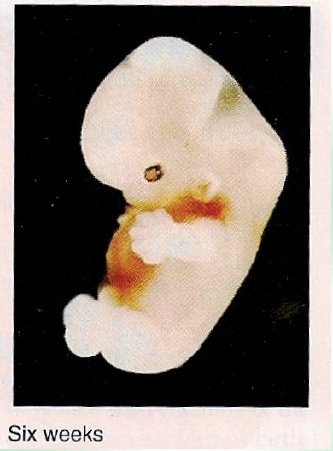
the most vulnerable period of development for many organs. If interrupted at this stage can cause congenital damage. a) Head is larger than trunk c) Brain undergoes marked differentiation d) Forelimbs lengthen/fingers
What Occurs During the 7th and 8th Weeks?
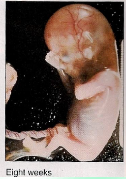
- Distinct human characteristics are noticable.
- Primitive body organs are formed
- Nervous system begins to coordinate body activity
- External genitalia are forming but not differentiated- can't tell if its a boy or girl yet
- Rudimentary body systems are developed at the end of the 8th week ( * rudimentary means not very advanced)
Extra- Embryonic Membranes provide ____ to the fetus
provide protection, respiration, excretion, and nutrition for the embryo to fetus. This is known as afterbirth at parturition.
Extra- Embryonic Membranes:
Amnion
membrane from the epiblast; surrounds embryo (amniotic sac); sac if filled with amniotic fluid. This fluid cushions and protects, maintains constant pressure and temperature; allows freedom of fetal development
Extra- Embryonic Membranes:
Yolk Sac
thin membrane from hypoblast ; produces blood for the embryo until the liver forms and produces germ cells migrate to primordial gonads.
Extra- Embryonic Membranes:
Allantois
out-pouching from yolk sac
Extra- Embryonic Membranes:
Chorion
originates from the trophoblast. Gives rise to an extra-embryonic mesoderm that develops into: a) villi- finger-like extensions form the chorion and penetrate into the uterine tissue b) villous chorion- villi that are associated with uterine wall and branch out around lacunae.