Anatomy
the science of body structures and the relationships among structures; derived from Greek means “to dissect” or “to cut up”.
ANA= up
Tomy= to cut
Morphology
the science of form
Physiology
the study of body function
Pathology
the study of diseases
Dissection
“cut apart”
Gross (large) Anatomy
examined by the eye
ex: EW thats gross! " you can see it with your eye"
Surface Anatomy
shapes and markings on the surface of the body that reveal the organs below (landmarks).
Regional Anatomy
structures in a single region
Systemic Anatomy
all the organs within a system
Microscopic Anatomy (Histology)
need a microscope to view tissues and cells.
Histology= the study of tissues
Body Organization – 6 levels

1. Chemical
2. Cellular
3. Tissue
4. Organ Level
5. System Level
6. Organismal Level
Chemical level

atoms > molecules > proteins, lipids, etc
Cellular level

most basic structural and functional living unit or component of life.
note: cells are made up of molecules
Tissue level

aggregates of similar cells working together for a particular function.
Organ level
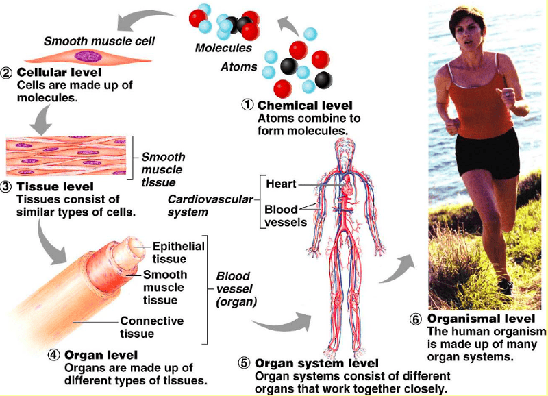
two or more different types of tissues working for a similar or specific function. Usually with a specific shape
System level

A system consists of different organs that have related functions, or are working together at a particular function
Organismal level

the whole body-all systems operate together and are interrelated to make up the complete (whole) organism.
Systemic Approach
examines the individual systems and the related organs.
Regional Approach
look at the structural relationships within a portion of the body and several systems. Specific body parts in a region will have similar names.
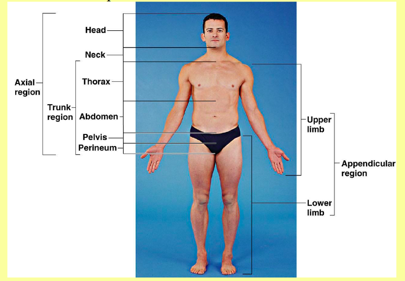
What are the 5 Major Body Regions?

1. Head (caput)- two subsystems 1. Facial 2. Cranial
2. Neck( cervix)
3. Trunk (torso)- divided into 3 subsections 1. Thorax 2. Abdomen 3. Pelvic
4. Upper Extremity ( limbs) : pectoral girdle, brachium, antebrachium, manus
5. Lower Extremity (limbs): pelvic girdle, femoral, crural, pes
Head

(caput)-two subdivisions: Facial and Cranial
Neck
(cervix) cervical region supports head and permits it to move.
Trunk

(torso)-divided into three subdivisions:
a. Thorax (thoracic region)
b. Abdomen-below the diaphragm
c. Pelvic-lower part of the trunk
Upper Extremity (Limbs)

- pectoral girdle: The shoulder girdle or pectoral girdle is the set of bones which connects the arm to the axial skeleton on each side
- Brachium: Upper Arm ( Shoulders-> Elbow)
- Antebrachium: forearm ( Elbow-> Wrist)
- Manus: hand
Lower Extremity ( Limbs)
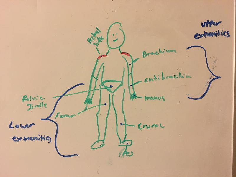
lower extremity refers to the human leg, including the gluteal or hip region, thigh and foot
- pelvic girdle: Defined to be the left and right ossa coxae.
- femoral: the thigh region
- crural:pertaining to the leg, between knee and ankle.
- pes: foot
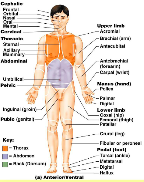
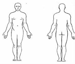
Anterior Ventral Diagram
FRONT VIEW OF BODY
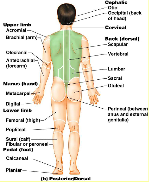
Posterior/ Dorsal Diagram
BACK VIEW OF BODY
Body Cavities and Membranes overall function
the body’s cavities and the membranes help protect, separate and support internal organs.
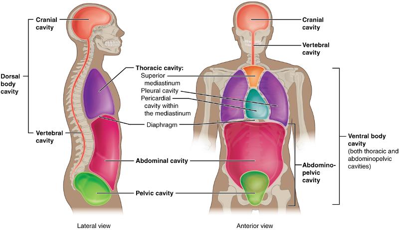
Two Major (Principal) Body Cavities Include:
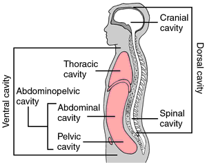
1. Dorsal - brain ( cranial cavity) and spinal chord ( vertebral canal)
2. Ventral - splits into two cavities: #1 Thoracic Cavity; #2 Abdominopelic Cavity
#1. Thoracic cavity (Chest Cavity)-subdivided into three cavities by membranes
A. pleural cavities (2) -Lungs
B. mediastinum- Area between the two lungs
C. pericardial cavity – Heart
#2. Abdominopelvic cavity-subdivided into two
A. abdominal-upper cavity
B. pelvic-lower cavity
Abdominal Regions and Quadrants- Abdominopelvic Cavity (4)
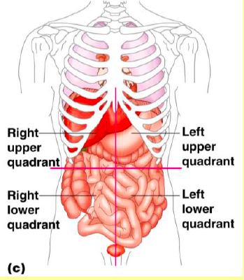
Most digestive organs are contained in the abdominopelvic cavity. To aid in the location of the abdominopelvic organs, the abdominopelvic cavity is divided into 4 quadrants (vertical and horizontal lines through the umbilicus
1. Right Upper Quadrant
2. Right Lower Quadrant
3. Left Upper Quadrant
4. Left Lower Quadrant
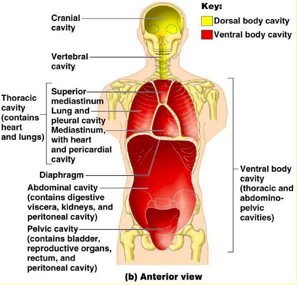
Anterior View of Doral and Ventral Body Cavities
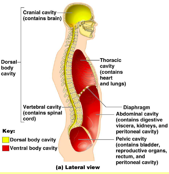
Lateral View of Doral and Ventral Body Cavities
Several smaller cavities exist within the head
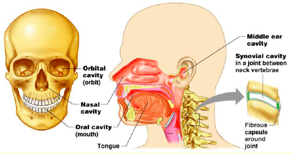
include the orbital cavity; nasal cavity; oral cavity; middle ear cavity
2 Types of Membranes
1. Mucous Membranes
2. Serous Membranes
Mucous Membranes
secretes a thick viscid substance mucus ( lubricates and protects). this mucus lines various cavities that enter or exist the body
Serous Membrane
lines the wall of the thoracic (chest cavity)and abdominopelivic cavities and covers visceral organs. secrets watery fluid.
A: Pleural Membrane
B: Pericardial Membrane
C: Perioneal Membrane
Pleural Membranes
LUNGS
HINT: PLURAL IS 2 -2 LUNGS
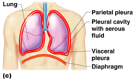
associated with Lungs ( right pleura and left pleura)
1. parietal pleura-lines thoracic walls
2. visceral pleura-adheres to lungs
3. pleural cavity- space between 2 pleurae
Pericardial membranes
HEART
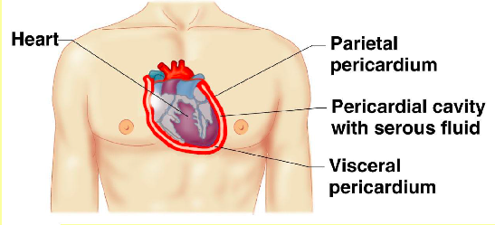
associated with Heart
1.parietal pericardium- covering surrounds the heart
2. visceral pericardium- c overs surface of the heart
3. pericardial cavity -space between the serous membranes
Peritoneal membranes
pronunciation:per·i·to·ne·al
ABDOMINAL
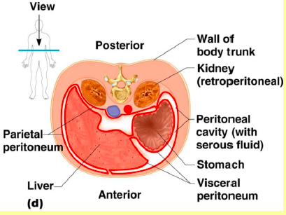
associated with the Abdominal
1. parietal peritoneum-lines the walls
2. visceral peritoneum-covers visceral organs
3. peritoneal cavity-space within the abdominopelvic cavity
a. retroperitoneal - kidneys, adrenal glands portion of pancreas that are outside of the peritoneal cavity

Planes of Reference-3 fundamental planes
* Anatomical Planes of the Body*
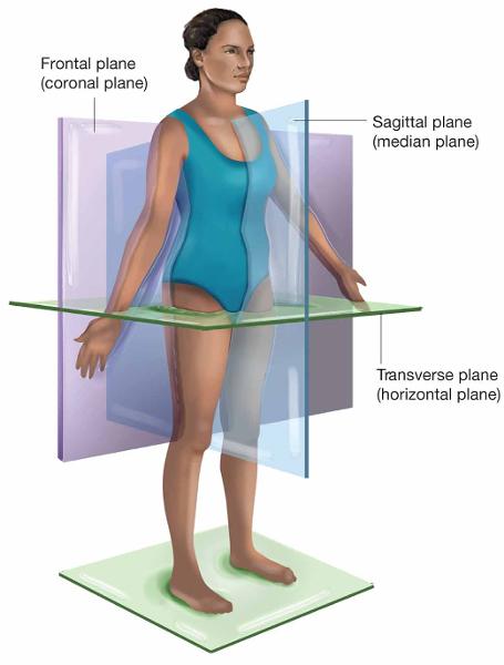
1. Midsagittal ( median)
2. Frontal (coronal)
3. Transverse ( horizontal)
Midsagittal (median) Plane
a vertical line which divides the body into a left section and a right section.
Frontal (Coronal) Plane
a vertical line which divides the body into a front (ventral, anterior) section and back (doral, posterior) section.
Transverse (horizontal) Plane
a horizontal line which divides the body into an upper (superior) section and a bottom (inferior) section.
Horizontal Line =
_________________
Vertical Line=
^
^
^
^
^
Medial and Lateral ( Directional Terms)
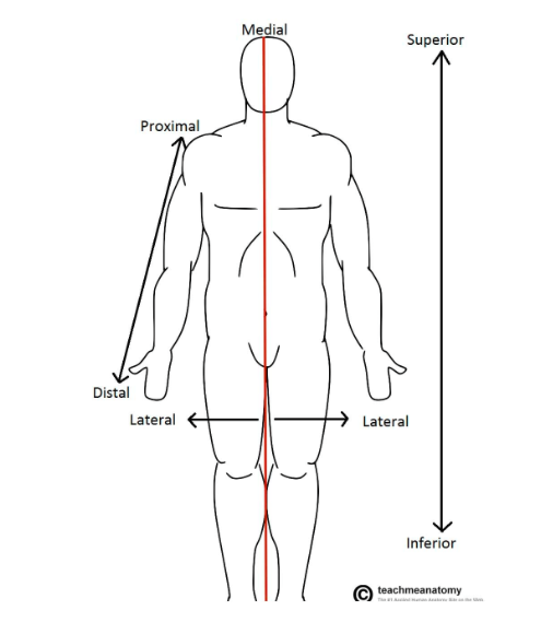
Imagine a line in the sagittal plane, splitting the right and left halves evenly. This is the midline. Medial means towards the midline, lateral means away from the midline.
Examples:
The eye is lateral to the nose.
The nose is medial to the ears.
Anterior and Posterior (Directional Terms)
Anterior (ventral) refers to the ‘front’, and posterior (dorsal) refers to the ‘back’. Putting this in context, the heart is posterior to the sternum because it lies behind it. Equally, the sternum is anterior to the heart because it lies in front of it
Superior and Inferior (Directional Terms)
These terms refer to the vertical axis. Superior means ‘higher’, inferior means ‘lower’. The head is superior to the neck; the umbilicus is inferior to the sternum.
Proximal and Distal (Directional Terms)
The terms proximal and distal are used in structures that are considered to have a beginning and an end (such as the upper limb, lower limb and blood vessels). They describe the position of a structure with reference to its origin – proximal means closer to its origin, distal means further away.
Anatomical position
body erect, feet parallel, eyes forward, arms to the side with palms forward. To be able to describe direction in relationship of one body part to another. Prone - a body face lying down Supine - a body facing up
Superficial vs Deep ( Directional Terms)
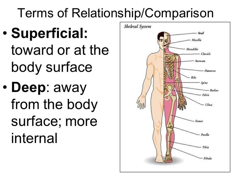
Superficial: toward or at body surface
Deep: away from the body surface; more internal
Disorder -Human body and Disease
is any abnormality of structure and/or function
Disease- Human Body and Disease
is an illness with a definite set of symptoms and signs
Symptoms- Human Body and Disease
subjective changes in body functions that are not apparent to an observer (ex. headache)
Signs- Human Body and Disease
objective changes that can be observed and measured (rash, fever)
Epidemiology- Human Body and Disease
is the science that deals with the why, when and where diseases occur and why they are transmitted among individuals in an area
Diagnosis- Human Body and Disease
is the science and skill of distinguishing one disorder or disease from another
Clinical procedures - important in determining anatomical structure and function in living individuals
1.observation-visual inspection
2. palpation-applying fingers firmly- using hands to examine the body
3. auscultation-listening (ex: heartbeat)
4. percussion –taping body parts with hands, or small instruments that produce listening to echo
5. medical imaging techniques
a. X-ray - radiographs
b. CT – radiograph computed tomography
c. PET – Positron Emission Tomography
d. Ultrasound - ultrasonography
e. MRI – magnetic resonance imaging