Name two functions of an articulation (joint)
1. Hold bones together
2. Allow rigid skeletal system some flexibility so gross body movements can occur
Structural classifications of joints
What type of tissue is present in the joint cavity? 3 types: fibrous, cartilaginous, synovial
Functional classifications of joints
How much movement is allowed by joint? 3 types: synarthroses (immovable), amphiarthroses (slightly movable), diarthroses (freely movable)
Fibrous joints
Made of dense regular connective tissue and no joint cavity. Joints are either synarthrotic or amphiarthrotic.
Examples: Suture (short fibers, located in skull), Gumphosis (periodontal ligament, tooth in bony socket)
Cartilaginous joints
Adjoining bones united by cartilage with no joint cavity. Joints are either synarthrotic or amphiarthrotic.
Examples: Intervertebral discs, Pubic symphysis (fibrocartilage)
Synovial joints
Most common type of joint in body; joints are freely movable. Remember, slick as snot
Several defining structural characteristics:
a. Joint cavity (filled with synovial fluid)
b. Articular cartilage (hyaline cartilage covering ends of bones forming joint)
c. Articular capsule (two layers enclosing joint cavity. Fibrous layer (dense irregular connective tissue) and inner layer (synovial membrane))
d. Synovial fluid (viscous fluid acting as lubricant)
e. Reinforcing ligaments
f. Nerves and blood vessels (sensory nerve fibers to detect pain and joint stretching, blood vessels supply synovial membrane)
g. Articular discs
h. Bursa and tendon sheath
Examples: Elbow joint, shoulder joint, hip joint, etc
Types of synovial joints: Plane
non-axial (gliding); between flat or slightly curved bones
examples: intertarsal, intercarpal joints
Types of synovial joints: Hinge
Uniaxial (flexion and extension); a rounded or cylindrical bone fits into a concave surface on the other bone
examples: elbow, interphalangeal joints, knee
Types of synovial joints: Pivot
Uniaxial (rotation); A rounded bone fits into a sleeve (a concave bone plus a ligament)
examples: proximal radioulnar, atlantoaxial joint
Types of synovial joints: Condylar
Biaxial (flexion, extension, adduction, abduction); An oval condyle fits into an oval depression on the other bone
examples: metacarpophalangeal (knuckle) and radiocarpal joints; metacarpal/carpal joints
Types of synovial joints: Saddle
Biaxial (flexion, extension, adduction, abduction); Articulating surfaces are saddle shaped; one surface is concave, the other is convex
examples: carpometacarpal joint of the thumb (this is mostly where this joint is)
Types of synovial joints: Ball-and-socket
Multiaxial (flexion, extension, abduction, adduction, rotation); the ball-shaped head of one bone fits into the cuplike depression of the other bone
examples: shoulder, hip joints
Origin
stationary, immovable, or less movable attachment of muscle to bone; anchor for part of body that moves
Insertion
More movable attachment site of muscle to bone; part of body that moves
Flexion
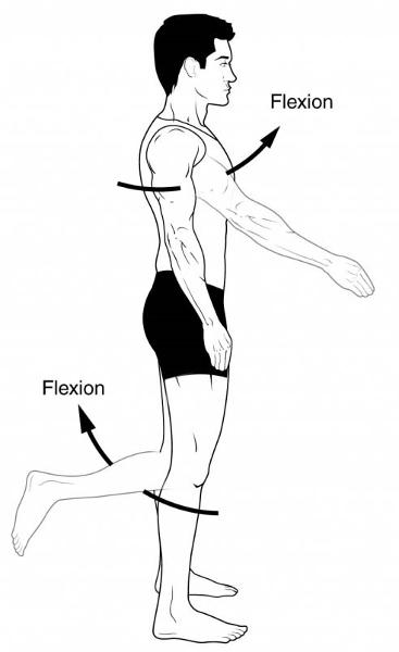
Movement (usually in sagittal plane) that decreases the angle of the joint and reduces the distance between two bones (example bending knee or elbow)
Extension
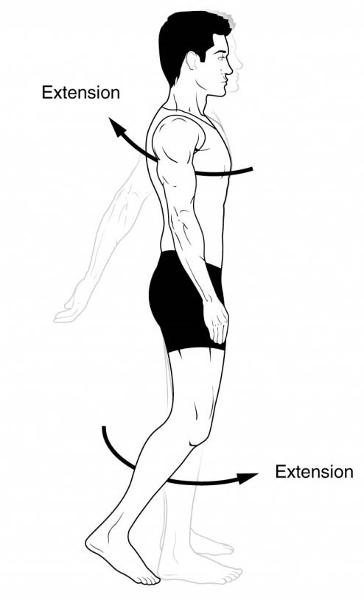
Movement that increases the angle of the joint and the distance between two bones or parts of body; opposite of flexion.
Abduction
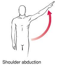
Movement of a limb away from the midline of the body, along the frontal plan, or the fanning movement of fingers or toes when they are spread apart. REMEMBER: Children are abducted from their parents...they are taken away
Adduction
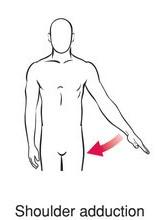
Movement of a limb towards the midline of the body or drawing the fingers or toes together; opposite of abduction.
Rotation
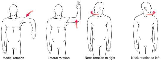
Movement of a bone around its longitudinal axis without lateral or medial displacement. Also describes movement of atlas around dens of axis.
Circumduction
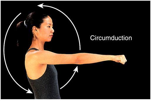
A combination of flexion, extension, abduction, and adduction commonly observed in ball and socket joints like the shoulder. The limb as a whole outlines a cone.
Pronation
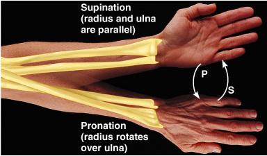
Movement of the palm of the hand from an anterior or upward-facing position to a posterior or downward facing position. The distal end of radius rotates over the ulna.
Supination

Movement of the palm from a posterior position to an anterior position. Radius and ulna are parallel.
Dorsiflexion
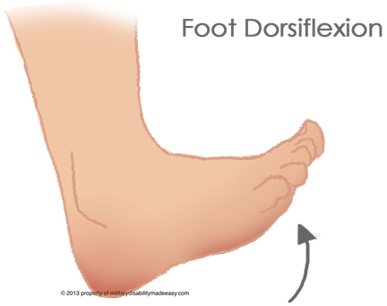
Movement of ankle joint that lifts foot so that its superior surface approaches the shin
Plantar flexion
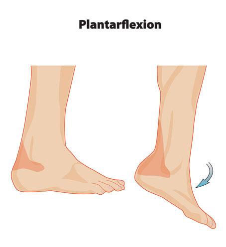
Movement of ankle joint in which foot flexes downward as if standing on one's toes or pointing the toes
Inversion
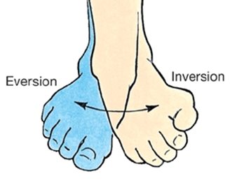
Movement that turns sole of the foot medially
Eversion

Movement that turns sole of foot laterally
Ligamentum teres
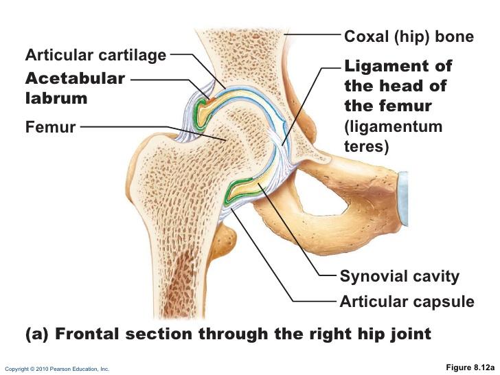
ligament in hip running from the fovea capitis on the femur head to the acetabulum, also called ligament of the head of the femur
Ligaments of the knee
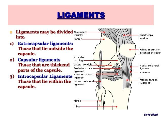
patellar, medial, lateral patellar (merged with articular capsule)
fibular and tibial collateral ligaments (prevent rotation during extension)
oblique popliteal and arcuate popliteal ligaments (reinforce knee)
posterior and anterior cruciate ligaments (intracapsular and prevent overflexion and hyperextension)
Glenohumeral joint
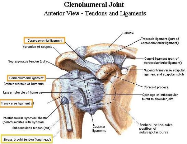
Shoulder joint