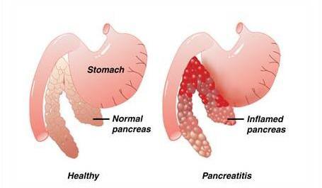
- Acute: pancreatic duct becomes obstructed, and enzymes back up, causing auto-digestion and inflammation of the pancreas.
- Chronic: progressive inflammatory disorder with destruction of the pancreas; cells are replaced by fibrous tissue; pressure within the pancreas increases, obstructing the pancreatic and common bile ducts
- Refer to Chart 50-3
PANCREAS
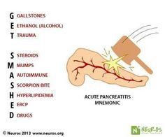
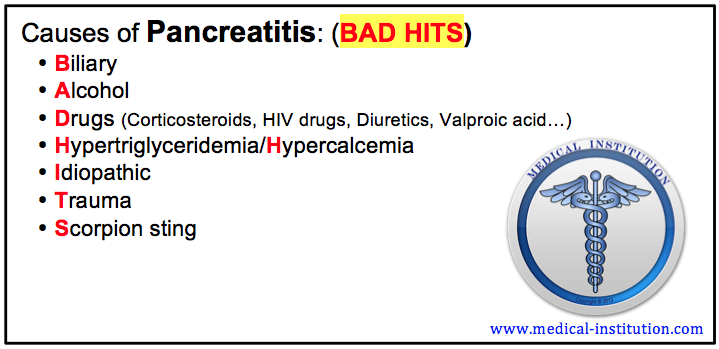
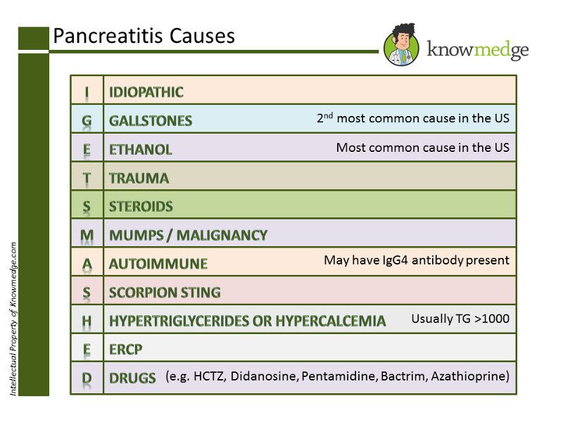
- Alcohol
- Billiary Disease
- Trauma
- Some Drugs ETC..
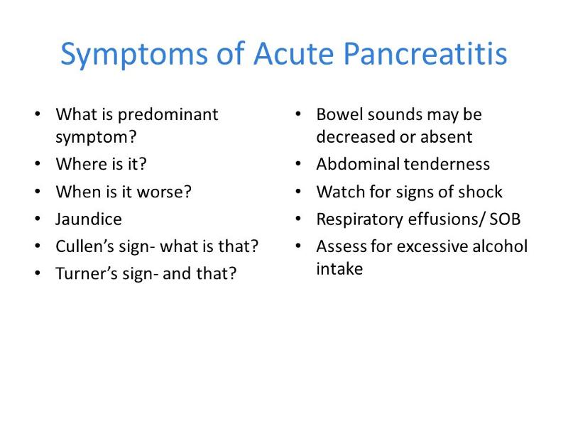
Acute Pancreatitis- Etiology

B

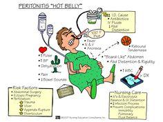
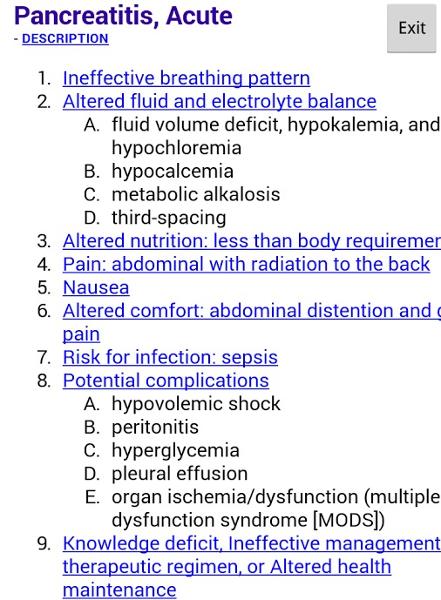
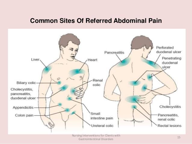
- Abdominal Pain
- Guarding
- Rigid board-like Abdomen (peritonitis)
- Hypotension or Shock
SIGNS AND SYMPTOMS OF PANCREAS
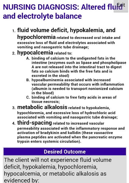
- Fluid and electrolyte disturbances
- Necrosis of the pancreas
- Shock
- Multiple organ dysfunction syndrome
- DIC
Collaborative Problems and Potential Complications PANCREAITIS
- Cardiovascular Failure
- ARDS
- Renal Failure
- Hemorrhage
- Infection
PANCREAITIS
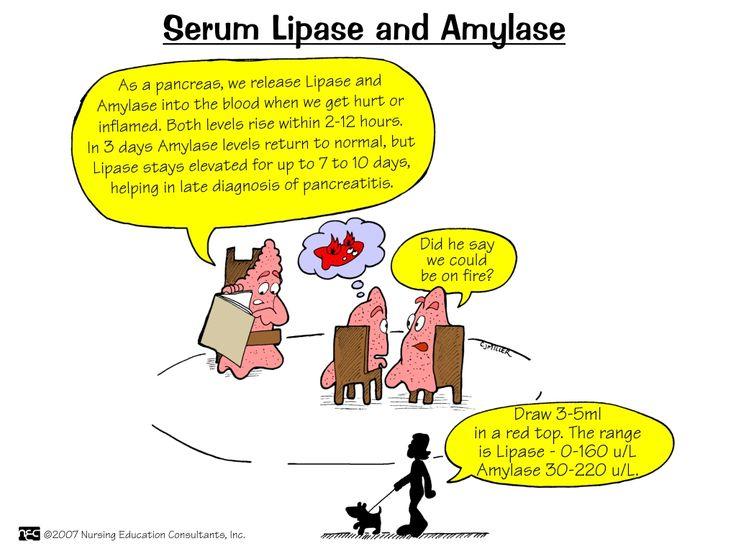
- Serum Amylase, Lipase
- X-Ray
- CT
- Ultrasound
DIAGNOSTIC TESTS
Chronic Pancreatitis
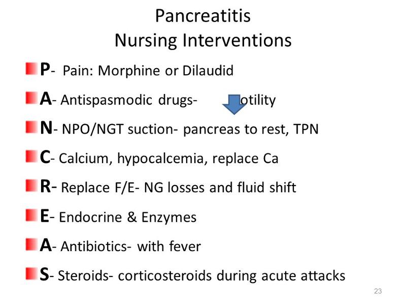
- IV Fluids/TPN
- Blood Products PRN
- Oxygen
- Analgesics, Antianxiety Agents
- NPO
- NG Suction
Histamine Antagonists
THERAPUTIC TX
Chronic Pancreatitis
- Pathophysiology
- Progressive Fibrosis
- Obstructed Ducts
- Ulceration
- Etiology
- Alcohol
- Biliary Disease
Chronic Pancreatitis
- Remissions and Exacerbations
- LUQ Pain and tenderness : “burn-out”
- Anorexia
- Malabsorption, Weight Loss, muscle wasting
- Steatorrhea and foul smelling stools
- Diabetes Mellitus
- Jaundice
Chronic Pancreatitis: SIGNS AND SYMPTOMS
- Pancreatic Enzymes Normal
- High Fecal Fat Level
- Changes on CT
DIAGNOSIS Chronic Pancreatitis
- Analgesics
- Pancreatic Enzyme Replacement
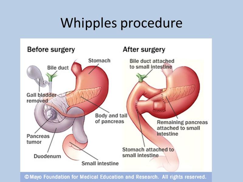
- Surgery
Chronic Pancreatitis TX
What is a major symptom of chronic pancreatitis?
A.Recurrent attacks of severe upper abdominal and back pain accompanied by vomiting
B.Fever, jaundice, confusion, and agitation
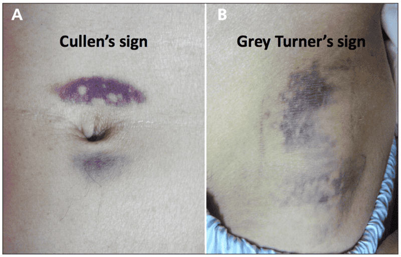
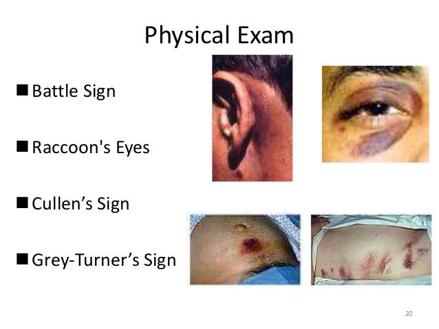
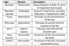
C.Ecchymosis in the flank or umbilical area
D.Abdominal guarding
A.Recurrent attacks of severe upper abdominal and back pain accompanied by vomiting
Chronic pancreatitis has recurrent attacks of severe upper abdominal and back pain accompanied by vomiting. Acute pancreatitis presents with fever, jaundice, confusion, agitation, ecchymosis in the flank or umbilical area, and abdominal guarding.
COPD
refers to diseases that obstruct air flow and commonly includes chronic bronchitis and emphysema (airflow limitation)
Exacerbation and co-morbidity contributes to the clinical manifestations
contributes to the clinical manifestations
Presents with 3 cardinal signs and symptoms
–Dyspnea
–Chronic cough
–Sputum production
Less common signs
- Wheezing
- Chest tightness
COPD SIGNS AND SYMPTOMS
cigarette, air pollution, allergy, autoimmunity, infection, occupational exposure, aging etc.
Excessive mucous production resulting in chronic cough is characteristic of chronic bronchitis.
RISK FACTOR FOR COPD
Inflammatory changes leads to mucociliary dysfunction & bronchial constriction
Increase production of mucus with a chronic cough that persists for at least 3 months of the year for 2 consecutive years
With continued irritation epithelial cells die, destroying their Celia
Goblet cells hypertrophy creating more tenacious mucus
The inability to clear the airway of excess mucus causes susceptibility to infections
CHRONIC BRONCHITIS
Repeated infections lead to airways showing scarring, stenosis and obstruction
- Because of the loss of alveolar walls and the capillaries surrounding them, there is increased pressure in the pulmonary circulation.
- This, along with thickening of the blood vessels, results in pulmonary hypertension.
- Which in turn leads to right side heart failure /cor pulmonale.
- Common characteristics
- Mucus hypersecretion
- Dysfunction of cilia
- Hyperinflation of lungs
- Gas exchange abnormalities
CHRONIC BRONCHITIS
Productive cough, exertional dyspnea, and wheezing
Over time, hypoxemia (PaO2 <60 mm Hg or O2 saturation <88%) may develop with hypercapnia (PaCO2 >45 mm Hg) leading to respiratory acidosis.
Because of their reduced oxygen saturation they start to retain fluid.
polycythemia develops as a result of increased production of red blood cells as the body attempts to compensate for chronic hypoxemia.
the resp. acidosis , polycythemia and < O2 leads to the constant cyanosis and bloated appearance and that’s why chronic bronchitis is referred to as “blue bloaters”
CLINICAL MANIFESTATIONS COPD
Degenerative, nonreversible disease characterized by
–Destruction of the alveoli
–Enlargement of the distal airspaces
–Breakdown of alveolar walls leads to loss of alveolar recoil+ decrease ventilation
Breathing out is the main problem
Pulmonary Emphysema
Thin, emaciated body frame
Dyspnea on exertion, later at rest
Shortness of breath and “air hunger”
Chronic productive cough
Classic “barrel chest” indicating constant lung over inflation and overwork of chest muscles used for breathing (diaphragm).
Dilatation and destruction of lung tissue distal to terminal bronchioles.
It leads to air trapping and an increase in total lung capacity.
There is also a loss of alveolar tissue, reducing capacity for gas exchange.
Carbon dioxide clearance is not impaired to the same extent as chronic bronchitis, usually because patients increase their respiratory effort and ventilation, the pt. Is not usually cyanotic
Instead you will observe a pinkish skin color and that is why they are called “pink puffers”
Clinical Manifestations:
Pulmonary Emphysema
H&P
CXR, CT scan
Pulmonary function tests
ABGs
Pulmonary test to rule out TB or malignancy
Diagnostic Eval. Of COPD
Elevated Hct - to low oxygenation levels
Hypoxemia-low PO2 less than 80mmHg
Hypercapnia- high PCO2 -45 mmHg
Resp acidosis
LABS COPD
The nurse reviews the arterial blood gases of a patient. Which result would indicate the patient has later stage COPD?
pH 7.32, PaCO2 58 mm Hg, PaO2 60 mm Hg, HCO3 30 mEq/LnpH 7.30, PaCO2 45 mm Hg, PaO2 55 mm Hg, HCO3 18 mEq/L
pH 7.40, PaCO2 40 mm Hg, PaO2 70 mm Hg, HCO3 25 mEq/L
pH 7.52, PaCO2 30 mm Hg, PaO2 80 mm Hg, HCO3 35 mEq/L
ABGS COPD
Answer: A
Rationale: In later stage COPD, the patient will have a low or low normal pH, a high normal or above normal PaCO2, and a high normal or above normal HCO3-. This indicates compensated respiratory acidosis, as the patient has chronically retained CO2 and the kidneys have conserved HCO3- to increase the pH to near or within the normal range.
This image shows multiple cystic areas caused by destruction of lung tissue typical of emphysema.
Lung Severe Emphysema:
The chest cavity is opened at autopsy to reveal numerous large bullae apparent on the surface of the lungs in a patient dying with emphysema.
Bullae are large dilated airspaces that bulge out from beneath the pleura.
Emphysema is characterized by a loss of lung parenchyma by destruction of alveoli so that there is permanent dilation of airspaces
BULLAE
nCan improve survival in patients with advanced COPD who have hypoxemia, (low blood oxygen levels)
nThis treatment can improve a patient's exercise tolerance and ability to perform on psychological tests which reflect different aspects of brain function and muscle coordination
HOME OXYGEN THERAPY COPD
Increasing the concentration of oxygen in blood also improves the function of the heart and prevents the development of Cor pulmonale.
Monitor amount of oxygen given. No more than 3L is recommended. Why?
Some patients cannot maintain this effort and their carbon dioxide levels rise. This stimulates respiratory drive for a time, but then they become desensitized to it and they depend on hypoxemia to drive ventilation.
Oxygen can also lessen sleeplessness, irritability, headaches, and the overproduction of red blood cells.
n Continuous oxygen therapy is recommended for patients with low oxygen levels at rest, during exercise, or while sleeping.
nReview Oxygen delivery system (NC, Mask etc..)
Suppressed respiratory drive and low O2
Fire
Oxygen toxicity
COMPLICATIONS OF OXYGEN THERAPY
COPD MEDICATIONS
Bronchodilators
Methylxanthines
Corticosteroids or Steroids
Antibiotics
Expectorants
Diuretics
Digitalis
Leukotriene Inhibitors
Tranquilizers
Pain meds
Cough suppressants (codeine, etc.)
Sleeping pills (barbiturates, etc.)
- The choice of bronchodilator depends on the patient’s response. However, when the patient has mild COPD or fewer symptoms, a short-acting bronchodilator is used as needed.
- Albuterol or ipratropium (Atrovent) may be used as single agents, but combining bronchodilators improves their effect and decreases the risk of adverse effects. These two agents (albuterol and ipratropium) can be nebulized together (DuoNeb) or delivered by one MDI (Combivent Respimat).
- In the moderate stages of COPD, a long-acting bronchodilator may be used in addition to a short-acting rescue bronchodilator, such as salmeterol (Serevent) or formoterol (Foradil).
Surgical removal of large air spaces called bullae that are filled with stagnant air
May be beneficial in selected patients
Recently, use of lasers to remove bullae has been suggested
BULLECTOMY
nHas been successfully employed in some patients with end-stage COPD.
n In the hands of an experienced team, the 1-year survival in patients with transplanted lungs is over 70 percent.
LUNG TRANSPLANT
Help to overcome the conditions which cause dyspnea, anxiety and allergic reactions
Smoking cessation
Oxygen therapy
Breathing retaining
Airway clearance techniques
Improve capacity for physical exercise and activities of daily living
Frequent rest periods
Diet : Offer small frequent meals;
high calorie, high protein
2-3L fluid unless contraindicated IV/PO
Daily weights
n Intermittent mechanical ventilatory support relieves dyspnea and rests respiratory muscles in selected patients.
nPurse lip breathing techniques
PULMONARY REHABILITATION
Continuous positive airway pressure (CPAP) is used as an adjunct to weaning from mechanical ventilation to minimize dyspnea during exercise.
Relaxation techniques may also reduce the perception of ventilatory effort and dyspnea
Breathing exercises and breathing techniques, such as pursed lips breathing and relaxation
Place in high Fowler’s position or seated on the bedside with the arms folded on the over bed table to promote full lung expansion (Tripod)
Keeping air passages reasonably clear of secretions is difficult for patients with advanced COPD.
Re: Some commonly used methods for mobilizing and removing secretions are already discussed.
nAvoid smoking environment
nBreathing toxic fumes such as glue, paint etc.
nAerosol products other than meds
nGoing to places with polluted atmosphere
nAvoid people with infections etc..
C: IS FOR CONTROLLED ENVIROMENT
Highly communicable disease
Caused by mycobacterium tuberculosis
–Rod shaped, gram+, acid fast bacillus (AFB)
http://en.wikipedia.org/wiki/Mycobacterium
WHAT IS TB?
Spread by airborne droplet from an affected person during
–Coughing
–Speaking
–Laughing
–Sneezing
Another person breathes in the bacteria & becomes infected
Close quarters
TRANSMISSION OF TB
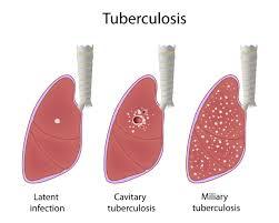
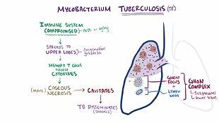
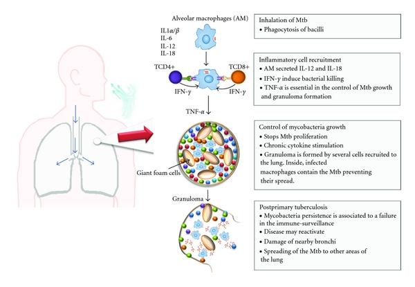
The tuberculosis bacterium invades the alveoli and begins to multiply forming a tubercle lesion
The body’s defense mechanism encapsulate the tubercle leaving a scar.
May continue to grow to form granulomas or cheese like mass called caseation
This is what is seen on a CXR and is called the Ghon tubercle or primary lesion
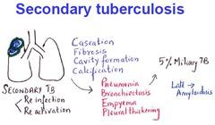
Primary lesions form, they may become dormant, but can be reactivated and become a secondary infections when re-exposed to the bacterium
Pathophysiology TB
www.anatomy.unimelb.edu.au/museum/collection.html
Dissected, preserved lung with tuberculosis TB
Close contact with infected person
Live in areas where TB in common
HIV infections, immune system diseases
The elderly
Homeless, poor, crowded living conditions
Poor access to heath care
Illicit drug use
Health care professional
WHOS AT RISK TB
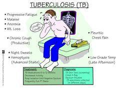
TB has an insidious onset and many pts are not aware of the symptoms until the disease is well advanced
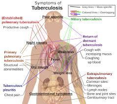
Fever, night sweats, malaise
Anorexia and weight loss
Cough non productive at first, but later produces yellow mucoid sputum
Hemoptysis
Pleuritic chest pain and tightness
Dyspnea if severe lung involvement
CLINICAL MANIFESTATIONS OF TB
H&P especially for pts at risk
Tests: Sputum smear and C&S
–AFB x 3 samples are usually obtained for an acid-fast smear . (May aspirate gastric fluid if unable to obtain specimen)
–The sputum test confirms the diagnosis of TB because
Chest X-rays
–Determines the presence of Ghon tubercles or lesions on the lungs and the extent of the disease (cheesy caseations)
–Indicates presence and extent of disease process but cannot differentiate active from inactive form
–
WBC and ESR increased
A + reaction is noted if the induration is greater than 5mm
This means that the patient has had contact with tubercle bacillus.
It does not mean that active disease is present in the body . (antibodies are now present)
Check for vaccination with BCG
DIAGNOSTIC TESTS TB
¨Goal of treatment is to prevent transmission, control symptoms, and prevent progression of the disease
¨6-12 months, may be as long as 24 months
¨2-4 meds Given concurrently
¨Mycobacteria grow slowly, resistance is common
vImproper or noncompliant use of treatment programs may cause the development of mutations in the tubercle bacilli resulting in a multidrug resistant strain of TB (MDR-TB
(+) Mantoux test, (-) CXR, and at risk for the disease are treated prophylactically to prevent development of active TB
Isoniazid (INH) for 9-12 months
Medical management TB
Combination drug therapy is most effective
–It destroys the organism and minimize development of multiple drug resistant strain of the bacilli
–After TB meds have been given for 2-3 weeks, the risk of transmission is reduced greatly
ACTIVE TB
Drug of choice
Take with meals if GI upset otherwise take one hr before or 2hr after meals. Absorption is decreased if given with food
Avoid antacids exp. Aluminum based – decrease absorption
Peripheral neuritis
– drug is usually given with Vitamin B6 (Pyridoxine) to prevent this side effect
Hepatitis
Nausea, vomiting, diarrhea, stomach pain
Avoid foods with tyramine
– aged cheese, smoked fish, tuna, sauerkraut, avocado etc.
Combined with INH will make you sicker
Drugs for TB : INH Isoniazid
150-300mg , maximum dose 600mg /day
S/S: hepatotoxicity, hepatitis, blood dyscrasias
Steven Johnson’s syndrome
Red/orange stain to urine, feces, saliva, skin, sputum, sweat and tears ..
Rifampin (+ INH)
15mg/kg po daily or 50mg /kg twice weekly when use in combination with other TB drugs. Max. 1200mg/day
S/S: optic neuritis leading to color blindness (exp.Red- Green)
Peripheral neuritis; GI irritation
Gouty arthritis from ^ uric acid levels
Ethambutol/myambutol
Bactericidal. Kills mycobacterium present in macrophages
S/S: hepatotoxicity, thrombocytopenia
Hyperuricemia- gout (hot painful, swollen big toe, ankle or knee)
Gi irritation
Pyrazinamide (PZA
Not used as much theses days; has now been replaced by Ethambutol
S/S GI upset, fever, rash, hepatitis
Fluid retention because of high sodium content
150mg/kg/day
Para-Aminosalicylic Acid
Antibiotics- aminoglycosides
IM/ IV
S/S: nephrotoxicity
8th cranial nerve damage- ototoxicity
Streptomycin
Maintain prescribed isolation precautions
Diet high carbohydrate, protein, iron & Vit. C.
Small frequent meals. Stress food & meds combinations
Check for vaccination with BCG vaccine (Bacille Calmette-Guerin Vaccine)
Lifestyle changes
Review other nursing measures in the Hinkle chapter.
NURSING MANAMGEMENT TB
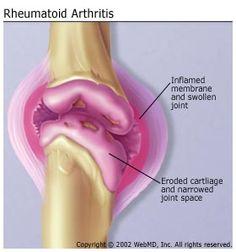
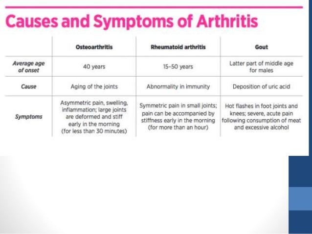
Rheumatoid arthritis (RA) is a chronic disease in which various joints in the body are inflamed, leading to swelling, pain, stiffness, and the possible loss of function.
Rheumatoid arthritis should not be confused with other forms of arthritis, such as osteoarthritis or arthritis associated with infections.
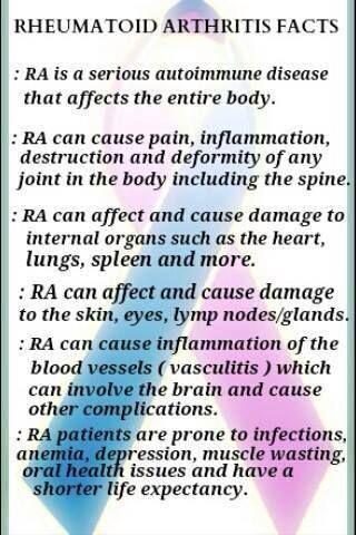
Rheumatoid arthritis is an autoimmune disease in which the body's immune system attacks joints and other tissues.
The pattern of joints affected is usually symmetrical, involves the hands and other joints, and is worse in the morning.
Rheumatoid arthritis is a systemic (body-wide) disease, involving other body organs, whereas osteoarthritis is limited to the joints. Both forms of arthritis can be crippling.
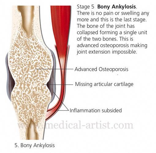
Synovitis
Synovium Thickens, Fluid Accumulates
Destructive Pannus Erodes Joint Cartilage, Destroys Joint Bone
Pannus Converted to Bony Tissue
Joint Deformity
Other Connective Tissue Affected
RHEUMATOID ARTHRITIS


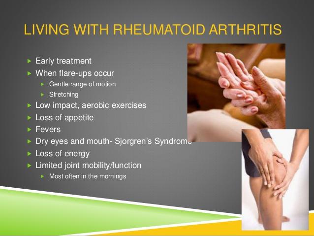


Early Symptoms
Bilateral, Symmetrical Joint Inflammation
Reddened, Warm, Swollen, Stiff, Painful
Stiffness After Resting
Activity Decreases Pain and Stiffness
Low Grade Fever, Weakness, Fatigue, Anorexia
Late Symptoms
Joint Deformity
Secondary Osteoporosis
SIGNS AND SYMPTOMS RA
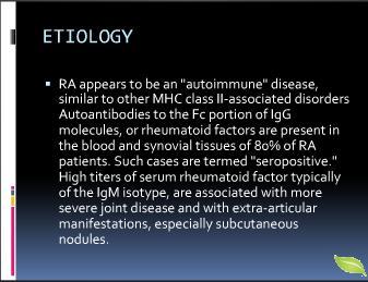
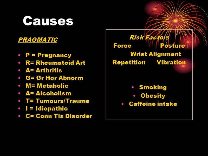
Unknown
Genetic Predisposition
Environmental
Abnormal Autoimmune Response – Antibodies (Rheumatoid Factor)
some environmental or biologic trigger, such as a viral infection or hormonal changes
ETIOLOGY RA
Health history: include onset of and evolution of symptoms, family history, past health history, and contributing factors
Functional assessment
Arthrocentesis
X-rays, bone scans, CTs, and MRIs
Tissue biopsy
Blood studies
Patient Assessment and Diagnostic Findings RA
Rheumatoid Factor (RF)
Red Blood Cell (RBC)
C4 Complement Decreased
Erythrocyte Sedimentation Rate (ESR)
Antinuclear Antibody (ANA)
C-reactive Protein (CRP)
DIAGNOSIS RA
Medication
Disease-modifying Antirheumatic Drugs (DMARDs)
Sulfasalazine (Azulfidine)
Leflunomide (Arava)
Etanercept (Enbrel)
Adalimumab (Humira)
Methotrexate -antineoplastic
Gold salts- antiflammatory effect
Prednisone
NSAIDs – Indocin, motrin, naprosyn etc.
Salicylates -ASA
COX -2 Inhibitor- Celebrex
Heat/Cold
Balanced Rest and Activity
Surgery – Total Joint Replacement
Therapeutic Interventions RA
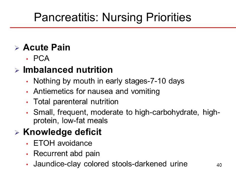
Acute Pain
Disturbed Body Image
Fatigue
Self-care Deficit
Impaired Physical Mobility
Deficient Knowledge
Ineffective coping
NURSING DIAGNOSIS RA
Disease Process
Medication Management
Rest and Exercise
Vocational Counselor
Community Resources
PATIENT EDUCATION HR
Major goals may include:
Relief of pain and discomfort
Relief of fatigue
Promotion of restorative sleep
Increased mobility
Maintenance of self-care
Improved body image
Effective coping
Absence of complications
Nursing Process: The Care of the Patient with a Rheumatic Disease—Planning
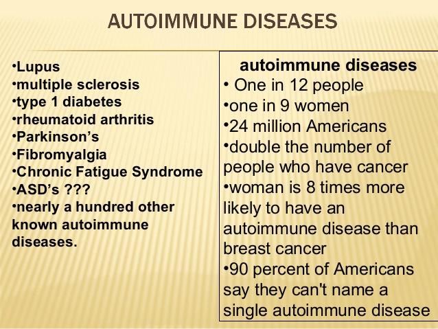
Autoimmune Disease
More than 80 autoimmune diseases
75% of autoimmune diseases affect women
One of the top 10 causes of death in women 65 and younger
Lupus: Wolf
Systemic Lupus Erythematosus (SLE)
1.4 million cases of lupus in the USA
90% are women
80% diagnosed between 15 and 45 years of age
50% have organ-threatening disease to kidneys, heart, lungs, liver, CNS, and hematopoetic system.
INCIDENCE OF LUPUS
Definition:
Chronic, inflammatory disturbance caused by an exaggerated production of “auto-antibodies.”
Immune complexes and fibrin deposit in blood vessels, collagen, and in organs
Can result in necrosis of glomerular capillaries
Systemic Lupus Erythematous
Abnormal Antibodies
Immune Complex Formation
Complement System Activation
Affects Connective Tissue
Pathophysiology
SYSTEMIC LUPUS ERYTHEMATOUS
Disturbance is probably caused by some combination factors of:
1. Genetic
2. Hormonal
3. Environmental
Immuno-Regulatory
SYSTEMIC LUPUS ERYTHEMATOUS
Genetics: Occurs in families by no gene isolated yet.
Hormonal: Estrogen metabolism is abnormal, flares with pregnancy and post-partum and post-menopausal HRT
Environmental: Sunlight, stress, burns, infection, antibiotics
SYSTEMIC LUPUS ERYTHEMATOUS
Drugs:
Hydralazine (Apresoline)
Procainamide (Pronestyl)
Isoniazid (INH)
Chloropromazine (Thorazine)
Anticonvulsants
NOTE: Usually if taken away, process goes away.
SYSTEMIC LUPUS ERYTHEMATOUS
Systemic Lupus Erythematous (SLE)
Multiple organs affected
1 test will not diagnose – Watch
signs/symptom
Discoid Lupus erythematous
Mild involving skin of face, neck, cheeks, etc..
Rarely progresses to Systemic Lupus
Drug induced lupus
Symptoms go away when triggering drug is
removed
TYPES OF LUPUS
NOTE: Not a specific syndrome – varies from patient to patient
- Can be insidious or acute
- Can be mild or rapidly progression to organ failure
- Characteristically has exacerbations (flares)
- Has times of remission
- Presents with vague, nonspecific symptoms
- Fever
- Fatigue
- Weight Loss
- Arthralgia
- Malaise Note: Symptoms usually precedes or precipitates an exacerbation Many patients are undiagnosed for years !!
HISTORY/PHYSICAL LUPUS
Note: 4 or more symptoms simultaneously or
serially
Most common sites are:
Cutaneous
Muscle Tissue
Lining of the lungs (pleurisy)
Pericarditis
Nervous system
Kidneys
CLINICAL MANIFESTATIONS LUPUS
80% have Skin Manifestations:
Skin: butterfly rash across the cheeks and bridge of the nose (40% of patients)
Diffuse maculopapular rash upon sun exposure (flat pimple like)
Discoid lesions (round like coins)
Both can itch, burn and cause “red palms”
Alopecia
Ulcers (Nasal or oral) - 35% of patients
Pruritus
ASSESSMENT LUPUS
Polyarthralgia
Morning stiffness (95%)
Migratory without
signs of overt
inflammation
Usually involves hands, feet,a
dn knees
Usually symmetrical
Deformity is rare
MUSCOSKELETAL LUPUS
Atherosclerosis
Pericarditis (25%)
Pleurisy
(50%)
Raynaud’s Syndrome (20%) – Patriotic fingers
Pleural Effusions
Cardiovascular disease – Cardiac Precautions
CARDIOPULMONARY LUPUS
Renal Disease (50% of patients)
50% with proteinuria, cellular casts
Microscopic
hematuria
Elevated serum creatinine
Ankle
edema,
Frequent UTI’s
10% develop renal failure
RENAL LUPUS
Lupus Nephritis – leading cause of death followed by cardiac involvement
Develop High Blood Pressure
Must monitor lab values (BUN/CR),
electrolytes, blood chemistry.
Lupus Nephritis
Seizures
(15%)
Headaches
Dizziness
Strokes
Organic Brain
Syndrome (memory loss, disorientation, depression)
Psychosis
CENTRAL NERVOUS SYSTEM LUPUS
Caused by antibodies attacking blood cells
Anemia (98%) with reticulocytosis (excessive RBC’s, but immature)
Leukopenia (80%) – Highly prone to infections (lymphs
<4000
Note: Carries a 30% mortality
Thrombocytopenia (36%) platelets <100,000
Hematologic LUPUS
Dysphagia
Anorexia
Diarrhea
Vomiting
Hemorrhage
(upper of lower GI Bleed)
Pain
GI SYMPTOM LUPUS
Biopsy
Erythrocyte Sedimentation Rate
Immunological Tests
Antinuclear Antibody Titers
Antibodies Against SR Proteins
DIAGNOSTIC TESTS LUPUS
Fatigue
Fever without infection
Weight Loss
ASSOCIATE SYMPTOMS LUPUS
ELEVATED:
Sed Rate: Erythrocyte Sedimentation Rate
(ESR)
(measures the rate of the fall of RNC suspended in plasma)
Note: Falls faster when lots of antibodies (gamma globulins) are attached.
Positive Serum Tests:
High + ANA (occurs when the body is fighting own DNA – in autoimmune diseases)
+LE Prep – 75% of those with lupus will have these cells-lots of false+ tests.
DIAGNOSTIC STUDIES LUPUS

- Infection or Injury
- Immunity
- Fatigue
- Stress
- Tissue Integrity & Clotting
- Thermoregulation
- Gas Exchange
- Inflammation