ACL
(Anterior Cruciate Ligament)
is attached to the posterior lateral condyle of the femur and to a notch in the midline of the tibia between the tibial condyles. prevents the femur from sliding posteriorly on the tibia, prevents hyperextension of the knee, and limits the medial rotation of the femur when the leg is in a fixed position with the foot planted. A common injury of the knee
Posterior Cruciate Ligament
is attached to the posterior midline surface of the tibia and passes anteriorly, attaching to the medial condyle of the femur. prevents the femur from sliding anteriorly on the tibia, especially when the knee is bent.
Bankart
is an avulsion (tear) injury of the anterior capsule and labrum of the glenoid rim; it is usually caused by subluxation or luxation of the joint. The tear affects that portion of the labrum called the inferior glenohumeral ligament;
Indications for a Total Shoulder Artroplasty
chronic pain from glenohumeral arthritis with significant loss of ROM and joint function the condition has not been resolved by conservative medical therapy. Complications include narrowing of the joint space, osteophyte formation, and cysts.
Acromion
is a bony process on the scapula (shoulder blade). Together with the coracoid process it extends laterally over the shoulder joint.
Acetabulum
is a concave surface of the pelvis. The head of the femur meets with the pelvis at the this, forming the hip joint.
Greater Trochanter
is located on the upper, lateral part of the upper shaft of the femur. It serves as the point of insertion for the gluteus medius and gluteus minimus.
Lesser Trochanter
The iliopsoas muscle inserts onto
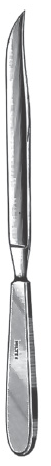
Liston amputating knife
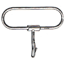
Gigli-Strully saw handle
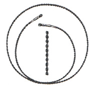
Gigli 12-in. saw
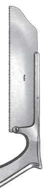
Satterlee bone saw
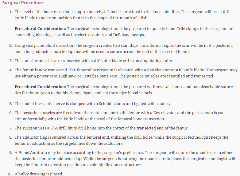
Above the Knee Amputation
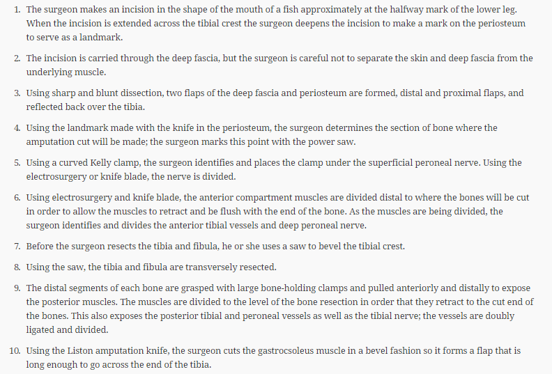
Below The Knee Amputation
Bunionectomy
medically referred to as hallux valgus, is a bony exostosis located on the medial side of the first metatarsal head of the great toe causing a lateral deviation of the toe.
Various types of surgical procedures are used to treat the condition, such as the Aken, Chevron, McKeever, Keller, and McBride techniques
Meniscus
A joint in which the two bony surfaces are joined by fibrocartilage The knee joint is cushioned to withstand activities such as walking, jumping, and running by a pair of are thick, crescent-shaped pads of cartilage that rest on the upper articular surface of the tibia. Injuries, particularly athletic injuries, are common and result in various types of tears in the cartilage.
Biters
Used in Arthroscopy Cases, Types include up, down, right, and left
Types of Arthroscopes
30 degree and 70 degree
How do you create a path for Screws and Nails?
Nontapping screws require that the drill hole be tapped with a tapping device prior to placement of the screw. Screws that are self-tapping can be identified by an angled notch near the tip of the screw.
Abduction
Moving a body part away from the midline of the body
Adduction
Moving a body part toward the midline of the body
Flexion
Bending a joint
Extension
Straightening a joint
Pronation
Pointing a body part downward (e.g., facing the palm of the hand downward)
Supination
Pointing a body part upward
Short arm cast
Applied from below the elbow to the metacarpal heads; wrist fracture
Long arm cast
Applied from axilla to metacarpal heads; fracture of forearm or elbow
Short leg cast
Applied from tibial tuberosity to metatarsal heads; ankle and foot fractures
Long leg cast
Applied from hip to metatarsal heads; fracture of femur, tibia, fibula, ankle
Cylinder cast
Applied from the groin to the ankle; required when complete knee immobilization is desired
Hip spica cast
Applied to trunk, complete leg of affected side, one-half of unaffected leg
Body jacket cast (Minerva jacket)
Applied to trunk of body to immobilize the spine
Who is responsible for implants
The FDA
What does an MRI diagnosis for Ortho?
This is a noninvasive imaging technique that relies on the body’s responses to a strong magnetic field.
CT Scan
is an X-ray of an organ or body detailing that structure at various depths. Multiple radiographs are taken at multiple angles, and the computer reconstructs these images to represent a cross-section or “slice” of the structure.
Polymethyl methacrylate (PMMA)
also referred to as bone cement, is routinely used during total joint arthroplasty. Bone cement stabilizes and keeps the implants in the correct anatomical position. The cement fills the cavity and spaces of the bone to form a bond between the implant and bone.
Tourniquet Times
should not be applied for more than 1 hour on an upper extremity or for more than 1½ hours on the thigh. After 1 hour of pressure, the surgeon should be notified, and again every 15 minutes thereafter.
fracture table
commonly used for surgery on a hip fracture and for femoral nailing. must be well understood by the personnel using it in order for it to be properly set up. have several moving parts and can cause injury to the patient and OR personnel if not correctly handled. The patient can be placed in the supine or lateral position
lateral position
is frequently used for operations on the hip and shoulder. The vacuum beanbag is often used to stabilize the patient in the lateral position, eliminating the need for roll towels and tape over the hips. The beanbag can be contoured to the body shape of the patient by adjusting the beanbag while the air is suctioned out.
reamers
The canal is reamed with subsequent larger sizes of reamers until the opening in the canal corresponds to the diameter of the shaft of the prosthesis.
Charnley
Self Retaining retractor used in total hip arthroplasty
Immovable Joints
synarthroses, the bones are in close contact with each other and separated by a thin layer of cartilage. An example is the suture lines of the cranial bones.
Slightly Movable Joint
amphiarthrosis -Lying between the bones of the joint is a disk of fibrous cartilage that connects the bones. Examples of this type of joint include cartilage that connects the vertebrae and the disk of cartilage called the symphysis pubis that connects the pubic bones. This type of joint allows some movement due to the limited flexibility of the cartilage.
Freely Movable Joints
A diarthrosis. All diarthroses are also referred to as synovial joints because these joints all contain a synovial membrane that secretes synovial fluid. Diarthroses are further classified according to the movements they allow
Ball-and-Socket Joints
This type of joint allows for the widest range of motion (ROM). It consists of a bone with a ball-shaped head that articulates with the cup-shaped socket in another bone. Movement in all planes is possible, including rotational. Examples include the shoulder and hip joints.
Condyloid Joints
allows for movement in only one plane with some lateral movement. The joint is composed of a condyle of one bone fitting into the fossa of another bone. An example is the temporomandibular joint in which the condyle of the mandible fits into the fossa of the temporal bone.
Gliding Joints
allow side-to-side and twisting movements. The articulating surfaces of the bones in the gliding joint are either flat or slightly curved. An example is the carpals of the wrist joint.
Hinge Joints
The elbow . This type of joint allows movement in only one plane, much like the motion permitted by the hinge on a door. is formed by the convex surface of one bone fitting into the concave surface of the adjacent bone.
Pivot Joints
allow only a rotational movement around a central axis. The joint formed at the proximal end of the radius
Saddle Joints
allow movement in a variety of planes. The articulating surfaces of the bones have both concave and convex regions. The surface of one bone fits into the equivalent surface of the other bone. An example is the joint formed by the trapezium of the wrist with the metacarpal of the thumb.
What is the thickest and strongest tendon in the body
The achillies Tendon
When to use Nails
- The type of nail to be used depends on the type and location of the fracture, whether ipsilateral trochanteric or condylar fractures are present, and whether bone fragments are present.
Names and Types of Nails
flexible nails such as the Rush and Ender; interlocking nails such as the Trigen; retrograde interlocking intramedullary nails; and standard nails such as the AO (surgical procedure description is for AO titanium femoral nail system).
Plates
- Surgical repairs that have been used in the past but have fallen out of favor include external fixation and plates and screws. The complications associated with plates and screws include infections and broken or bent screws and plates, which have contributed to femoral refracture.
Patella
Sesamoid (round) bones are found within tendons. Another example is the two sesamoid bones found on the head of the metatarsal in the foot forming what is referred to as the “ball” of the foot.
long bones
include bones of the arm (humerus), legs (femur), hands, and feet (phalanges)
Short bones
are the bones of the wrists (carpals) and ankles (tarsals). As evidenced by the wrist and ankle bones, short bones usually occur in clusters and aid in the movement of an extremity.
Esmarch
Used to exsanguinate the limb before use of the Tourniquet
Type of suture used for tendon to tendo
Fiberwire or Ethibond
Spinal Needle
Used for positioning
Distraction
s a term used to describe bone fragments that are separated so that bone contact does not occur.
Avascular necrosis
occurs when the capillary network or collateral circulation cannot be reestablished following a traumatic injury or when the vascular system is disrupted by other means.
compound fracture
compromises the integrity of the skin and allows for the possible entry of microorganisms, which may cause infection of the bone and injury to surrounding soft tissues
Delayed union
is a term used to describe an increase in the healing time of fractures. The reasons for are pathological (e.g., osteoporosis), mechanical (e.g., distraction of the fracture site or inadequate immobilization), or traumatic, referring to the type of injury sustained (e.g., comminuted fractures).
Nonunion
is when the fractured bone ends do not unite.
Malunion
occurs when the fracture heals in a position that does not resemble the original anatomical form of the bone and alters the mechanical function of the bone.
Compartmental syndrome
is an increase in pressure within a closed space that usually occurs in the forearm and tibia.
Stages of Healing
Inflammation
begins at the time of injury and lasts approximately 2 days.
Stages of Healing
Cellular Proliferation
begins approximately on the second day following the traumatic event. Macrophages debride the area and allow for the formation of a fibrin mesh that seals the approximated edges of the fracture site. The fibrin mesh serves as the foundation for capillary and fibroblastic ingrowth. A soft tissue or periosteal callus is formed on the outer surface or cortex of the fractured bone by the collagen-producing fibroblasts and osteoblasts.
Stages of Healing
callus formation
stage lasts 3–4 weeks. The soft tissue growth continues and the bone fragments grow toward one another, bridging the gap. Osteoblasts form a matrix of collagen that invades the periosteal callus, bridging the fracture site and uniting the two ends of the bone. Fibrous tissue, cartilage, and immature bone stabilize the fracture site.
Stages of Healing
Ossification
begins 2 or 3 weeks following the injury and can last 3–4 months. The matrix of osteoblasts, now called the osteoid, calcifies, firmly uniting the bone. The bone is now able to accept mineral deposits.
Stages of Healing
Remodeling
is the maintenance state of normal bone. Following a fracture, any devitalized tissue is removed and the new bone is organized to provide maximum support and function.
Colles Fracture
- a fracture of the lower end of the radius in the wrist with a characteristic backward displacement of the hand.
Active Bone Growth
epiphyseal plate
epiphyses
The proximal portion of a long bone
diaphysis
is composed of compact bone that surrounds the medullary cavity.
Cortical Bone
Type of bone tissue that is hard and dense, and that surrounds the marrow cavity; also referred to as compact bone
cancellous bone
A type of bone tissue found at the ends of bone and lining the medullary marrow cavity; composed of columns of trabeculae with large spaces in between; also referred to as spongy bone due to its appearance
Para Thyroid Hormone
Stimulates Bone Growth
Unicompartmental implants
are used to replace either the medial or lateral side of the corresponding articular surface of the femur and tibia.
Bicompartmental implants
replace the medial and lateral surfaces of the femur and tibia.
Tricompartmental implants
replace the medial and lateral surfaces of the femur and tibia, including the patella.
Unconstrained implant:
Requires minimal resurfacing of the tibia and femur, and good collateral and posterior cruciate ligaments;
semiconstrained implant:
Used when there is a difficulty with ligament balance
fully constrained implant
Implant is jointed together by hinges and only allows motion in a sagittal plane.
ACL Grafts
Autografts
patellar tendon, hamstring, quadriceps tendon,
allograft
Taken from another person
Most common Cause for a distal Radius Fracture
Fall onto an outstretched Arm
Ringers Solution
Used in Arthroscopy cases when using the ESU
Softening bone in children
Osteomalacia also called Rickets
Bucket Handle Tear
consists of an incomplete longitudinal tear with displacement of the inner portion of the meniscus. When a tear of this type is encountered, an arthroscopic partial meniscectomy or repair can be completed.
What instrument is commonly called Turkey Foot or Eagle Talon
Lowman Bone Holding Clamp