Three parts of the Axial skeleton

1.The Skull
2.The Vertebral Column
3.The Thoracic Cage
Cranial Bones
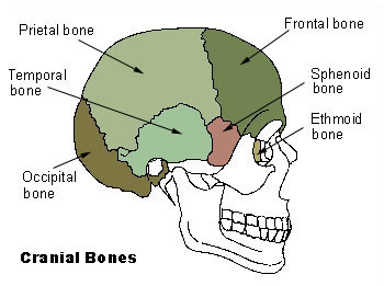
-encloses and protects the fragile brain tissue
Facial Bones
-supports the eyes and position them anteriorly
-provides attachment sites for facial muscles
All but one of the bones of the skull are joined by interlocking joints called ____________.

Sutures
The mandible, or lower jawbone is attached to the rest of the skull by a _______________.
freely movable joint
Cranial Vault/Calvaria

-the superior, lateral, and posterior walls of the skull
Cranial Base
-forms the skull bottom
What are the three distinct depressions of the cranial base?

1.Anterior cranial fossae
2.Middle cranial fossae
3.Posterior cranial fossae
Frontal Bone

-anterior portion of cranium
-forms the forehead
Supraorbital foramen (notch)
-opening above each orbit allowing blood vessels and nerves to pass
Glabella
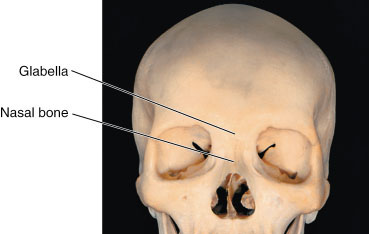
-smooth area between the eyes
Parietal Bone (2)
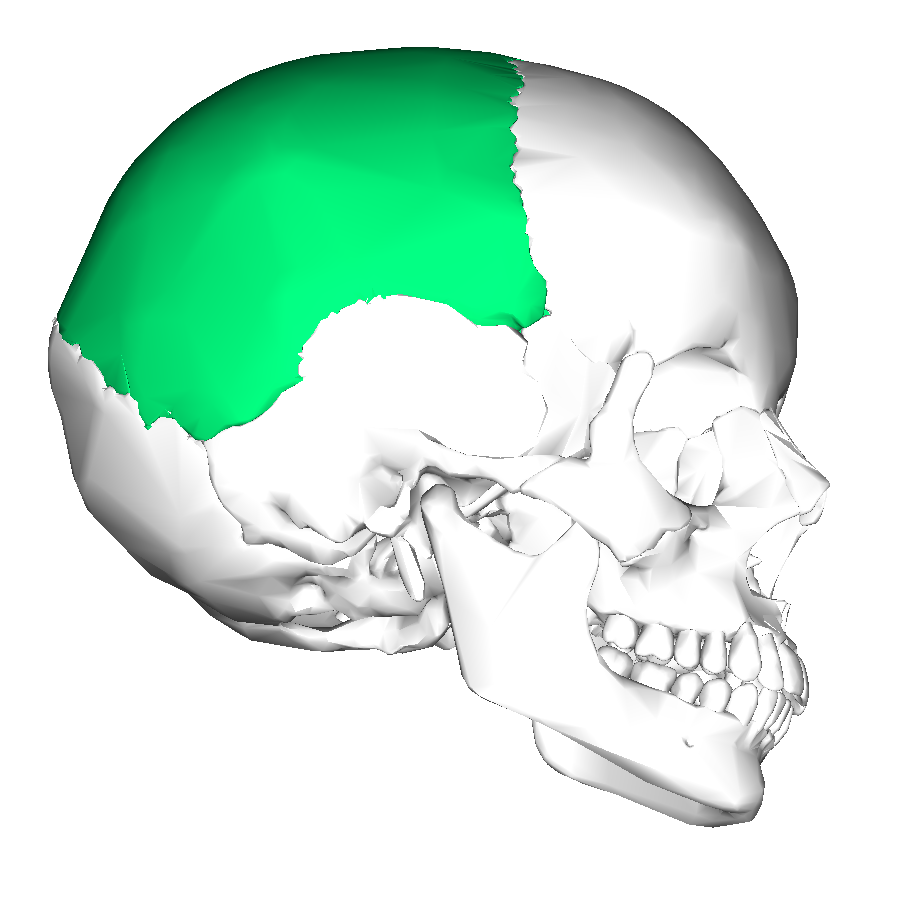
-posterolateral to the frontal bone, forming sides of the cranium
Sagittal Suture
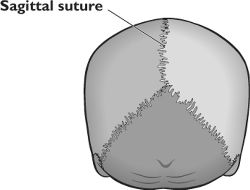
-midline articulation point of the two parietal bones
Coronal Suture
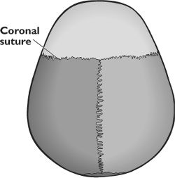
-point of articulation of parietals with frontal bone
Temporal Bone

-inferior to parietal bone on lateral skull
What are the three major parts of the temporal bone?

1. Squamous part
-border the parietals
2.Tympanic part
-surrounds the external ear opening
3.Petrous
-forms the lateral portion of the skull base and contains the mastoid process
Squamous Suture

-point of articulation of the temporal bone with the parietal bone
Zygomatic Process

- bridgelike projection joining the zygomatic bone (cheekbone) anteriorly
Mandibular Fossa

-rounded depression on the inferior surface of the zygomatic process
-forms the socket for the condylar process of the mandible
-where the mandible joins the lower cranium
External Acoustic Meatus

-canal leading to eardrum and middle ear
Styloid Process

-needlelike projection inferior to external acoustic meatus
-attachment point for muscles and ligaments of the neck
Jugular Foramen

-opening medial to the styloid process through which the internal jugular vein and cranial nerves IX, X, and XI pass
Carotid Canal

-opening medial to the styloid through which the internal carotid artery passes into the cranial cavity
Internal Acoustic Meatus

-opening on posterior aspect (or petrous part) of temporal bone allowing passage of cranial nerves
Foramen Lacerum

-jagged opening between the petrous temporal bone and the sphenoid providing passage for a number of small nerves and for the internal carotid artery to enter the middle cranial fossa
Stylomastoid Foramen

-tiny opening between the mastoid and styloid processes through which cranial nerve VII leaves the cranium
Mastoid Process

-Rough projection inferior and posterior to external acoustic meatus
-attachment site for muscles
Occipital Bone
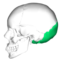
-most posterior bone of the cranium
Lambdoid Suture

-site of articulation of occipital bone and parietal bones
Foramen Magnum

-large opening in base of occipital, which allows the spinal cord to join with the brain
Occipital Condyles

-rounded projections lateral to the foramen magnum that articulate with the first cervical vertebra (atlas)
Hypoglossal Canal

-opening medial and superior to the occipital condyle through which the hypoglossal nerve pass
External Occipital Crest and Protuberance
.jpg)
-midline prominences posterior to the foramen magnum
Sphenoid Bone
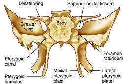
Bat-shaped bone forming the anterior plateau of the middle cranial fossa across the width of the skull.
-articulates with all the other cranial bones
Greater Wings

-portions of the sphenoid seen exteriorly anterior to the temporal and forming a part of the eye orbits
Pterygoid Processes
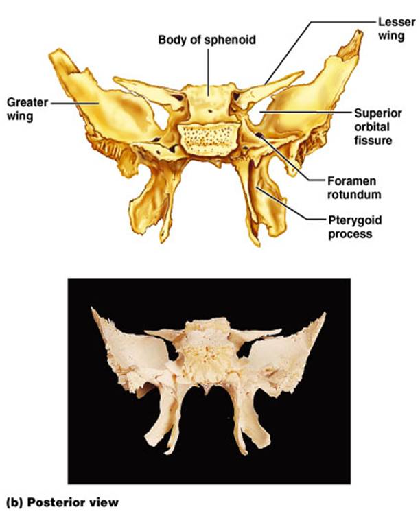
-inferiorly directed trough-shaped projections from the junction of the body and the greater wings
Superior Orbital Fissures

-jagged openings in orbits provding passages for cranial nerves III, IV, V, and VI to enter the orbit where they serve the eye
Sella Turcica
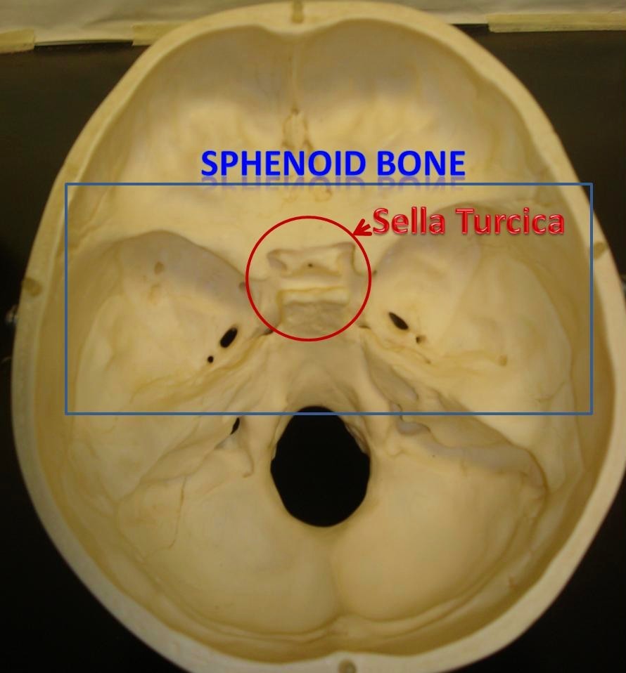
- saddle shaped region in the sphenoid midline
Lesser Wings
-bat shaped portions of the sphenoid bone anterior to the sella turcica
Optic Canals

- openings in the bases of the lesser wings through which the optic nerve (cranial nerve II) enter the orbits to serve the eyes
Foramen Rotundum

- opening lateral to the sella turcica providing passage for a branch of the fifth cranial nerve
Foramen Ovale

opening posterior to the sella turcica that allows passage of the fifth cranial nerve.
Foramen Spinosum

opening lateral to the foramen ovale which the meningeal artery passes
Ethmoid Bone
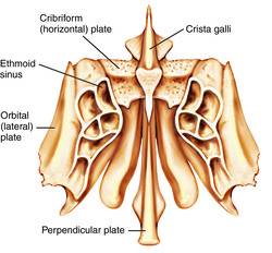
irregularly shaped bone anterior to the sphenoid
Crista Galli

vertical projection providing a point of attachment for the dura meter, helping to secure the brain within the skull
Cribriform Plates

bony plates lateral to the crista galli through which olfactory fibers pass to the brain
Perpendicular Plate

inferior projection of the ethmoid bone that forms the superior part of the nasal septum
Lateral Masses
irregularly shaped and thin-walled bony regions flanking the perpendicular plate laterally
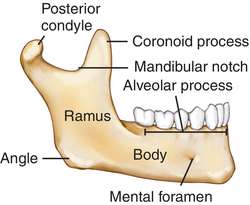
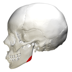
Mandible
the lower jawbone, which articulates with the temporals in the only freely movable joints of the skull
Mandibular body
horizontal portion; forms the chin
Mandibular Ramus
vertical extension of the body on either side
Condylar Process

articulation point of the mandible with the mandibular fossa of the temporal bone
Coronoid Process

jutting anterior portion off the ramus
-site of muscle attachment
Mandibular Angle
posterior point at which ramus meets the body
Mental Foramen

-prominent opening on the body that transmits the mental blood vessels and nerve to the lower jaw
Mandibular Foramen

-permis passage of the nerve involved with tooth sensation
Alveolar Process

superior margin of the mandible, contains sockets in which the teeth lie
Maxillae
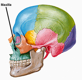
-two bones fused in a median suture
-forms the upper jawbone and parts of the orbits
Palatine Processes

-from the anterior hard palate; meets medially in the intermaxillary suture
Infraorbital Foramen

-opening under the prbit carrying the infraorbital nerves and blood vessels to the nasal region
Lacrimal Bone

-fingernail sized bones forming part of the medial orbit walls between the maxilla and the ethmoid bone
Palatine Bone
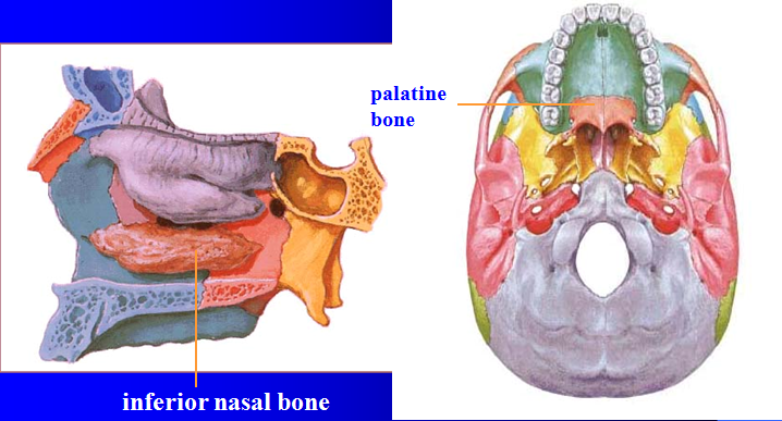
-paired bones posterior to the palatine processes