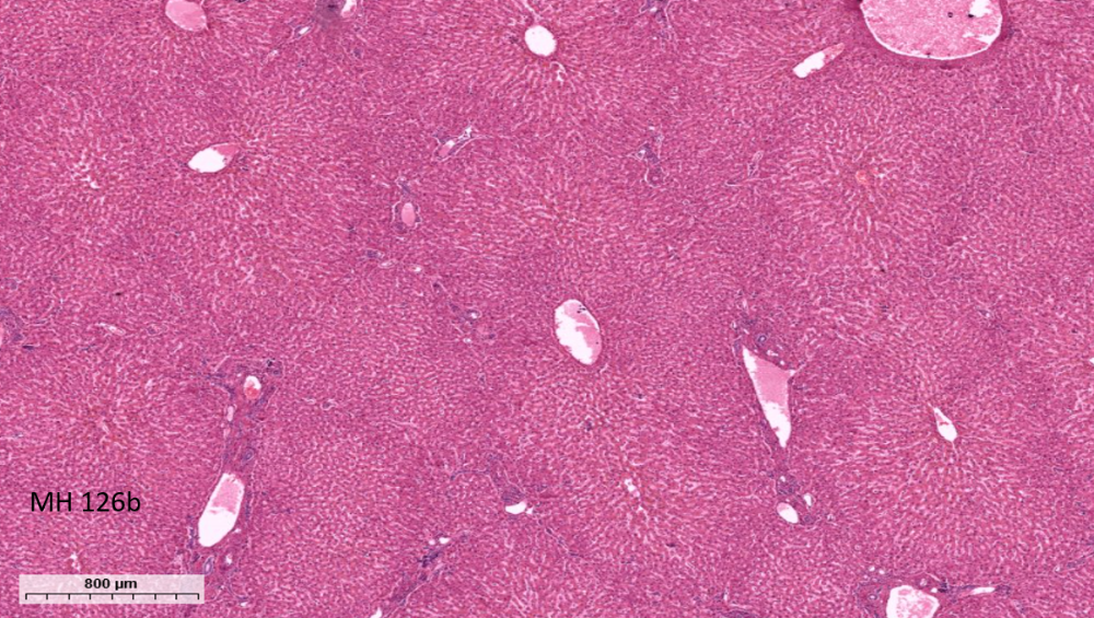
What organ is this?
Liver
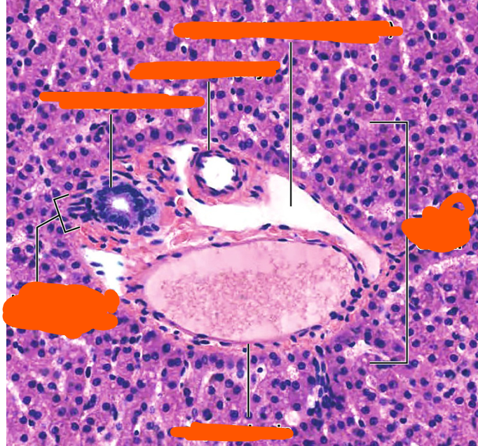
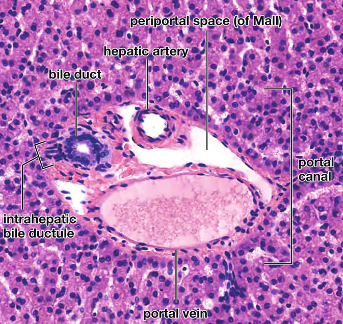
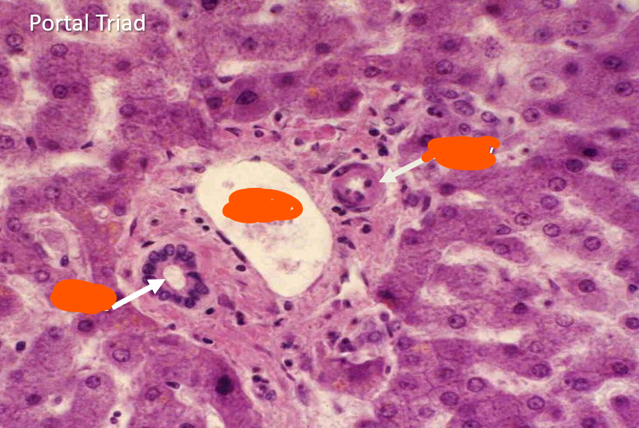
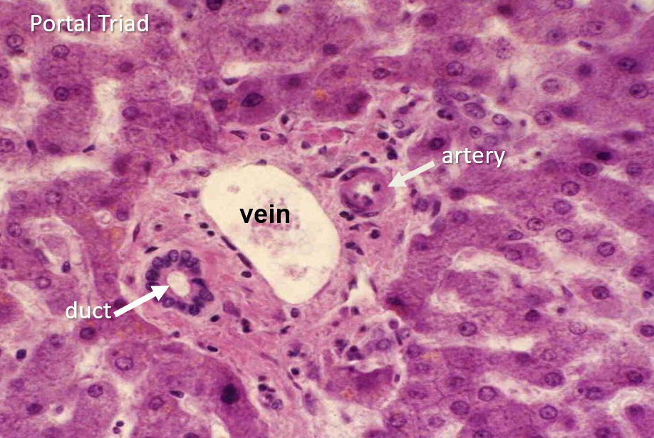
Which statement best describes the liver relative to other glands in the body?
A. Largest gland in the body
B. Largest endocrine gland
C. Smallest digestive gland
D. Largest lymphoid gland
A. Largest gland in the body
A major metabolic role of the liver is to process absorbed nutrients and store excess carbohydrate in which form?
A. Triglyceride
B. Glycogen
C. Cholesterol
D. Lactate
B. Glycogen
Which liver function helps protect the body from potentially harmful medications and environmental chemicals?
A. Bile storage
B. Lymph formation
C. Detoxification
D. Hematopoiesis
C. Detoxification
Which group consists entirely of blood proteins synthesized by the liver according to this source?
A. Albumin, fibrinogen, lipoproteins
B. Hemoglobin, insulin, albumin
C. Immunoglobulins, fibrinogen, renin
D. Myoglobin, albumin, glucagon
A. Albumin, fibrinogen, lipoproteins
Why is hepatic secretion of albumin, fibrinogen, and lipoproteins considered an endocrine function?
A. They enter bile canaliculi
B. They are secreted into blood
C. They remain in hepatocytes
D. They enter lymphatics
B. They are secreted into blood
Which product of the liver is released through an exocrine pathway rather than directly into the bloodstream?
A. Albumin
B. Fibrinogen
C. Bile
D. Lipoproteins
C. Bile
Which term refers to the connective tissue framework that supports the functional cells of the liver?
A. Parenchyma
B. Sinusoid
C. Portal triad
D. Stroma
D. Stroma
The connective tissue capsule of the liver is composed predominantly of which collagen type?
A. Type I collagen
B. Type II collagen
C. Type III collagen
D. Type IV collagen
A. Type I collagen
The reticular fiber framework within the liver stroma is composed predominantly of which collagen type?
A. Type I
B. Type III
C. Type II
D. Type IV
B. Type III
What is the principal role of the liver stroma?
A. Produce bile
B. Store glycogen
C. Support hepatocytes
D. Form portal blood
C. Support hepatocytes
Which term refers to the functional tissue of the liver rather than its supporting connective tissue?
A. Stroma
B. Capsule
C. Reticulum
D. Parenchyma
D. Parenchyma
What cell type makes up the majority of the liver parenchyma?
A. Hepatocytes
B. Kupffer cells
C. Cholangiocytes
D. Endothelial cells
A. Hepatocytes
Embryologically, hepatocytes are derived from which tissue?
A. Neural crest ectoderm
B. Foregut endoderm
C. Intermediate mesoderm
D. Surface ectoderm
B. Foregut endoderm
How are hepatocytes organized structurally within the liver parenchyma?
A. Solid spherical nests
B. Stratified epithelial sheets
C. Interconnected plates 1–2 cells thick
D. Isolated single cells
C. Interconnected plates 1–2 cells thick
Which vessel provides most of the blood volume reaching the liver?
A. Portal vein
B. Hepatic artery
C. Hepatic vein
D. Inferior vena cava
A. Portal vein
What type of blood is delivered to the liver by the portal vein?
A. Oxygen-rich, nutrient-poor blood
B. Oxygen-poor, nutrient-rich blood
C. Oxygen-rich, nutrient-rich blood
D. Oxygen-poor, nutrient-poor blood
B. Oxygen-poor, nutrient-rich blood
What is the primary contribution of the hepatic artery to hepatic circulation?
A. Nutrient-rich venous blood
B. Lymphatic fluid
C. Bile
D. Oxygenated blood
D. Oxygenated blood
Where are portal triads located within a classic hepatic lobule?
A. Around the central vein
B. At the corners of the lobule
C. Within the Space of Disse
D. Along bile canaliculi
B. At the corners of the lobule
Which three structures form the classic portal triad?
A. Hepatic vein, artery, lymphatic
B. Portal vein, central vein, bile duct
C. Central vein, sinusoid, lymphatic
D. Portal venule, hepatic arteriole, bile duct
D. Portal venule, hepatic arteriole, bile duct
Branches of the portal vein within a portal triad give rise to portal venules, which then lead to:
A. Inlet venules
B. Central veins
C. Bile ductules
D. Hepatic veins
A. Inlet venules
Where do the inlet venules derived from portal venules ultimately empty?
A. Bile canaliculi
B. Sinusoids
C. Central ducts
D. Lymphatics
B. Sinusoids
Branches of the hepatic artery within a portal triad give rise to hepatic arterioles, which then form:
A. Portal venules
B. Central venules
C. Inlet arterioles
D. Bile ductules
C. Inlet arterioles
The blood within hepatic sinusoids is best described as a mixture of approximately:
A. 75% arterial, 25% venous
B. 50% arterial, 50% venous
C. 100% venous
D. 25% arterial, 75% venous
D. 25% arterial, 75% venous
Which sequence correctly traces bile drainage toward the bile duct component of the portal triad?
A. Bile canaliculi → canals of Hering → bile ducts
B. Bile ducts → canaliculi → canals of Hering
C. Sinusoids → canaliculi → bile ducts
D. Canals of Hering → sinusoids → bile ducts
A. Bile canaliculi → canals of Hering → bile ducts
Which statement about lymphatics in the portal region is correct?
A. They replace the bile duct
B. They are present but not part of the triad
C. They are one of three triad components
D. They drain directly into sinusoids
B. They are present but not part of the triad
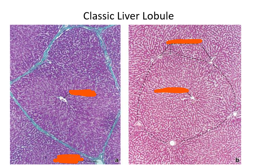
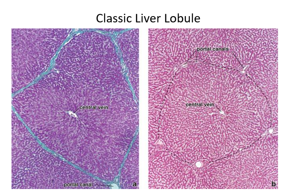
Within a classic hepatic lobule, mixed blood from the portal vein and hepatic artery flows through sinusoids toward which structure?
A. Central vein
B. Bile duct
C. Portal triad
D. Canal of Hering
A. Central vein
How does the direction of bile flow compare with the direction of blood flow within a classic hepatic lobule?
A. Both flow toward central vein
B. Bile flows opposite blood
C. Both flow toward portal triads
D. Bile enters sinusoids
B. Bile flows opposite blood
Bile produced by hepatocytes travels toward the portal region primarily through which microscopic channels?
A. Sinusoids
B. Central venules
C. Bile canaliculi
D. Space of Disse
C. Bile canaliculi
Which liver structural model is based primarily on gross microscopic morphology?
A. Portal lobule
B. Liver acinus
C. Hepatic sinusoid
D. Classic lobule
D. Classic lobule
What shape is classically used to describe the classic hepatic lobule?
A. Polygonal
B. Diamond-shaped
C. Circular
D. Triangular
A. Polygonal
The classic hepatic lobule is organized conceptually around the direction of blood flow toward the:
A. Portal triad
B. Bile duct
C. Central vein
D. Hepatic artery
C. Central vein
Which combination best describes the major components of a classic hepatic lobule?
A. Central vein, bile canaliculi, capsule
B. Portal vein, hepatic vein, gallbladder
C. Bile ducts, lymphatics, connective tissue
D. Central vein, hepatocyte plates, sinusoids, portal triads
D. Central vein, hepatocyte plates, sinusoids, portal triads
Within a classic hepatic lobule, hepatocytes are arranged in plates that radiate outward from the:
A. Central vein
B. Portal triad
C. Bile duct
D. Hepatic artery
A. Central vein
Which liver structural model is based on the exocrine function of the liver but is considered of little clinical usefulness in these notes?
A. Classic lobule
B. Portal lobule
C. Liver acinus
D. Hepatic sinusoid
B. Portal lobule
Which structural model is considered the functional structural unit of the liver because it best correlates perfusion, metabolism, and pathology?
A. Portal lobule
B. Classic lobule
C. Liver acinus
D. Bile canaliculus
C. Liver acinus
What is the characteristic geometric shape of the liver acinus?
A. Hexagonal
B. Circular
C. Triangular
D. Diamond-shaped
D. Diamond-shaped
Which structures define the four corners of a liver acinus?
A. Two central veins and two portal triads
B. Four central veins
C. Four portal triads
D. Two bile ducts and two sinusoids
A. Two central veins and two portal triads
What structures are located near the center of the liver acinus?
A. Central veins and bile ducts
B. Terminal portal venules, hepatic arterioles, bile ducts
C. Hepatic veins and lymphatics
D. Gallbladder ducts and sinusoids
B. Terminal portal venules, hepatic arterioles, bile ducts
Which hepatic acinar zone lies closest to the distributing vessels?
A. Zone 3
B. Zone 2
C. Zone 1
D. Zone 1
Where is Zone 1 located relative to the classic hepatic lobule?
A. Near its periphery
B. Around the central vein
C. Inside the central vein
D. Outside the liver
A. Near its periphery
Because Zone 1 is closest to incoming blood, hepatocytes there are exposed to relatively high levels of:
A. Carbon dioxide
B. Oxygen, nutrients, and toxins
C. Bile pigments
D. Lymphatic fluid
B. Oxygen, nutrients, and toxins
How does the metabolic activity of Zone 1 hepatocytes compare with more distal acinar cells?
A. Completely absent
B. Lower
C. Higher
D. Identical
C. Higher
Following bile duct occlusion, which acinar zone is expected to show the earliest cellular changes?
A. Zone 1
B. Zone 2
C. Zone 3
A. Zone 1
During impaired hepatic circulation, which acinar zone is the last to undergo cell death?
A. Zone 1
B. Zone 2
C. Zone 3
A. Zone 1
Following hepatic injury, which acinar zone is described as the first to regenerate?
A. Zone 1
B. Zone 2
C. Zone 3
A. Zone 1
Which hepatic acinar zone lies farthest from the distributing vessels and closest to the terminal hepatic vein?
A. Zone 1
B. Zone 2
C. Zone 3
C. Zone 3
Which acinar zone is the first to undergo ischemic injury because it receives the least favorable blood supply?
A. Zone 1
B. Zone 2
C. Zone 3
C. Zone 3
Ischemic necrosis occurring in Zone 3 is commonly referred to as:
A. Periportal necrosis
B. Centrilobular necrosis
C. Portal fibrosis
D. Lobular hyperplasia
B. Centrilobular necrosis
Which hepatic acinar zone is the first to show fatty accumulation according to these notes?
A. Zone 1
B. Zone 2
C. Zone 3
D. Portal zone
C. Zone 3
Zone 3 hepatocytes are relatively late to respond to direct toxins but may be especially vulnerable to:
A. Portal hypertension
B. Bile pigments
C. Nutrient excess
D. Toxic metabolic products
D. Toxic metabolic products
If Zone 3 hepatocytes are damaged by acetaminophen metabolites, which zone may subsequently become injured as its cells take over more metabolic function?
A. Zone 1
B. Portal triad
C. Zone 2
D. Central vein
C. Zone 2
When liver pathology is described as “centrilobular,” which anatomic region is being referenced?
A. Around the portal triad
B. At the lobular periphery
C. Within Zone 1
D. Near the central vein in Zone 3
D. Near the central vein in Zone 3
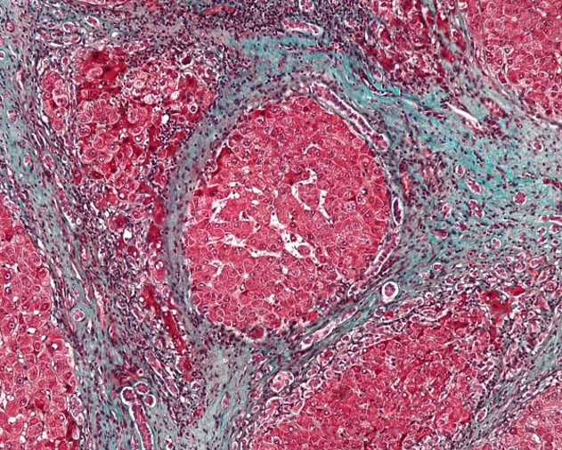
What does this pt have?
Cirrhosis
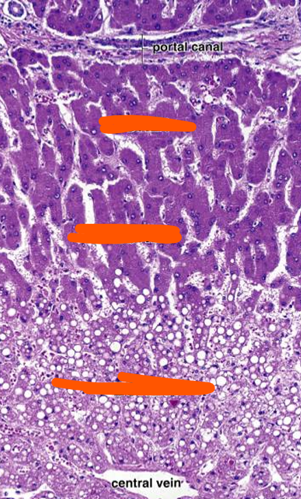
zone 1
zone 2
zone 3
Alcohol-related liver disease classically progresses through which three major phases?
A. Steatosis, hepatitis, cirrhosis
B. Hepatitis, fibrosis, cholangitis
C. Steatosis, cholangitis, carcinoma
D. Necrosis, hepatitis, steatosis
A. Steatosis, hepatitis, cirrhosis
According to this guide, chronic alcohol use initially causes accumulation of what substance within hepatocytes?
A. Glycogen
B. Fat
C. Iron
D. Bilirubin
B. Fat
In severe alcoholic fatty liver, the liver may enlarge to approximately what proportion of its normal weight?
A. Twice normal
B. Four times normal
C. Three times normal
D. Five times normal
C. Three times normal
Which hepatocytes are affected first during alcoholic fatty liver disease?
A. Zone 1 hepatocytes
B. Zone 2 hepatocytes
C. Periportal hepatocytes
D. Zone 3 hepatocytes
D. Zone 3 hepatocytes
A patient with alcoholic fatty liver stops drinking completely. According to this guide, what is the expected response?
A. Immediate cirrhosis
B. Fatty change is reversible
C. Progressive portal hypertension
D. Permanent hepatocyte loss
B. Fatty change is reversible
Compared with later alcohol-related liver disease, uncomplicated alcoholic fatty liver usually produces:
A. Severe hepatic failure
B. Marked portal hypertension
C. Few functional consequences
D. Extensive fibrous septa
C. Few functional consequences
Which alcohol-related liver disease is described as more severe than simple fatty liver?
A. Cardiac cirrhosis
B. Alcoholic steatosis
C. Portal hypertension
D. Alcoholic hepatitis
D. Alcoholic hepatitis
Alcoholic hepatitis is clinically important partly because it carries:
A. Significant mortality
B. No mortality risk
C. Only cosmetic effects
D. Minimal hepatic injury
A. Significant mortality
Which description best characterizes alcoholic hepatitis histologically?
A. Pure fatty accumulation
B. Inflammatory necrotizing disease
C. Isolated bile duct obstruction
D. Noninflammatory fibrosis
B. Inflammatory necrotizing disease
Which inflammatory cell predominates in the inflammatory response of alcoholic hepatitis according to this guide?
A. Eosinophil
B. Lymphocyte
C. Neutrophil
D. Plasma cell
C. Neutrophil
In alcoholic hepatitis, hepatocyte necrosis is especially prominent in which region?
A. Zone 1
B. Zone 2
C. Portal triads
D. Zone 3
D. Zone 3
Which pathologic process defines hepatic cirrhosis in this guide?
A. Scarring of the liver
B. Isolated steatosis
C. Acute neutrophilic inflammation
D. Bile duct dilation
A. Scarring of the liver
A patient with advanced cirrhosis may develop which major vascular complication?
A. Systemic hypotension
B. Portal hypertension
C. Pulmonary stenosis
D. Coronary vasospasm
B. Portal hypertension
Besides portal hypertension, cirrhosis can ultimately result in:
A. Renal artery stenosis
B. Splenic infarction
C. Liver failure
D. Pancreatitis
C. Liver failure
What is the characteristic histologic arrangement of fibrosis in cirrhosis?
A. Fibers around central veins
B. Collagen inside hepatocytes
C. Isolated portal inflammation
D. Fibrous septa around hepatocellular nodules
D. Fibrous septa around hepatocellular nodules
Why does extensive fibrosis impair hepatic function in cirrhosis?
A. Normal architecture becomes disrupted
B. Hepatocytes produce excess oxygen
C. Portal veins disappear completely
D. Sinusoids become bile ducts
A. Normal architecture becomes disrupted
A liver biopsy stained with trichrome shows blue bands surrounding islands of liver tissue. What does the blue staining represent?
A. Hepatocyte glycogen
B. Connective tissue scarring
C. Accumulated intracellular fat
D. Neutrophilic infiltration
B. Connective tissue scarring
On trichrome staining of cirrhotic liver, islands surrounded by blue connective tissue represent remaining:
A. Bile duct epithelium
B. Portal venules
C. Liver parenchyma
D. Lymphatic tissue
C. Liver parenchyma
Which cardiovascular disorder can chronically damage the liver and eventually produce cardiac cirrhosis?
A. Hypertension
B. Aortic stenosis
C. Endocarditis
D. Heart failure
D. Heart failure
Why are Zone 3 hepatocytes especially vulnerable when cardiac output and hepatic perfusion decrease?
A. They normally receive least oxygen
B. They receive highest oxygen levels
C. They lack mitochondria
D. They surround hepatic arteries
A. They normally receive least oxygen
In addition to receiving relatively little oxygen, Zone 3 hepatocytes normally receive relatively low levels of:
A. Bile
B. Nutrients
C. Collagen
D. Lymph
B. Nutrients
Which type of injury especially threatens Zone 3 hepatocytes during heart failure?
A. Autoimmune injury
B. Hyperglycemic injury
C. Ischemia and hypoxia
D. Bile salt toxicity
C. Ischemia and hypoxia
According to this guide, which finding is expected in Zones 1 and 2 during the described cardiac cirrhosis changes?
A. Severe necrosis
B. Fatty accumulation
C. Fibrous nodules
D. No noticeable changes
D. No noticeable changes
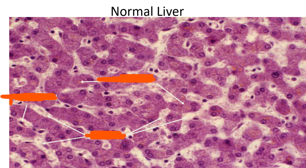
Kupffer cell, lipofuscin, sinusoids
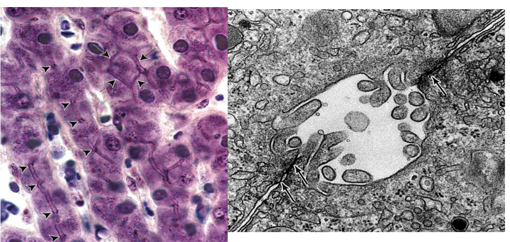
_____ _____
Bile Canaliculi
Where are hepatic sinusoids located within the liver parenchyma?
A. Between hepatic plates
B. Inside portal venules
C. Within bile ducts
D. Beneath the capsule
A. Between hepatic plates
Hepatic sinusoids receive blood originating from which structures?
A. Central veins
B. Portal triads
C. Bile canaliculi
D. Hepatic veins
B. Portal triads
Hepatic sinusoids are specialized capillaries adapted to permit what type of blood flow?
A. Very slow blood flow
B. Rapid pulsatile flow
C. Unidirectional lymph flow
D. High-pressure arterial flow
A. Very slow blood flow
Which structural feature is characteristic of hepatic sinusoids compared with ordinary capillaries?
A. Narrow rigid lumens
B. Dilated lumens
C. Continuous thick walls
D. Stratified epithelium
B. Dilated lumens
Which combination best describes the endothelial lining and basal lamina of hepatic sinusoids?
A. Continuous endothelium, continuous basal lamina
B. Fenestrated endothelium, thick basal lamina
C. Discontinuous endothelium, discontinuous basal lamina
D. Stratified endothelium, absent lumen
C. Discontinuous endothelium, discontinuous basal lamina
The fenestrae of hepatic sinusoidal endothelial cells characteristically:
A. Contain thick diaphragms
B. Open into bile ducts
C. Are lined by hepatocytes
D. Lack diaphragms
D. Lack diaphragms
Which resident liver cells are mononuclear phagocytes associated with hepatic sinusoids?
A. Kupffer cells
B. Ito cells
C. Cholangiocytes
D. Hepatocytes
A. Kupffer cells
Where are Kupffer cells located within the hepatic sinusoid?
A. Only within bile canaliculi
B. In sinusoidal walls and luminal surfaces
C. Exclusively in portal triads
D. Inside the Space of Disse only
B. In sinusoidal walls and luminal surfaces
One important function of Kupffer cells is phagocytosis of:
A. Bile salts
B. Hepatocyte nuclei
C. Aged red blood cells
D. Cholesterol particles
C. Aged red blood cells
Which hepatic cell normally serves as an important storage site for vitamin A?
A. Kupffer cell
B. Cholangiocyte
C. Sinusoidal endothelial cell
D. Ito cell
D. Ito cell
Where are Ito cells normally located?
A. Space of Disse
B. Portal vein lumen
C. Bile canaliculus
D. Central vein
A. Space of Disse
In cirrhosis, Ito cells contribute to hepatic fibrosis by transforming into:
A. Cholangiocytes
B. Myofibroblasts
C. Kupffer cells
D. Endothelial cells
B. Myofibroblasts
Why are activated Ito cells important in the pathogenesis of cirrhosis?
A. They destroy erythrocytes
B. They secrete bile
C. They participate in fibrosis
D. They form portal blood
C. They participate in fibrosis
The Space of Disse is located between which two structures?
A. Two adjacent hepatocytes
B. Portal venule and arteriole
C. Bile duct and lymphatic
D. Sinusoidal endothelium and hepatocytes
D. Sinusoidal endothelium and hepatocytes
What important hepatocyte surface specialization projects into the Space of Disse?
A. Microvilli
B. Cilia
C. Flagella
D. Stereocilia
A. Microvilli
The discontinuous sinusoidal barrier allows blood fluid and macromolecules to contact hepatocyte microvilli within the:
A. Bile canaliculus
B. Space of Disse
C. Portal venule
D. Central vein
B. Space of Disse
What major physiologic process occurs across the Space of Disse?
A. Formation of bile ducts
B. Erythrocyte production
C. Exchange between blood and hepatocytes
D. Direct mixing of bile and blood
C. Exchange between blood and hepatocytes
Which component of bile helps emulsify dietary fats?
A. Bilirubin
B. Cholesterol
C. Electrolytes
D. Bile acids
D. Bile acids
Which group contains substances normally present in bile according to this source?
A. Bile acids, phospholipids, cholesterol, bilirubin
B. Hemoglobin, insulin, albumin, glycogen
C. Fibrinogen, glucose, IgG, calcium
D. Urea, creatinine, albumin, insulin
A. Bile acids, phospholipids, cholesterol, bilirubin
In addition to bile salts, which substances are largely reabsorbed from the intestine according to this source?
A. Bilirubin and hemoglobin
B. Albumin and fibrinogen
C. Cholesterol, lecithin, and electrolytes
D. Glycogen and triglycerides
C. Cholesterol, lecithin, and electrolytes
Bilirubin glucuronide represents the detoxified end product of the breakdown of:
A. Cholesterol
B. Bile acids
C. Albumin
D. Hemoglobin
D. Hemoglobin
What is the normal route of elimination of bilirubin glucuronide according to this source?
A. Excretion in feces
B. Filtration into sweat
C. Storage in hepatocytes
D. Secretion into blood
A. Excretion in feces
In cirrhosis, bilirubin may leak into the circulation and accumulate in tissues. What clinical finding can result?
A. Cyanosis
B. Jaundice
C. Erythema
D. Pallor
B. Jaundice
Which tissues are specifically described as becoming visibly stained by excess bilirubin?
A. Liver and spleen
B. Heart and lungs
C. Skin and sclera
D. Kidney and bladder
C. Skin and sclera
Which structure is a narrow intercellular channel formed between adjacent hepatocytes?
A. Sinusoid
B. Canal of Hering
C. Space of Disse
D. Bile canaliculus
D. Bile canaliculus
How are bile canaliculi physically formed?
A. Shallow grooves between adjacent hepatocytes
B. Tubes lined by endothelial cells
C. Ducts formed by Kupffer cells
D. Channels within the Space of Disse
A. Shallow grooves between adjacent hepatocytes
Why are bile canaliculi said to have no independent wall of their own?
A. They lack tight junctions
B. Their walls are adjacent hepatocytes
C. They are surrounded by sinusoids
D. They are formed from collagen
B. Their walls are adjacent hepatocytes
What prevents bile within canaliculi from leaking freely between adjacent hepatocytes?
A. Desmosomes
B. Basement membrane
C. Tight junctions
D. Reticular fibers
C. Tight junctions
Which sequence correctly traces bile flow from hepatocytes toward the portal triad?
A. Sinusoids → canaliculi → bile ducts
B. Central vein → Hering canals → ducts
C. Canaliculi → sinusoids → ducts
D. Canaliculi → canals of Hering → bile ducts
D. Canaliculi → canals of Hering → bile ducts
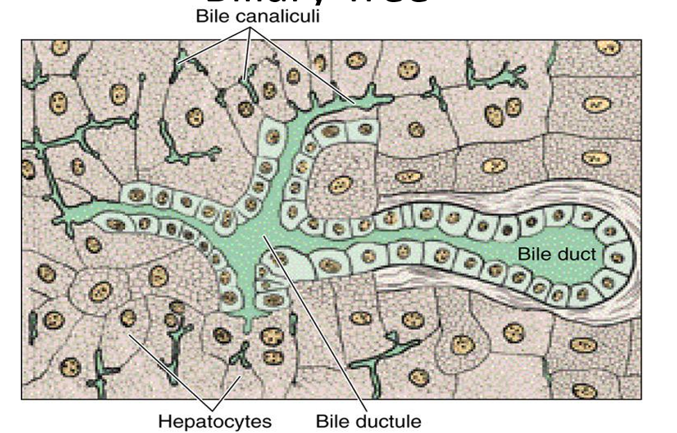
_____ _____
Biliary Tree
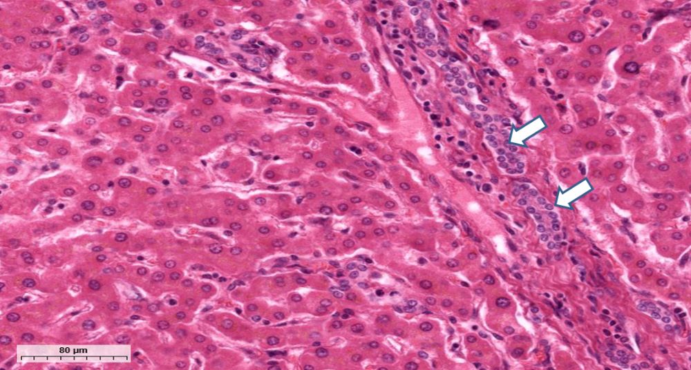
_____ _____
bile ducts
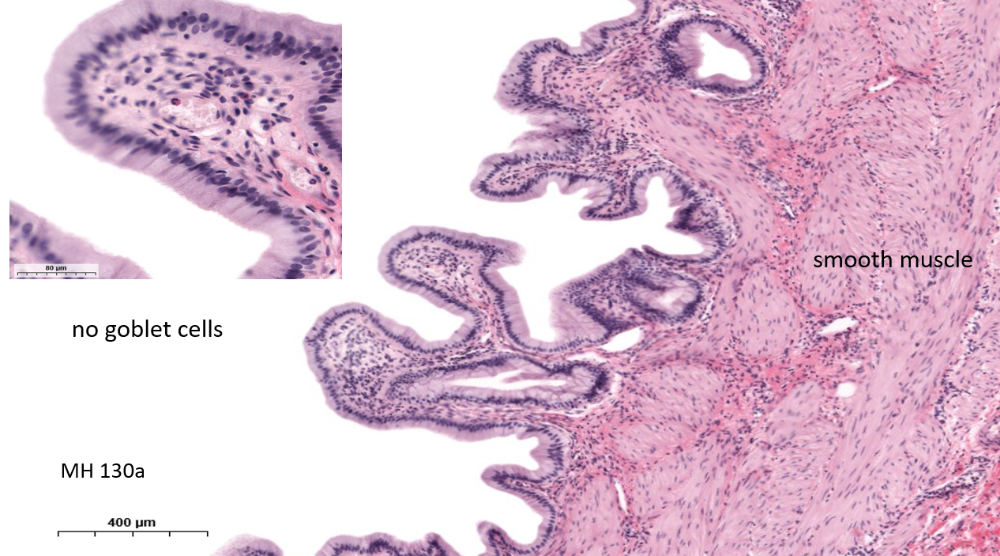
what is this organ?
gallbladder
Which structure represents the first portion of the biliary tree after bile leaves the canaliculi?
A. Bile ductules
B. Common hepatic duct
C. Cystic duct
D. Common bile duct
A. Bile ductules
Bile ductules, also called canals of Hering, are lined primarily by which type of cells according to this source?
A. Hepatocytes
B. Cuboidal nonhepatocytes
C. Squamous cells
D. Endothelial cells
B. Cuboidal nonhepatocytes
Bile ductules ultimately drain into which structures?
A. Hepatic sinusoids
B. Central veins
C. Bile ducts
D. Lymphatics
C. Bile ducts
Which epithelial types may line the bile ducts within portal triads?
A. Squamous or cuboidal
B. Transitional or cuboidal
C. Pseudostratified or columnar
D. Cuboidal or columnar
D. Cuboidal or columnar
As intrahepatic bile ducts converge, they form which larger structures?
A. Cystic ducts
B. Hepatic ducts
C. Pancreatic ducts
D. Central ducts
B. Hepatic ducts
The right and left hepatic ducts unite to form the:
A. Common bile duct
B. Cystic duct
C. Common hepatic duct
D. Pancreatic duct
C. Common hepatic duct
The common hepatic duct joins which structure to form the common bile duct?
A. Right hepatic duct
B. Left hepatic duct
C. Pancreatic duct
D. Cystic duct
D. Cystic duct
Which sequence correctly traces bile flow from the liver toward the intestine?
A. Hepatic ducts → common hepatic duct → common bile duct
B. Cystic duct → hepatic ducts → bile ductules
C. Common bile duct → hepatic ducts → duodenum
D. Hepatic duct → cystic duct → canaliculi
A. Hepatic ducts → common hepatic duct → common bile duct
The common bile duct empties into which portion of the gastrointestinal tract?
A. Stomach
B. Duodenum
C. Jejunum
D. Ileum
B. Duodenum
The common bile duct enters the duodenum at which named structure?
A. Pylorus
B. Ligament of Treitz
C. Ampulla of Vater
D. Ileocecal valve
C. Ampulla of Vater
Which sphincter regulates flow through the ampulla of Vater?
A. Pyloric sphincter
B. Lower esophageal sphincter
C. Ileocecal sphincter
D. Sphincter of Oddi
D. Sphincter of Oddi
What is the primary function of the gallbladder between meals?
A. Store and concentrate bile
B. Synthesize bile acids
C. Produce bilirubin
D. Secrete pancreatic enzymes
A. Store and concentrate bile
Which hormone stimulates the gallbladder to release stored bile into the duodenum?
A. Gastrin
B. CCK
C. Secretin
D. Somatostatin
B. CCK
What type of epithelium lines the gallbladder mucosa?
A. Stratified squamous
B. Simple cuboidal
C. Simple columnar
D. Transitional
C. Simple columnar
Immediately beneath the gallbladder epithelium is which connective-tissue layer?
A. Submucosa
B. Muscularis mucosae
C. Serosa
D. Lamina propria
D. Lamina propria
Gallbladder epithelial cells contain which apical specialization that aids absorption?
A. Microvilli
B. Cilia
C. Stereocilia
D. Flagella
A. Microvilli
How does the gallbladder concentrate bile while it is being stored?
A. Secretes additional cholesterol
B. Absorbs water
C. Removes bilirubin
D. Adds bile acids
B. Absorbs water
Which description best characterizes the mucosal folds of the gallbladder?
A. Sparse and straight
B. Flat and unbranched
C. Numerous and branched
D. Keratinized and rigid
C. Numerous and branched
Near which part of the gallbladder do mucosal invaginations form mucus-secreting glands or sinuses?
A. Fundus
B. Body
C. Neck
D. Cystic duct
D. Cystic duct
What do the gland-like mucosal invaginations near the cystic duct secrete?
A. Mucus
B. Bile salts
C. Digestive enzymes
D. Bicarbonate
A. Mucus
A histology specimen shows simple columnar mucosa with numerous folds but no submucosa. Which organ is favored over ileum?
A. Stomach
B. Gallbladder
C. Jejunum
D. Colon
B. Gallbladder
Which histologic feature helps distinguish gallbladder from ileum according to this source?
A. Presence of villi
B. Presence of goblet cells
C. Absence of submucosa
D. Presence of lymphatics
C. Absence of submucosa
According to this source, gallstones occur approximately how much more often in females than males?
A. Twice as often
B. Three times as often
C. Four times as often
D. Equally often
A. Twice as often
A patient with gallstones develops symptomatic biliary pain. Which locations are typical according to this source?
A. Left lower quadrant
B. Suprapubic region
C. Epigastrium or right upper quadrant
D. Left shoulder only
C. Epigastrium or right upper quadrant
Gallbladder pain may be referred to the right shoulder primarily because of irritation of which structure?
A. Pancreas
B. Liver capsule
C. Right kidney
D. Inferior diaphragm
D. Inferior diaphragm
A patient has a large gallstone causing obstruction of bile flow. Which complication can result?
A. Obstructive jaundice
B. Nephrotic syndrome
C. Hemolytic anemia
D. Respiratory alkalosis
A. Obstructive jaundice
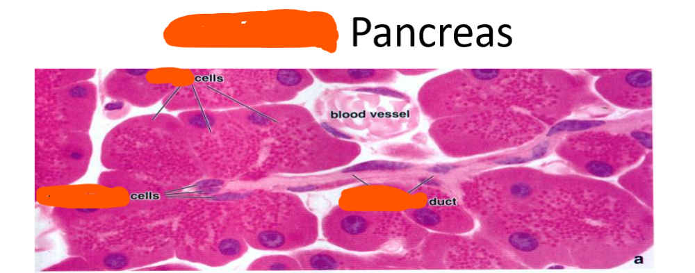
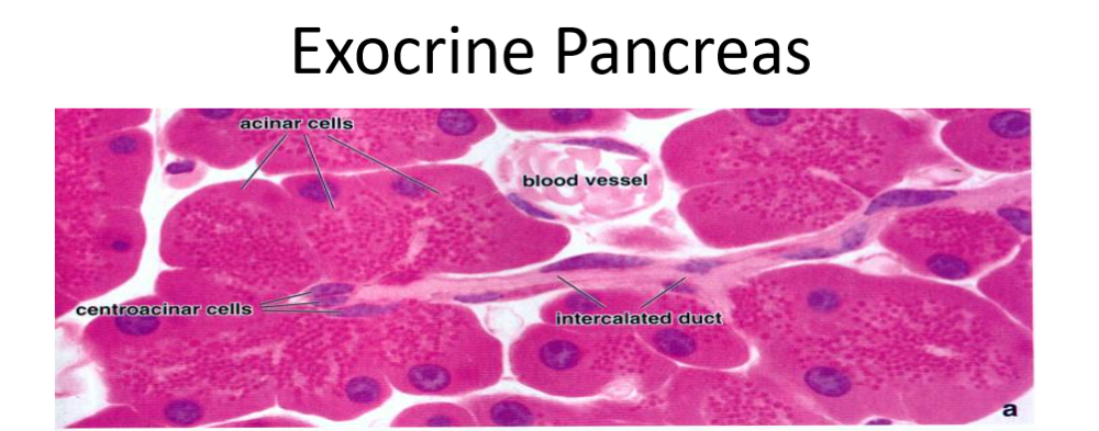
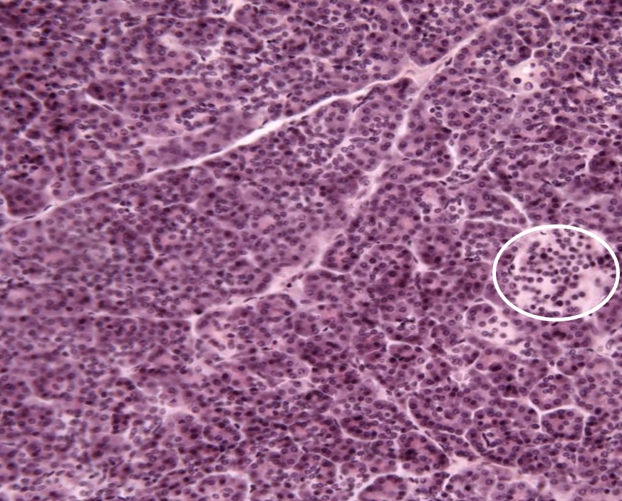
islet of langerhans
A patient with recurrent bacterial infection of the biliary tree develops pigment gallstones. Which stone type is most strongly associated with bacterial cholangitis?
A. Brown pigment stones
B. Cholesterol stones
C. Black pigment stones
D. Mixed cholesterol stones
A. Brown pigment stones
Formation of brown pigment gallstones is associated with an increased concentration of which substance in bile?
A. Conjugated bilirubin
B. Unconjugated bilirubin
C. Cholesterol esters
D. Bile phospholipids
B. Unconjugated bilirubin
Which cell type forms the secretory acini of the exocrine pancreas?
A. Mucous cells
B. Endocrine cells
C. Serous cells
D. Squamous cells
C. Serous cells
The basal cytoplasm of a pancreatic acinar cell stains basophilic because it contains abundant:
A. Rough endoplasmic reticulum
B. Zymogen granules
C. Lipid droplets
D. Smooth endoplasmic reticulum
A. Rough endoplasmic reticulum
The polarized organization of a pancreatic acinar cell is best described by which arrangement?
A. Apical RER, basal granules
B. Basal nuclei, apical RER
C. Basal RER, apical zymogen granules
D. Basal granules, apical mitochondria
C. Basal RER, apical zymogen granules
Which group contains digestive enzymes secreted by pancreatic acinar cells?
A. Amylase, insulin, glucagon
B. Bile acids, lipases, nucleases
C. Pepsin, gastrin, trypsin
D. Proteases, lipases, nucleases
D. Proteases, lipases, nucleases
Which gastrointestinal hormone directly stimulates secretion of digestive enzymes from pancreatic acinar cells?
A. Cholecystokinin
B. Secretin
C. Gastrin
D. Somatostatin
A. Cholecystokinin
Cholecystokinin that stimulates pancreatic acinar secretion is released by enteroendocrine cells located primarily in the:
A. Stomach
B. Duodenum
C. Ileum
D. Colon
B. Duodenum
Besides CCK, pancreatic acinar enzyme secretion can be stimulated by activation of which autonomic pathway?
A. Sympathetic fibers
B. Pelvic splanchnics
C. Parasympathetic vagal fibers
D. Lumbar splanchnics
C. Parasympathetic vagal fibers
Which hormone stimulates pancreatic duct cells to secrete bicarbonate-rich fluid?
A. Secretin
B. CCK
C. Gastrin
D. Insulin
A. Secretin
Secretin that stimulates pancreatic ductal secretion is released from enteroendocrine cells of the:
A. Jejunum
B. Duodenum
C. Stomach
D. Colon
B. Duodenum
On pancreatic histology, pale-staining cells located within the lumen of an acinus are most likely:
A. Islet cells
B. Acinar cells
C. Centroacinar cells
D. Kupffer cells
C. Centroacinar cells
Centroacinar cells are continuous with which portion of the pancreatic duct system?
A. Interlobular duct
B. Main pancreatic duct
C. Accessory duct
D. Intercalated duct
D. Intercalated duct
Which epithelial type lines pancreatic intercalated ducts?
A. Cuboidal epithelium
B. Simple squamous epithelium
C. Columnar epithelium
D. Transitional epithelium
A. Cuboidal epithelium
Which sequence correctly traces pancreatic secretions through the smaller ducts?
A. Interlobular → intercalated → intralobular
B. Intercalated → intralobular → interlobular
C. Intralobular → interlobular → intercalated
D. Intercalated → interlobular → intralobular
B. Intercalated → intralobular → interlobular
Which epithelial type lines the larger interlobular pancreatic ducts according to this source?
A. Stratified squamous
B. Cuboidal
C. Columnar
D. Transitional
C. Columnar
According to this source, interlobular ducts ultimately drain into the main or accessory pancreatic ducts, and the terminal ducts empty into which structure?
A. Stomach
B. Jejunum
C. Common hepatic duct
D. Duodenum
D. Duodenum
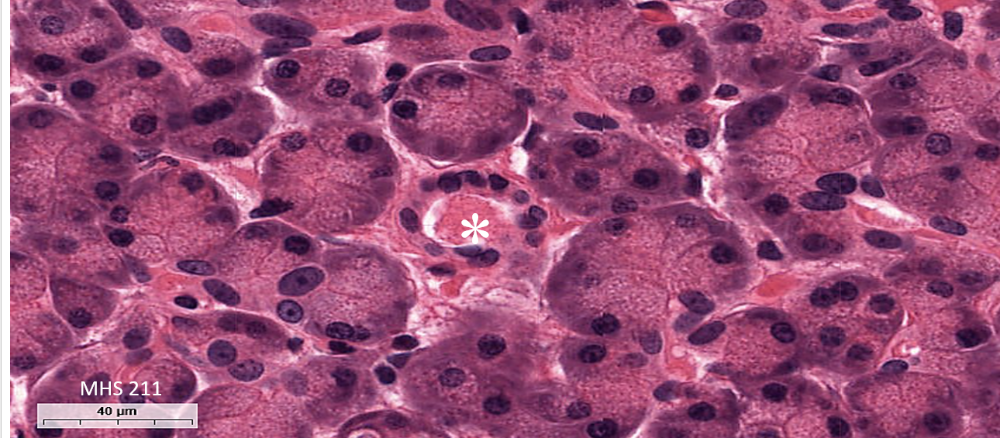
star is _____ duct
Intralobular duct
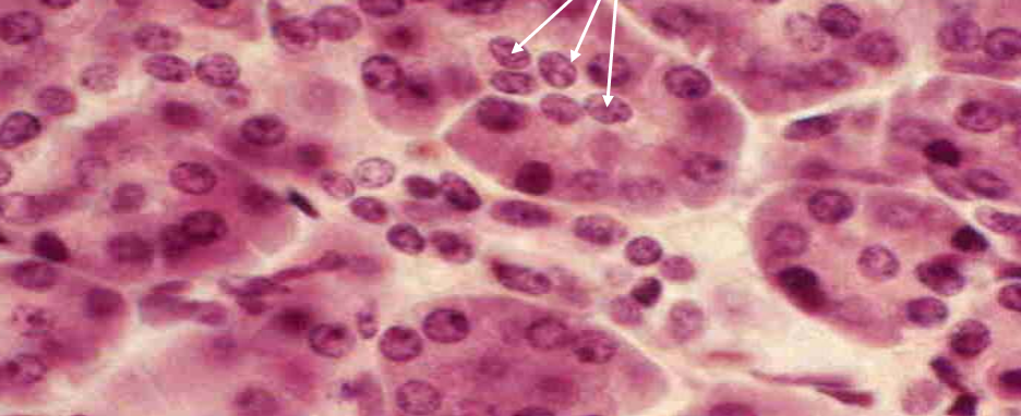
_____ cells
Centroacinar cells