What are the 3 PET biomarkers?
1. 18F-FDG
2. 18F-FLT
3. 18F-FMISO
FLT PET measures what?
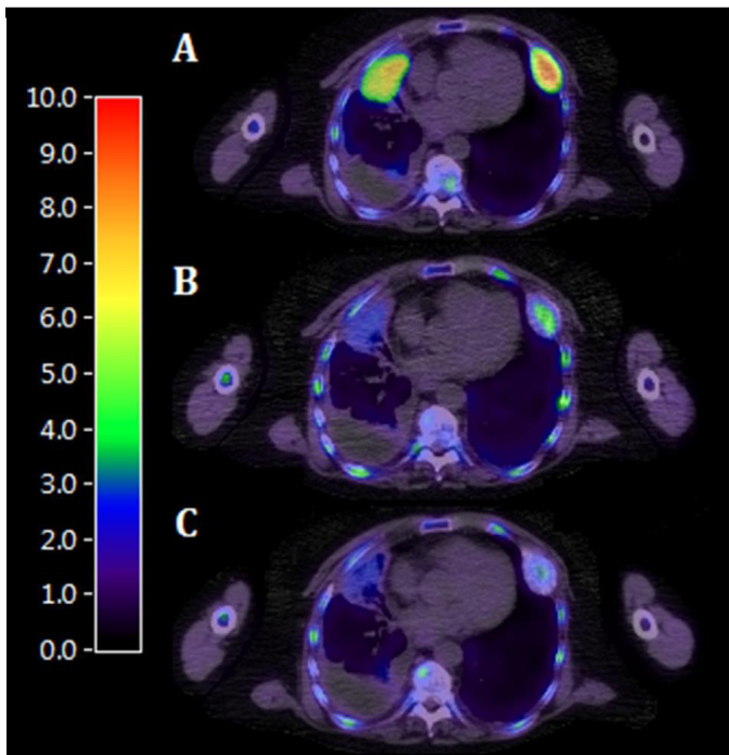
Cell proliferation (half life 109)
Uses the TK1 pathway
FDG PET measures what?
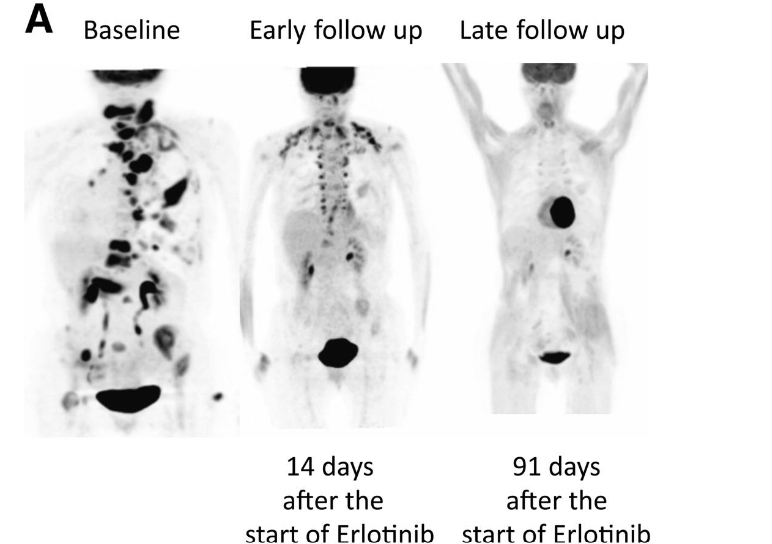
Glucose metabolism (half life 109)
Uses GLUT transport pathway
FMISO PET measures what?
Hypoxia
What can be used to measure apoptosis?
Annexin V and caspase-based PET
What can be used to measure necrosis?
DWI MRI
CEST MRI
contrast-enhanced ultrasound/CT
DCE MRI
What can be used to measure cellularity/tumor microenvironment?
DWI MRI
What can be used to measure perfusion?
DSC MRI
DCE MRI
IVIM
Doppler
contrast ultrasound
What can be used to measure cell proliferation?
FLT PET
EGFR/HER2 targeted PET
amino acid pathways(LAT1/systemL/FET PET)
DWI MRI (indirectly-restricted diffusion)
What can be used to measure angiogenesis?
DCE MRI
DSC MRI
IVIM MRI
contrast enhanced MRI/CT
Doppler ultrasound (pulsed/color)
Arterial spin loading (ASL)
Vascularized tumor burden
What can be used to measure blood flow?
Doppler ultrasound (pulsed/color)
DSC MRI
IVIM
What growth factor is associated with angiogenesis?
VEGF
Increased vascular permeability in tumors is due to what?
Abnormal blood vessel growth (angiogenesis)
What can be used to measure invasion and metastasis?
DWI MRI
FDG PET (staging)
CT/MRI (bone and brain metastasis)
MR spectroscopy
EGFR/HER2 targeted PET
What can be used to measure amino acid transport?
Radiolabeled amino acid PET
What imaging can be used to measure receptor expression?
EGFR and HER2 targeted PET imaging
What imaging can be used to measure tissue structure and anatomy?
T1/T2 weighted MRI
Fluorescence imaging can be used to measure what?
cell death and angiogenesis
What does RECIST help us measure?
Tumor size and diameter for disease progression
What is PET?
Positron emission tomography
uses radiotracers to visualize changes in metabolic activity
What is the main quantitative metric for PET?
SUV (standardized uptake value)
What is SUVpeak?
The average uptake of tracer in a specific region of the hottest spot of the tumor
What is SUVmax?
maximum tracer uptake in a region of interest
What is SUVmean?
average amount of tracer in a region of interest
How do you calculate SUV?
tissue concentration/injected dose*body weight
What is caspase-based PET?
Caspases are enzymes that are involved in cell death. Radiotracers are designed to bind to active caspases.
What is Annexin V imaging?
Annexin V binds to PS (phosphatidylserine) which sits on the inner cell membrane. During apoptosis, PS becomes exposed and Annexin V binds to it.
What is DWI MRI imaging?
Diffusion-weighted imaging measures the movement of water molecules.
What is f (perfusion fraction)?
looks at microvascular perfusion from a single voxel
What is K (kurtosis)?
The diffusion behavior from gaussian distribution (heterogeneity)
What is the main quantitative metric for DWI MRI?
ADC (apparent diffusion coefficent)
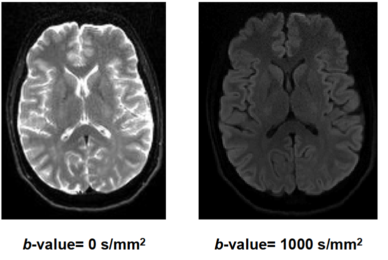
DWI MRI requires how many b values?
At least 2
High ADC...
low cellularity (more diffusion)
Increased cellularity can be associated with increased....
cell proliferation
What could be the causes of ADC increasing after treatment?
cell death and breakdown of cellular membranes, causing an increase in extracellular space and water movement
Low ADC...
high cellularity (less diffusion)
What can influence ADC?
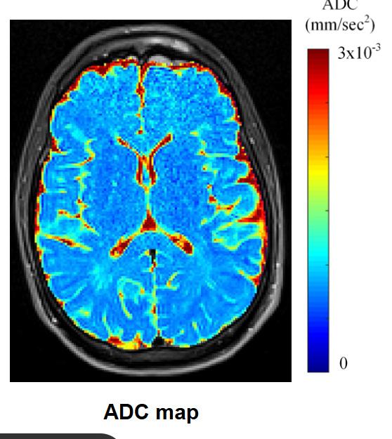
cellularity, necrosis, edema, extracellular space
What can a T1 MRI measure?
Anatomical structure and contrast between tissues
longitudinal recovery
What does flair (T2W) imaging show?
edema
What can a T2 MRI measure?
Inflammation, edema, necrosis
transverse recovery
What is gadolinium-enhanced MRI?
Gadolinium is a contrast agent (used in T1)
It can tell us about the delivery of contrast from blood to tissue
What does ultrasound use for in imaging?
High-frequency sound waves
What contrast agent is used for a T2 MRI?
Iron oxide
What contrast agent is used for a T1 MRI?
Gadolinium (increases T1 signal intensity)
What can gadolinium-enhanced MRI measure?
Vascularity, perfusion, permeability
What is DCE MRI?
Dynamic contrast-enhanced MRI
dynamic so it needs multiple images (b values)
What does DCE MRI measure?
blood flow, blood volume, vascular permeability
What are the quantitative metrics used in DCE MRI?
Ktrans (transfer of contrast from blood plasma to extracellular space)
Kep (rate of movement (washout) back from extracellular space to blood)
Ve (extravascular volume fraction)
Vp (fractional plasma volume)
What does the Tofts model show?
Describes how contrast moves between compartments
(blood plasma => tissue/extracellular space)
What is K1 and K2 in the tofts model?
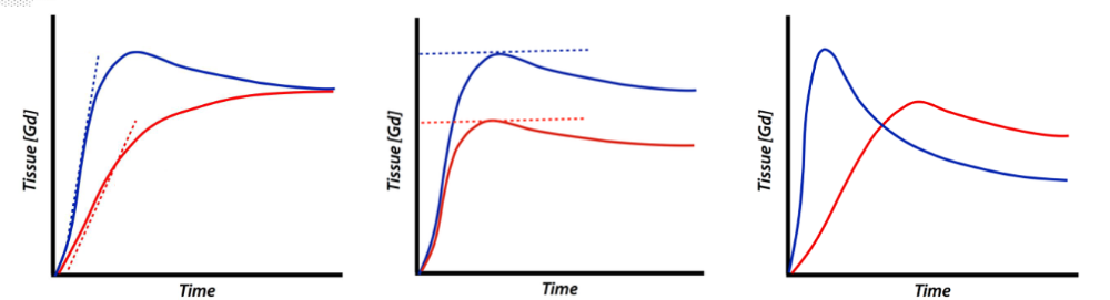
K1 - transfer from blood to plasma
K2 - return from tissue to plasma
Gadolinium decreases the signal in which MRI (T1 or T2)?
T2 because Gadolinium changes the local magnetic field around the blood vessels
What is DSC MRI?
Dynamic susceptibility contrast
uses gandolinium
What does DSC MRI measure?
perfusion (mostly used in the brain)
What are the quantitative metrics used in DSC MRI?
rCBV (cerebral blood volume)
rCBF (cerebral blood flow)
What is IVIM MRI?
Intravoxel incoherent motion
What can IVIM MRI measure?
Water diffusion and microvascular perfusion using b values
What are the quantitative metrics for IVIM MRI?
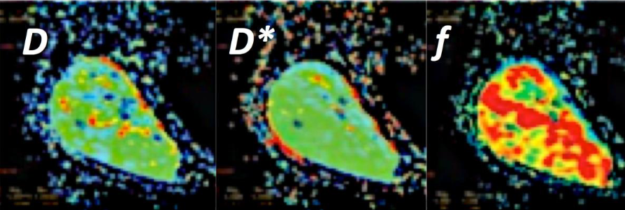
f (perfusion), D (diffusion), D* (pseudodiffusion)
What is pseudodiffusion?
bulk motion that produces a signal attenuation that mimics diffusion
What does doppler ultrasound measure?
blood movement caused by frequency shifts
What does pulsed doppler measure?
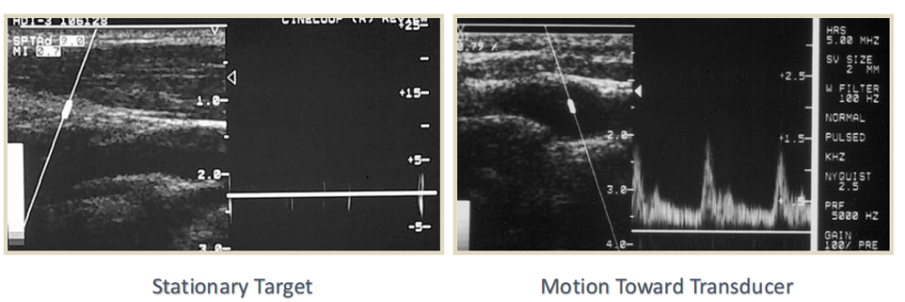
blood flow velocity (how fast the blood is moving)
What does color doppler measure?
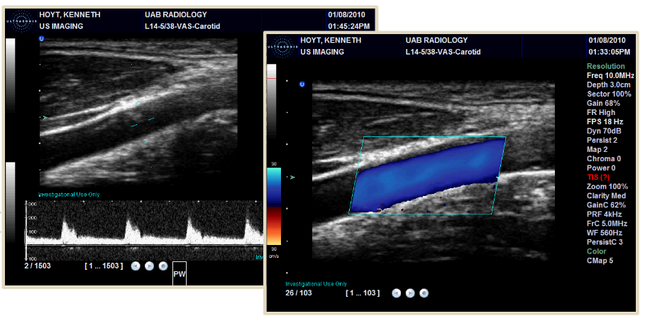
blood flow direction and location
(red vs blue is opposite directions)
What contrast agent is used in ultrasound?
Microbubbles
What is microbubble contrast ultrasound (CEUS)?
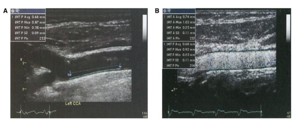
Using microbubbles to examine blood flow, perfusion, and vascularity
Microbubbles stay in the blood
What is contrast enhanced CT?
When iodinated contrast is put into the bloodstream allowing us to see blood flow, blood volume, vascularity, and permeability
CT is commonly used to determine what?
Bone metastasis
CT uses what quantitative metric?
Hounsfield units (measures radiodensity to quantify tissue attenuation in x-rays)
What increases and decreases the radiodensity of tissues in CT contrast?
iodinated contrast increases, air decreases
What can X-ray and CT tell us about a tumor?
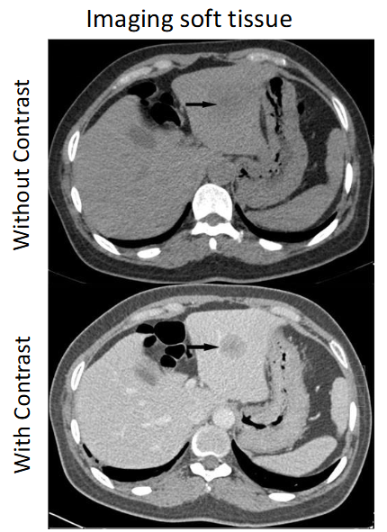
Anatomical/ tissue structure
x-ray = bone (white)/ air (dark)
What is SPECT?
Single photon emission computed tomography
Using gamma rays for radiotracers that can indetify biological function
Which has higher sensitivity and better spatial resolution, PET or SPECT?
PET
What is fluorescence imaging?
Using a fluorophore that absorbs light and emits it at a different wavelength.
What can fluorescence imaging show us?
receptor expression, tissue visualization, tumor margins
can also be used for targeted imaging
What is CEST MRI?
Chemical Exchange Saturation Transfer
How does CEST MRI work?
Saturated protons can be exchanged with water, which affects the water signal.
What is the main quantitative metric for CEST MRI?
APT (amide protein transfer)
Can tell us pH and protein concentration (increased APT = increased protein)
What is magnetization transfer?
free water protons are exchanged with macromolecule bound protons
What can magnetization transfer imaging tell us?
Fibrosis, tissue structure, and collagen
What is the quantitative metric for magnetization transfer?
Magnetization Transfer Ratio (MTR)
What is MR spectroscopy?
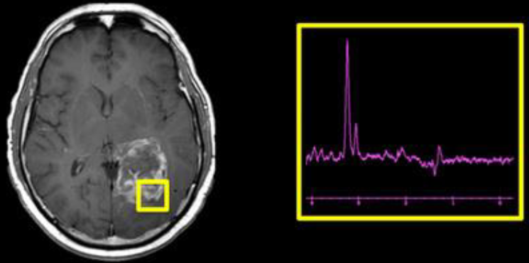
Tells us about the metabolites present in tissue (creatine, lactate, lipids)
can show us chemical shifts related to metastasis
do amino acids support cell proliferation?
They provide building blocks for protein synthesis and contribute to the energy requirements of rapidly dividing cells.
How can radiolabeled amino acids be used in imaging?
For PET because they are deisgned to follow amino acids transport
What amino acid transports are used?
SystemL (can cross blood brain barrier) and LAT1
helps increase amino acid transport for FET PET uptake
Why is FET PET used?
Because tumor cells have increased expression/activity of amino-acid transporters, particularly transport systems
Where is FET PET mostly used in the body?
The brain because normal brain tissue has relatively high glucose metabolism which is not useful for FDG
How is FET PET uptake increased?
When there is more amino acids transport through pathways like systemL and LAT1
What is EGFR-targeted PET?
A molecular imaging of a receptor (epidermal growth factor-uncontrolled cell divison)
downstream signaling can drive tumor proliferation
What is HER2 targeted imaging?
A molecular imaging of a receptor (breast or gastric cancer with agressive tumor growth)
Can lead to high rates of proliferation and tumor aggressiveness
When can historgrams be useful in imaging?
Using ADC or SUV, specifically looking at the distribution of voxels. Can be very useful for heterogeneity
Static vs dynamic imaging
Static - taking one image at a time (ex: PET - SUV)
Dynamic - repeated imaging over time (ex: DCE MRI - looking at washout)
What is RECIST?
A tumor response assessment framework (need at least 2 time points)
What is complete response for RECIST?
All target lesions disappear
What is partial response for RECIST?
>30 decrease in lesions
What is stable response for RECIST?
no progressive disease or partial response
What is progressive disease for RECIST?
>20 increase in lesions
RECIST can be used for what imaging types?
CT, MRI, ultrasound, PET
What are targeted approaches to activate cell death?
p53 signaling, annexin V, caspases, TNF
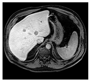
What imaging modality is this?
CT (computed tomography)
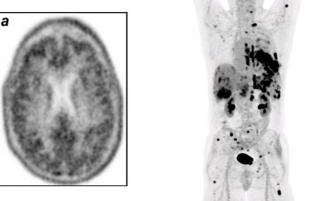
What imaging modality is this?
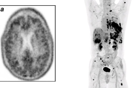
nuclear (PET/SPECT)
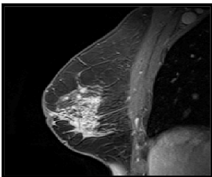
What imaging modality is this?
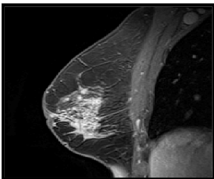
MRI
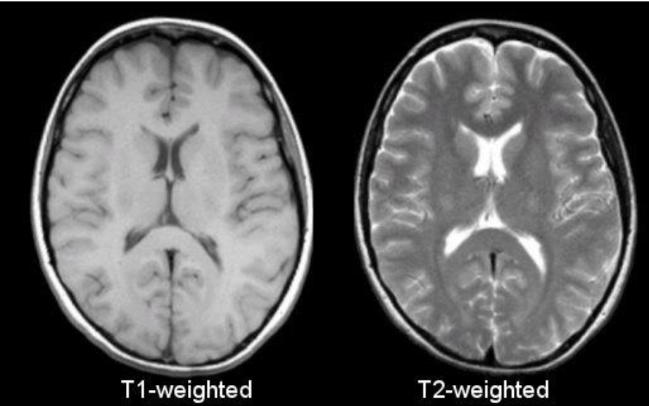
What imaging modality is this?
T1/T2 MRI
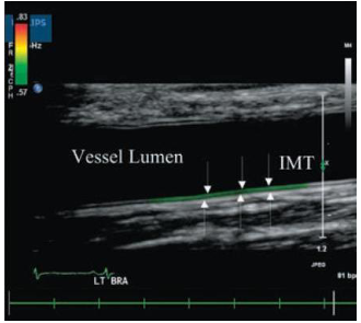
What imaging modality is this?
ultrasound
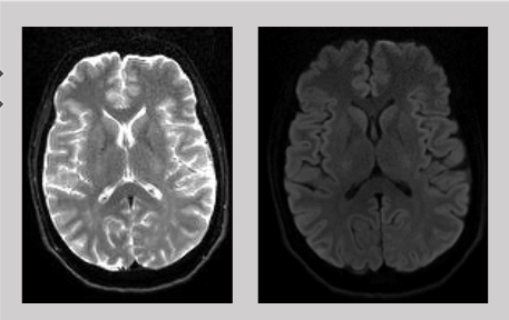
What imaging modality is this?
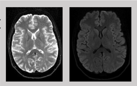
DWI MRI
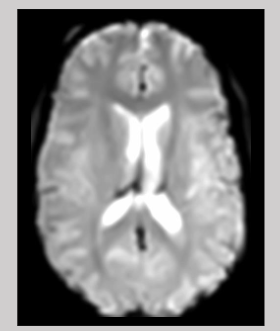
What imaging modality is this?
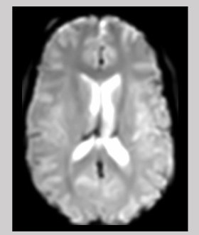
DSC MRI
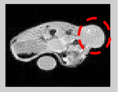
What imaging modality is this?
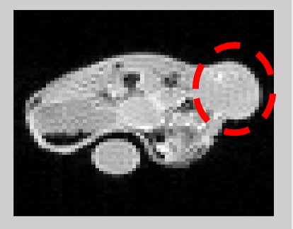
DCE MRI