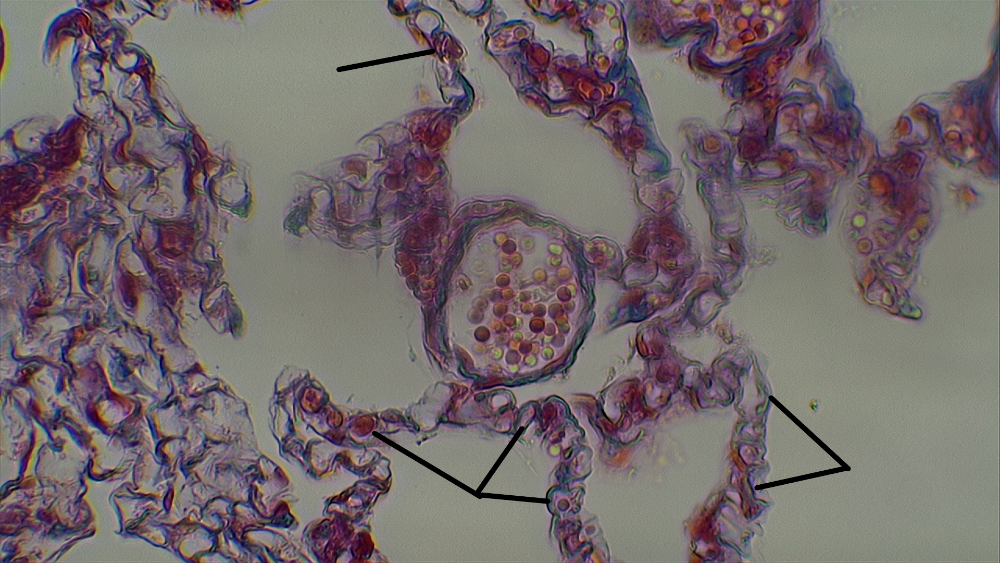
Identify the cells indicated at the end of the pointer (in the lung)
simple squamous epithelium
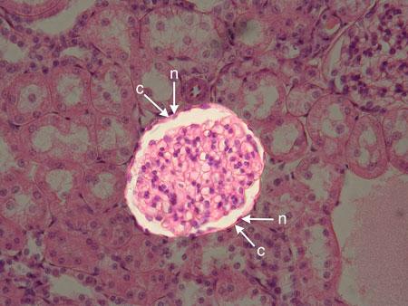
Identify the cell type shown at the arrows 'c' and 'n' for cell and nucleus. What tissue is this?
simple squamous epithelium
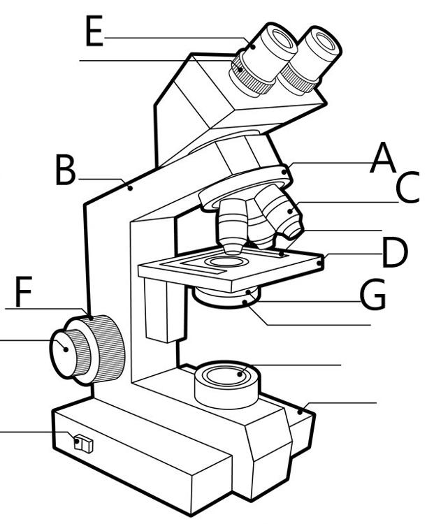
Identify the part of the microscope indicated at 'A' above.
Rotating Nosepiece
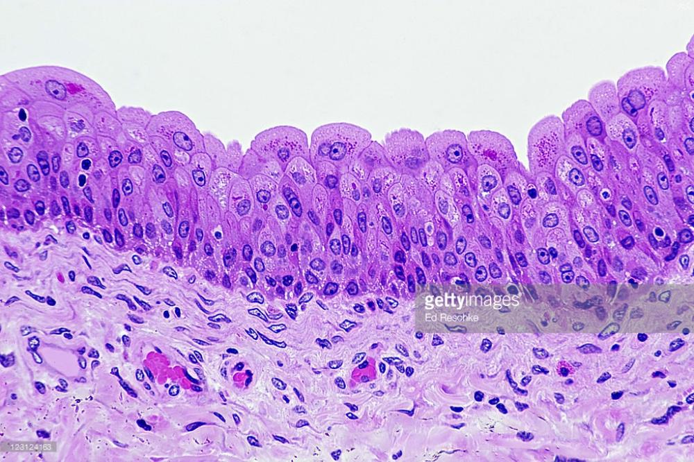
Identify this tissue above.
Transitional Epithelium
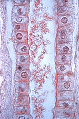
Identify the cells lining the tubule above. Be as specific as you can.
simple cuboidal
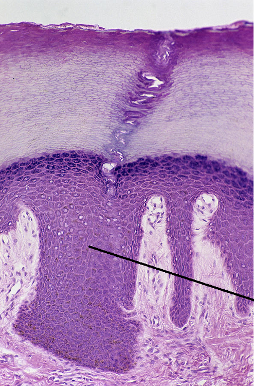
Identify the tissue type above.
Identify the specific abdominopelvic region indicated with the letter #4 above.
Stratified squamous Epithelium-keratinized
Some charged ions (K+) or (Na+) or (Cl-) or polar molecules are simply not compatible with the non-polar phospholipid tail groups embedded inside every cell membrane, and must use a protein to cross the membrane. The use of a protein channel to cross a membrane from high concentration to low concentration on the other side, is the definition of ___________________.
Facilitated Diffusion
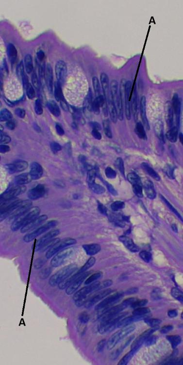
Identify the cell type indicated at 'A' above.
Simple Columnar Epithelium
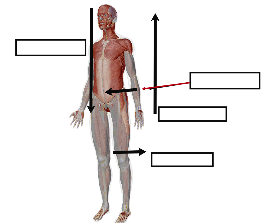
The term which means above something else, or towards the head, and is used only in the Axial Body is called ______________.
Superior
Passive diffusion of water across a membrane towards a region of higher salt (solute) is the definition of _____________.
Osmosis
Which objective lens on the microscope is considered 'low power'?
10x
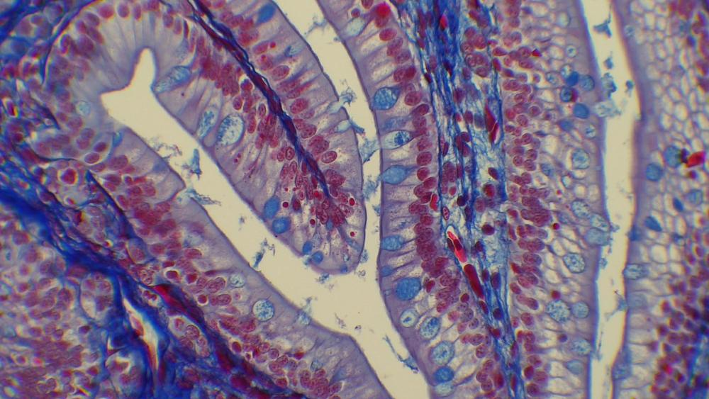
The cells with the cranberry colored nuclei and the greyish-violet brushborder microvilli are examples of of which cell type? What tissue is this?
Simple Columnar Epithelium
If a museum were to take a circular saw and cut the human body straight down the middle, producing equal left and right halves, we call this a ____________ section.
mid-sagittal
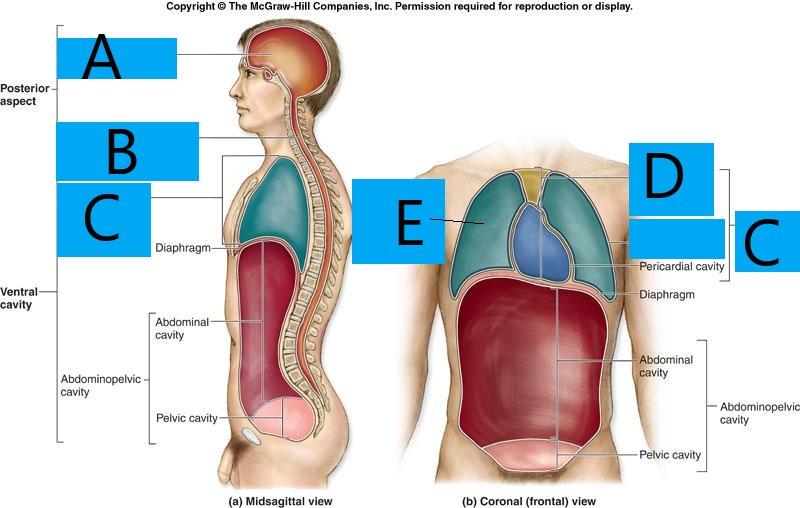
Identify the body cavity at 'C'
thoracic cavity
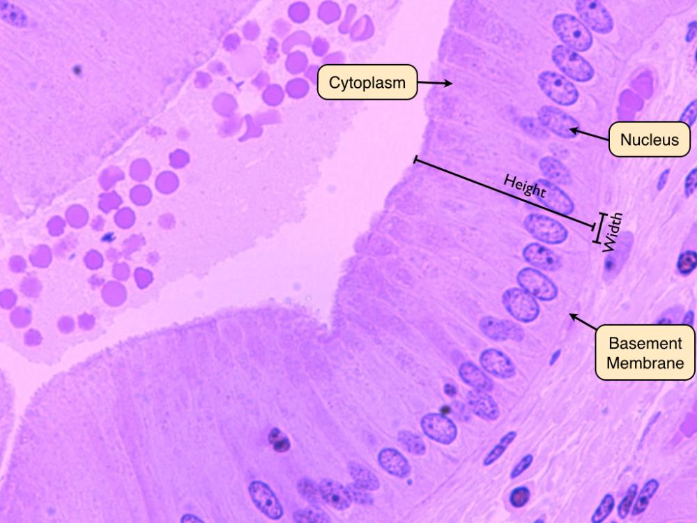
Identify the tissue indicated at the arrows and the labels above.
Simple Columnar epithelium
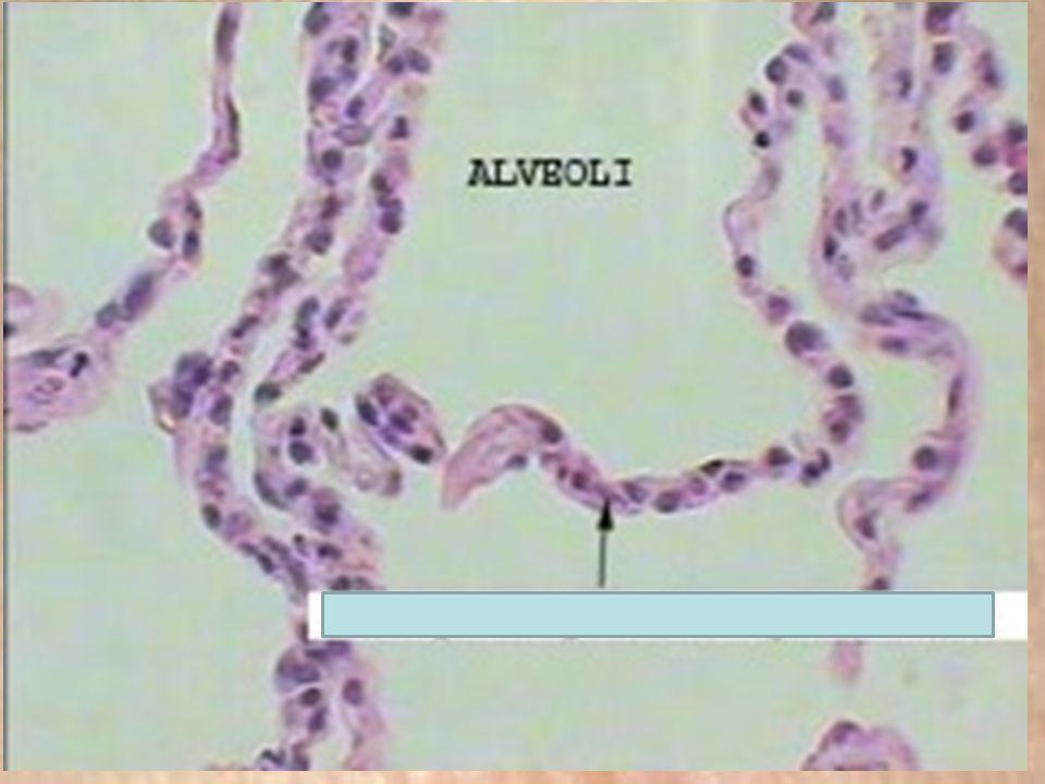
Identify the tissue type indicated above.
Simple squamous epithelium
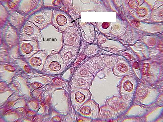
Identify the cell type or tissue type at the arrow.
Simple columnar epithelium
Simple squamous epithelium
Transitional epithelium
Simple cuboidal epithelium
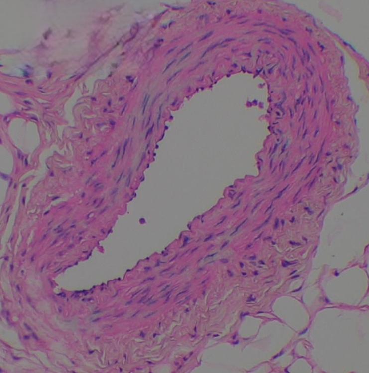
Identify the cell type above lining the inside of the blood vessel
simple squamous epithelium
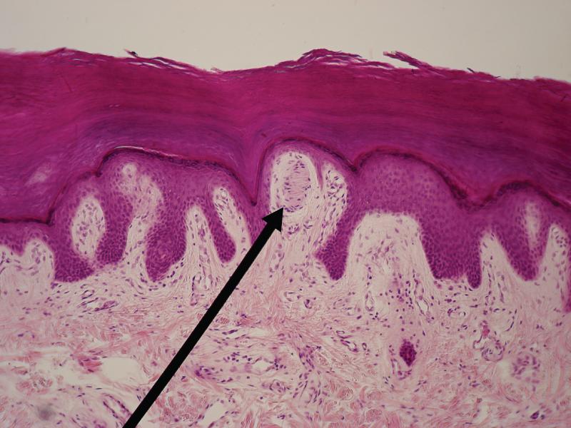
This image is showing a pointer, pointing at a sensory receptor in the skin of the finger. What is the tissue immediately above the sensory receptor in dark purple?
Stratified Squamous Epithelium-Keratinized
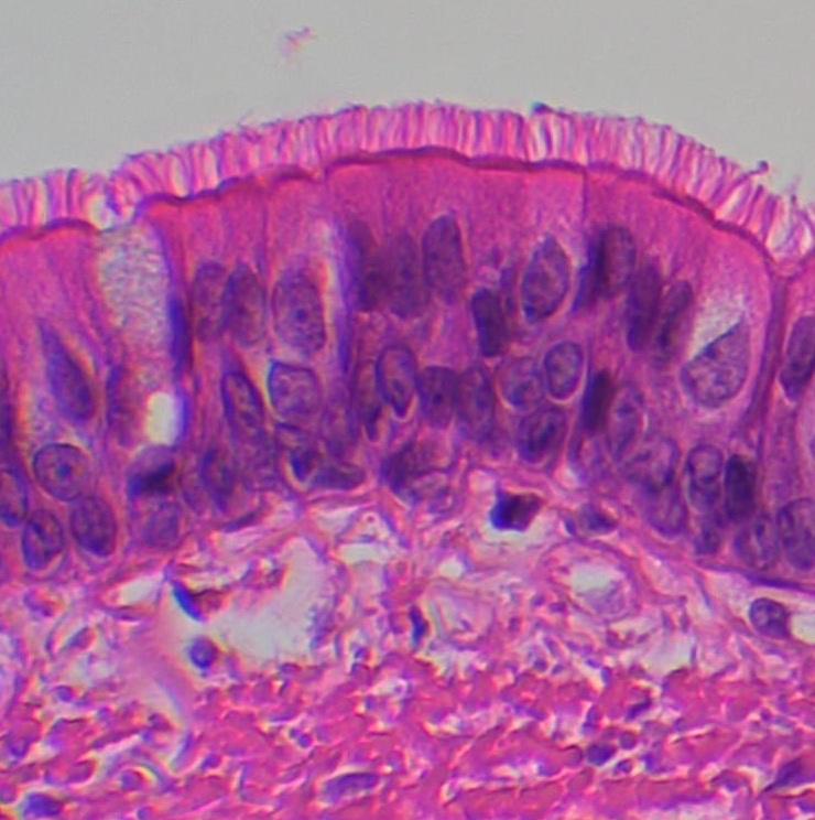
Name this tissue. Be as specific as you can be.
Pseudostratified Ciliated Columnar Epithelium
Simple cuboidal Epithelium
Simple Squamous Epithelium
Stratified Squamous Epithelium
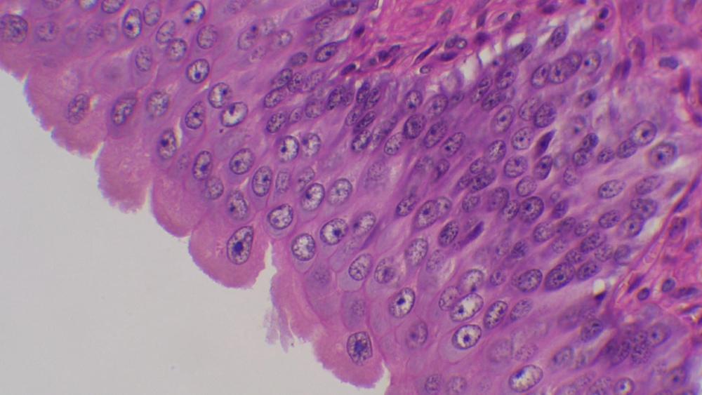
Identify the large globular cells above.
Transitional epithelium
What is the optical phenomenon we observed in the microscope that flips the letter 'e' upside down and backward when we observe it under the oculars?
Inversion
When we increase magnification, by switching to a higher power objective lens, what happens to your field of view?
shrinks or decreases
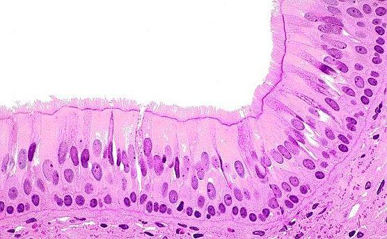
Identify the tissue type above.
Pseudostratified Columnar Ciliated Epithelium
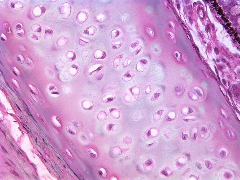
Identify the specific type of cartilage above.
Hyaline Cartilage
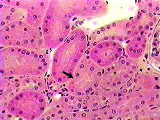
Identify the cell type indicated above.
Simple cuboidal epithelium
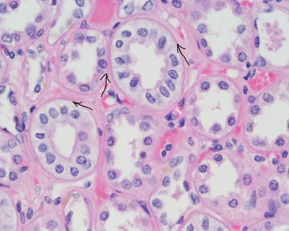
Identify the tissue and the feature indicated with the arrows.
Simple cuboidal epithelium with basement membrane
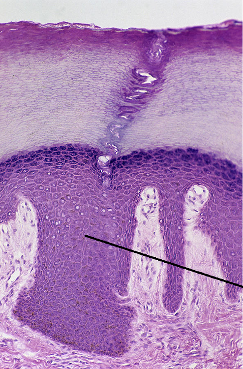
Identify the type of tissue at the end of this pointer above. (Be specific)
Stratified Squamous
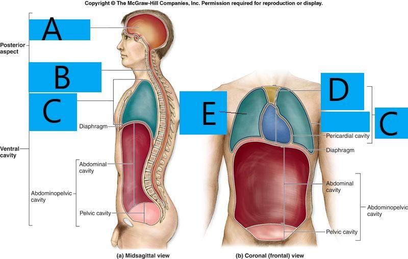
Identify the serous membrane which would coat the inside of the abdominopelvic cavity.
parietal peritoneum
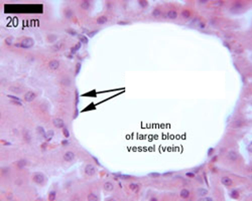
Identify the tissue indicated with the arrow on the inside of the blood vessel
simple squamous epithelium
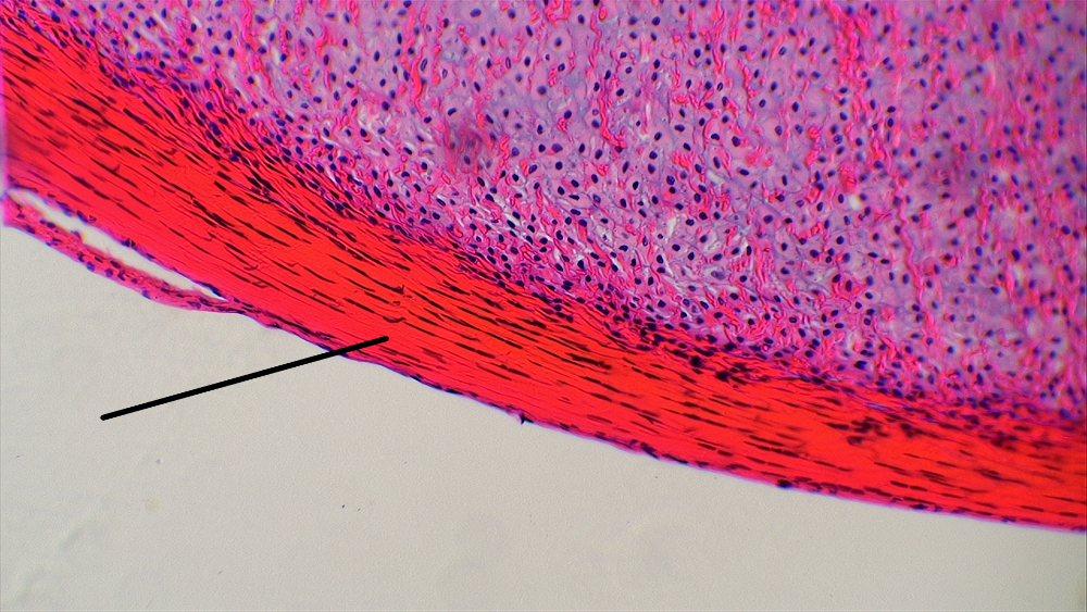
Identify the type of tissue at the end of the pointer.
Dense Regular Connective Tissue
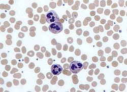
Identify the type of tissue shown above. Identify which of the four main types of tissue we discussed, then tell me as specifically as you can, what this tissue is called.
Connective Tissue; blood
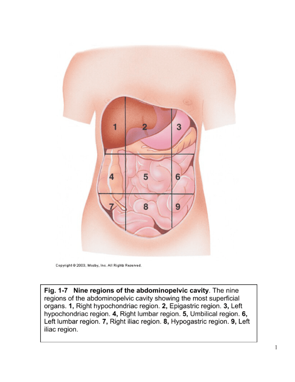
Look at the image above: Which region of the abdominopelvic cavity is directionally superior to the region labeled '5'.
Epigastric
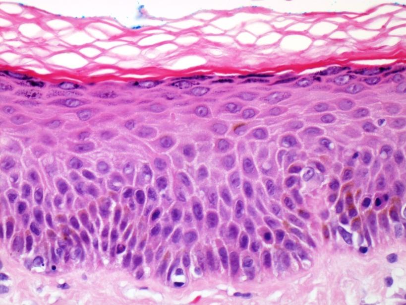
Identify the tissue type above.
Stratified Squamous Epithelium
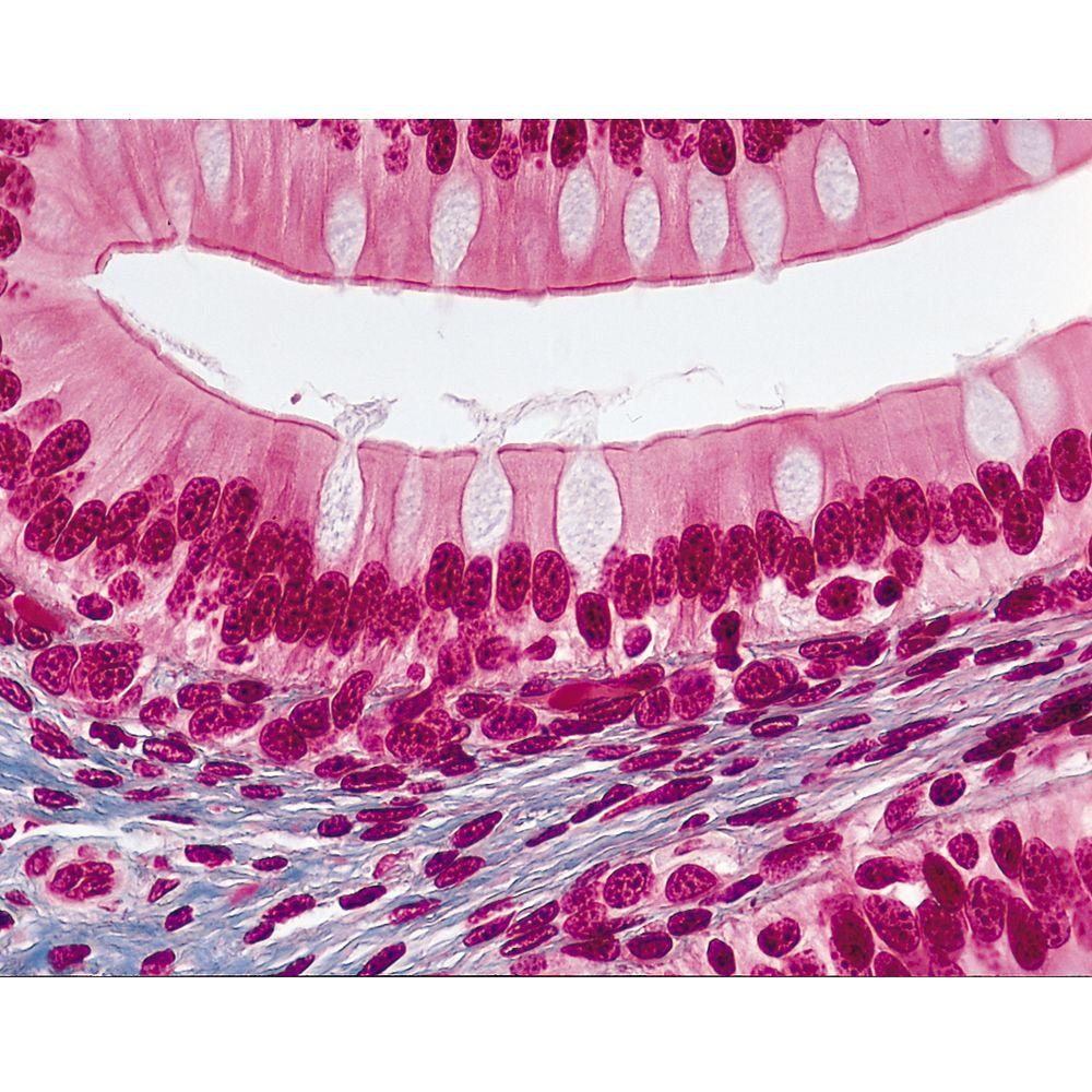
Identify the tissue type above, and the feature in silver globules nestled amongst the cells
Simple columnar epithelium with Goblet cells
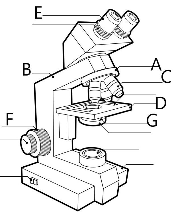
Identify the part of the microscope indicated at 'F' above.
coarse focus adjustment knob
The part of the cell which is responsible for acting like a 'gate-keeper' and regulating what goes in or comes out of the cell.
plasma membrane
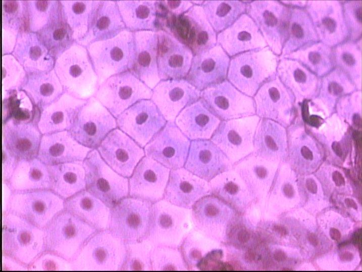
Identify the type of cells above, and their function.
simple squamous; gas exchange, filtration
When a body is divided by a plane of section dividing the body into posterior and anterior sections, we call this plane of section ____________________.
frontal or coronal plane of section