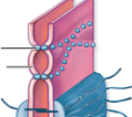
Name
Location
Function
Tight Junction of epithelial tissue
Above adhering junctions
Seal off intercellular space and prevent mixing of membrane proteins and lipids
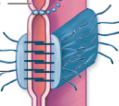
Name
Location
Function
Adhering Junction of epithelial tissue
Below tight junctions
Hold neighboring cells together and support tight junctions
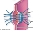
Name
Location
Function
Desmosome of epithelial tissue
Near basement membrane
Holds cells together and supports them during mechanical stress
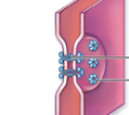
Gap Junction of epithelial tissue
Near basement membrane
Provides a passageway for ions, glucose, amino acids, and small solutes between cells
Organization of epithelial tissue classes
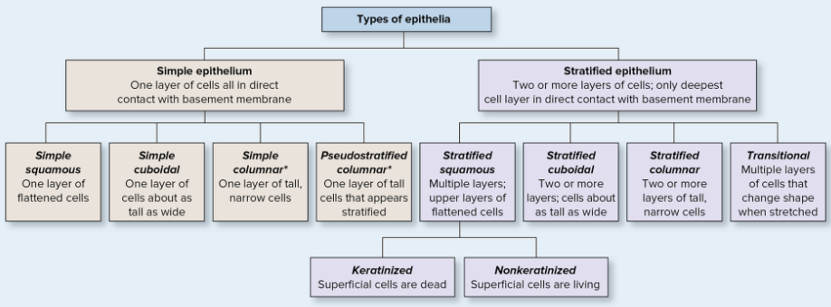
Simple epithelium: simple squamous, simple cuboidal, simple ciliated and non-ciliated columnar, pseudostratified columnar
Stratified epithelium: stratified keratinized and non-keratinized squamous, stratified cuboidal, stratified columnar, transitional (umbrella)
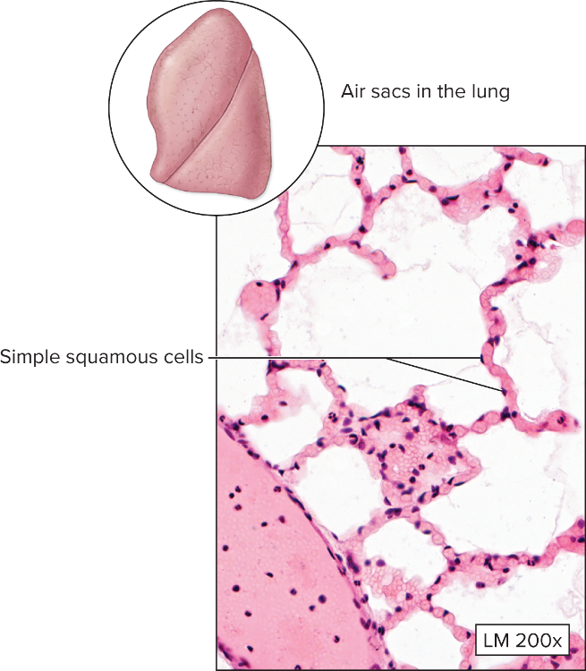
Name
Function
Location
Simple squamous epithelium
rapid absorption and filtration
ex:) lining of the heart/chambers, lymphatic vessels, and air sacs in lung
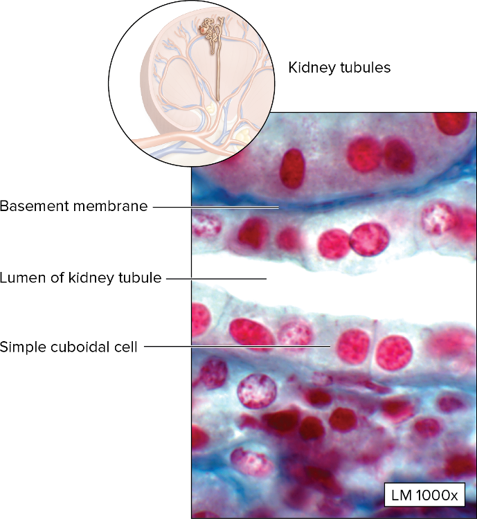
Name
Function
Location
Simple cuboidal epithelium
absorption and secretion
ex:) walls of the kidney tubules, ducts of exocrine glands, surface of the ovary
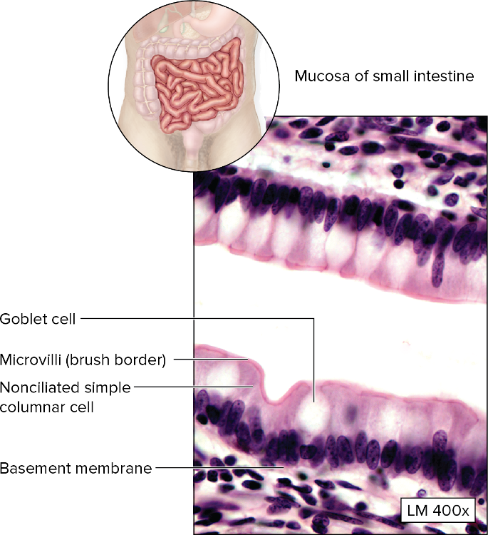
Name
Function
Location
Non-ciliated simple columnar epithelium with microvilli
secretion and absorption with help of microvilli
ex:) lining of most of the digestive tract
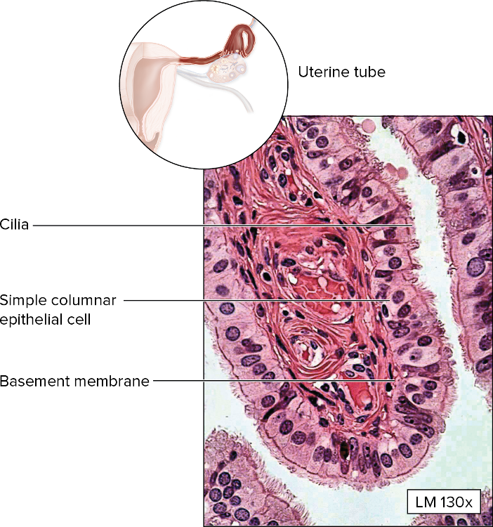
Name
Function
Location
Ciliated simple columnar epithelium
Secretion and movement of mucus
ex:) lining of uterine tubes and larger lung bronchioles
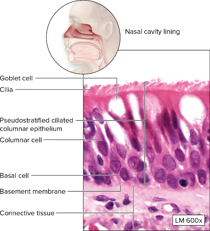
Name
Function
Location
Pseudostratified (simple) columnar epithelium with cilia - can be ciliated or nonciliated with microvilli
Cilia move dust and other particles
ex:) lining of most of the respiratory tract, non-ciliated version is very rare : epidymis and part of male urethra
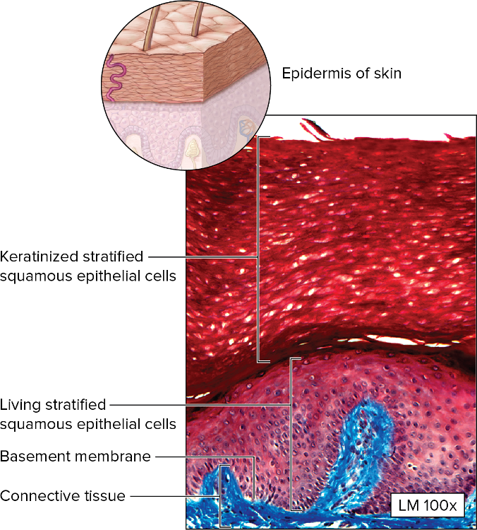
Name
Function
Location
Keratinized stratified squamous epithelium
Protection of underlying tissues
ex:) epidermis of skin
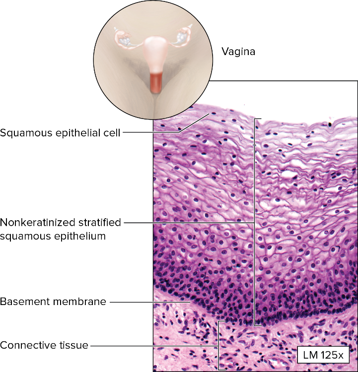
Name
Function
Location
Nonkeratinized stratified squamous epithelium
Protection of underlying tissues
ex:) lining of vagina, oral cavity, anus, and esophagus
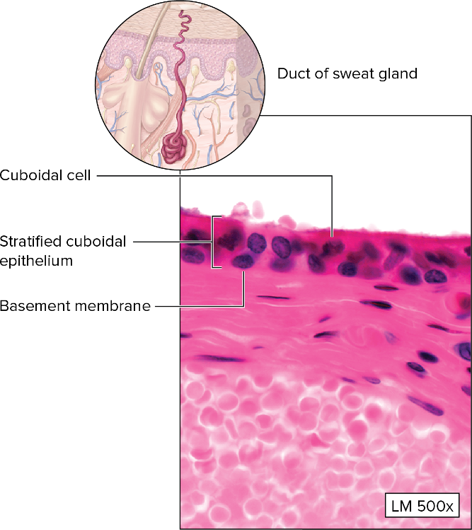
Name
Function
Location
Stratified cuboidal epithelium
Protection and secretion
ex:) large ducts in exocrine glands
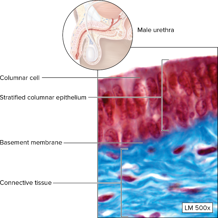
Name
Function
Location
Stratified columnar epithelium
Protection and secretion
ex:) rare - some parts of male urethra
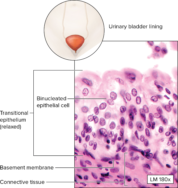
Name
Location
Function
Transitional (stratified) epithelium - some cells are binucleated
Accommodation of stretching and relaxing of tissue
ex:) lining of urinary bladder, ureter
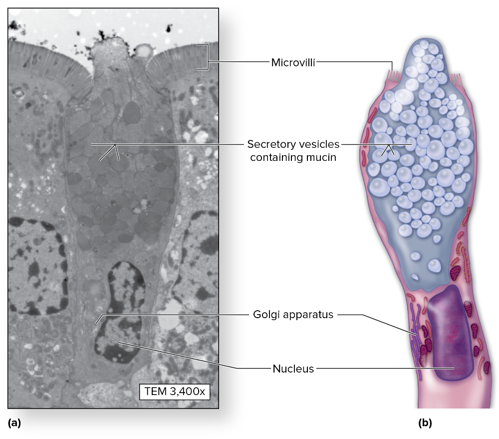
Name
Structure
Function
Unicellular exocrine gland (goblet cell)
Microvilli, secretory vesicles containing mucin, golgi, and nucleus
Mucin secretion for protection, warmth, moisture
Name
Structure
Function
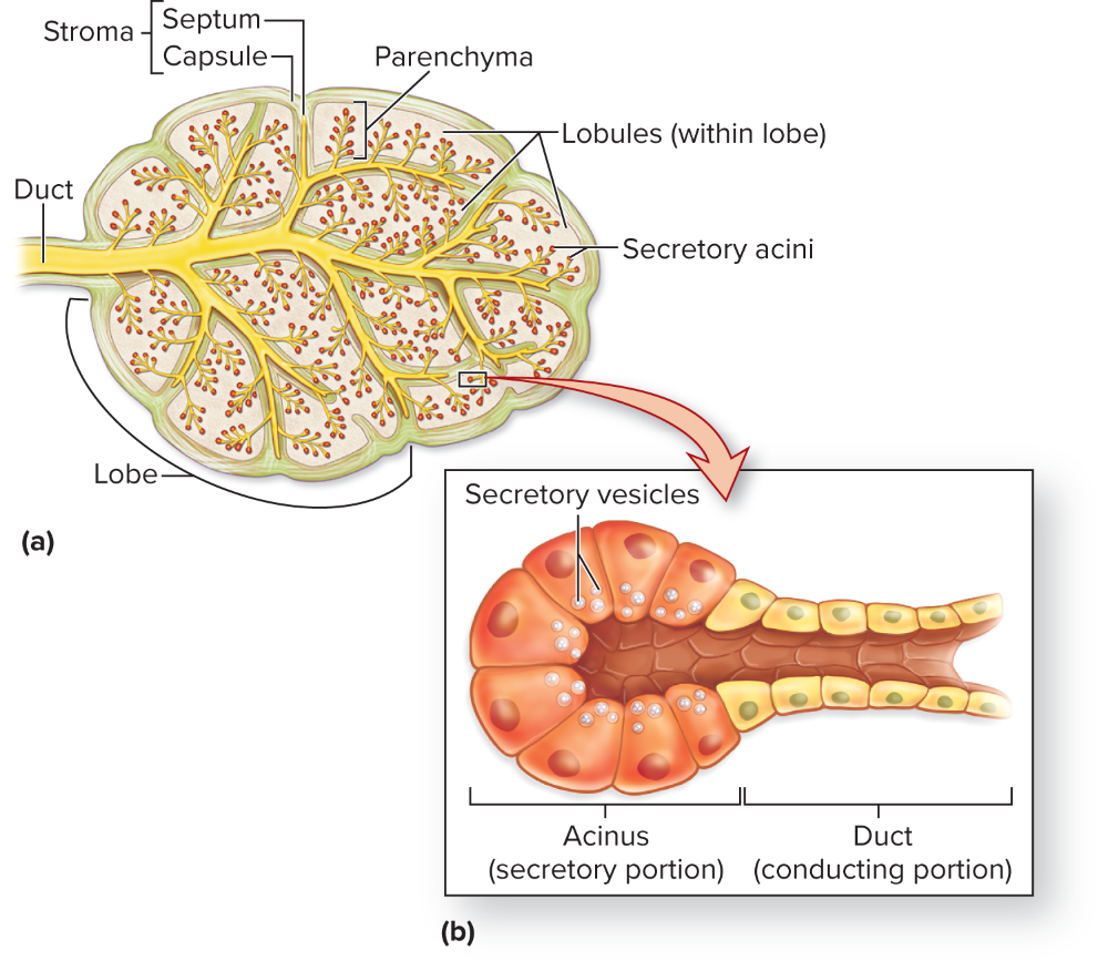
Multicellular exocrine gland
Numerous acini containing secretory vesicles and ducts
ex:) mammary glands, salivary glands, and pancreas secretions
Name
Function
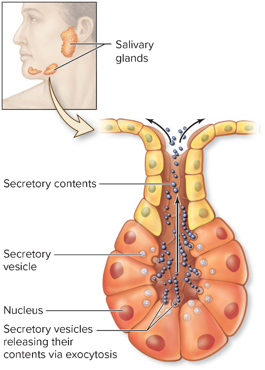
Merocrine multicellular gland
Secretion by exocytosis
ex:) lacrimal glands, eccrine glands
Name
Function
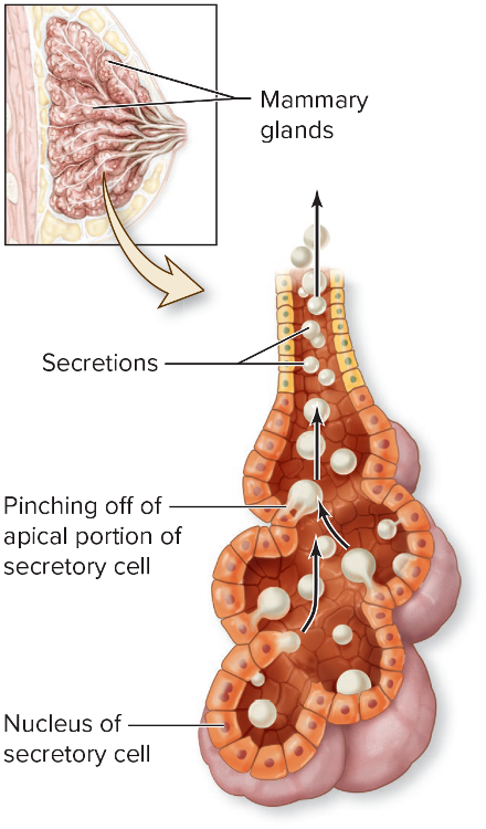
Apocrine multicellular gland
Secretion occurs when cell's apical portion pinches off
ex:) mammary and ceruminous glands
Name
Function
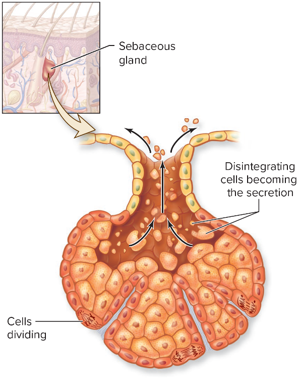
Holocrine multicellular gland
Cell ruptures and secretes viscous mixture of cell fragments
ex:) sebaceous glands in the skin
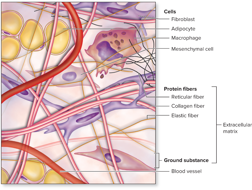
Name
Connective tissue
Organization of connective tissue classes
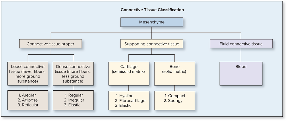
CT Proper:
-Loose: areolar, adipose, reticular
-Dense: regular, irregular, elastic
Supportive CT: cartilage and bone
Fluid CT: blood
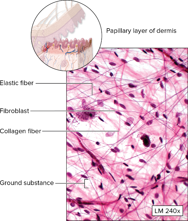
Name
Function
Location
Loose areolar CT proper
Surrounds and protects tissues and organs
ex:) papillary layer of dermis, subQ layer under skin, surrounds organs
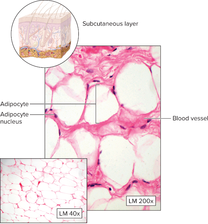
Name
Function
Location
Loose adipose CT proper
Stores energy, protects/cushions/insulates
ex:) subQ layer, covers and surrounds some organs
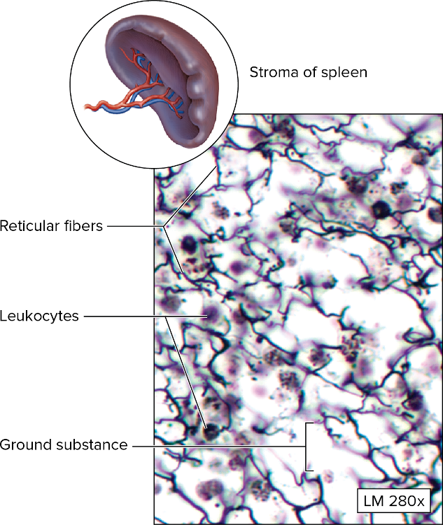
Name
Location
Function
Loose reticular CT proper
Supportive framework for spleen, lymph nodes, thymus, bone marrow
ex:) stroma of spleen, lymph nodes, bone marrow
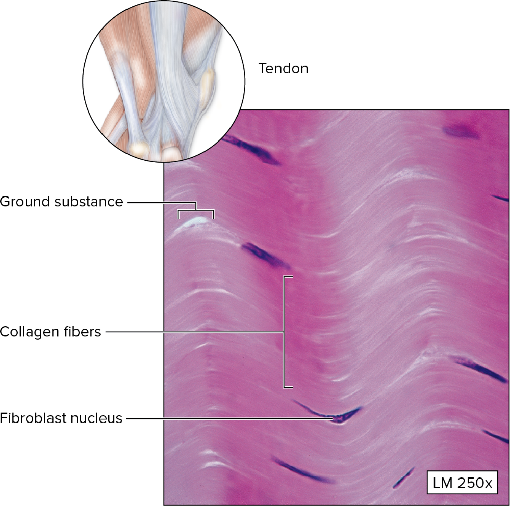
Name
Function
Location
Dense regular CT proper
Attaches muscle to bone and bone-bone, resists stress in one direction
ex:) tendons and most ligaments
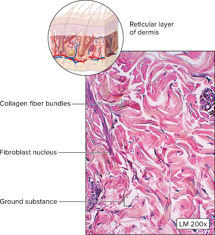
Name
Function
Location
Dense irregular CT proper
Withstands stresses applied in all directions
ex:) dermis, organ capsules
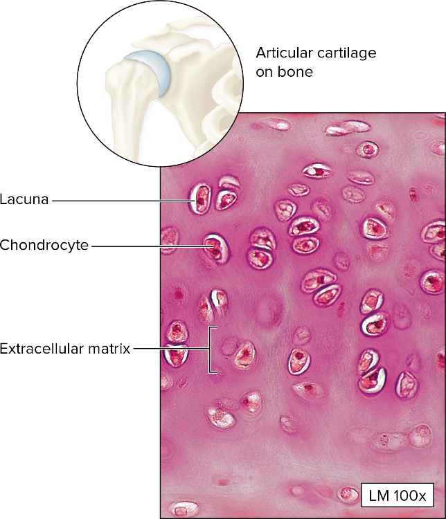
Name
Function
Location
Hyaline cartilage supporting CT
Smooth surfaces for movement at joints, supports soft tissue, model for bone growth
ex:) articular ends of long bones, costal cartilage, most of the trachea/larynx/nose
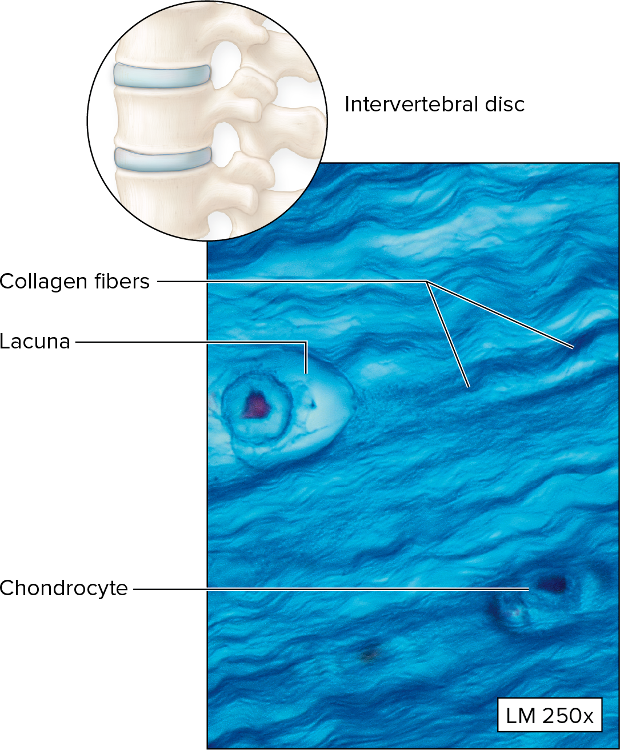
Name
Function
Location
Fibrocartilage supporting CT
Resisted compression and absorbs shock
ex:) intervertebral discs, pubic symphysis, menisci of knee joints
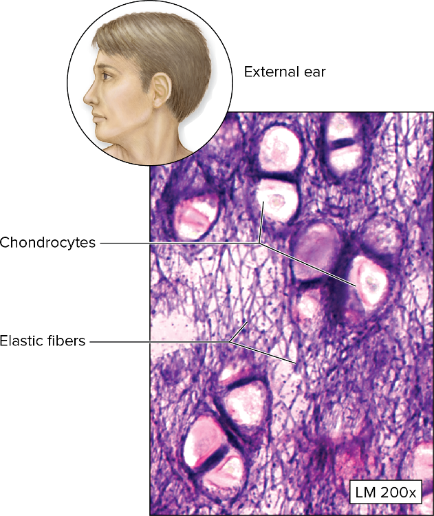
Name
Function
Location
Elastic cartilage supporting CT
Maintains structure and shape while permitting flexibility
ex:) epiglottis of larynx
Name
Function
Location
Osseus compact bone supporting CT
Supports soft structures and protects organs
ex:) all bones in the body
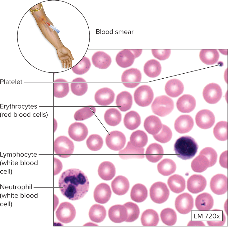
Name
Function
Location
Fluid blood connective tissue
Erythrocytes transport O2 and CO2, plasma and platelets/thrombocytes contain clotting agents, plasma transports nutrients and waste
ex:) blood vessels and the heart
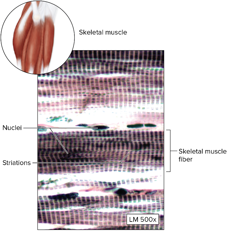
Name
Function
Location
Skeletal muscle CT - striated
Moves skeleton, voluntary movements, heat production
ex:) attached to bones and found in voluntary anus, urethra, and lips
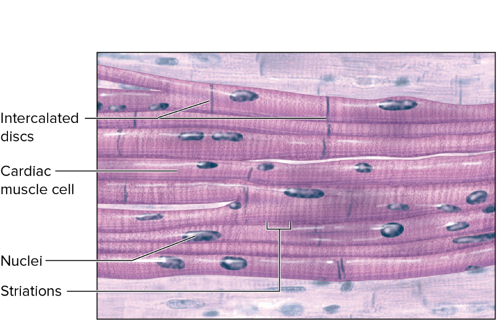
Name
Function
Location
Cardiac muscle CT - striated
Involuntary contraction and relaxation, pumps blood in heart
ex:) heart wall
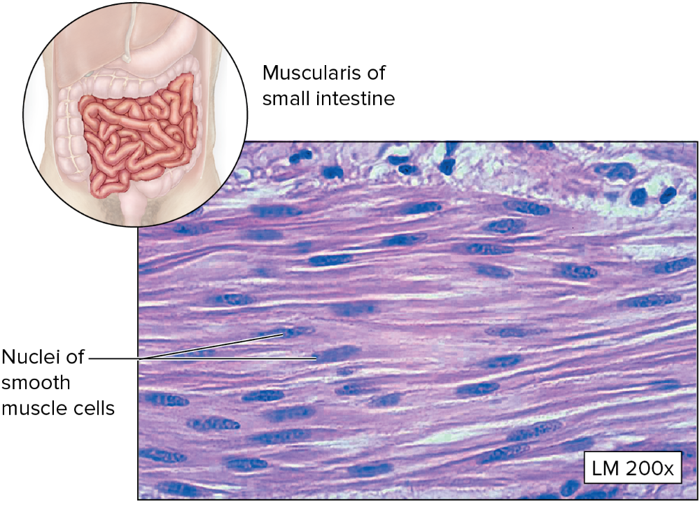
Name
Function
Location
Smooth muscle CT - nonstriated
Involuntary movements and motion, moves materials through internal organs
ex:) walls of internal organs; vessels, airways, stomach, bladder, uterus
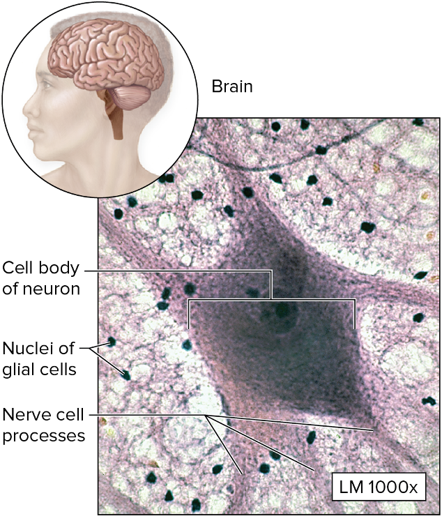
Name
Function
Location
Nervous tissue CT -
Control, info processing, storage and retrieval, and internal communication
ex:) brain, spinal cord, nerves