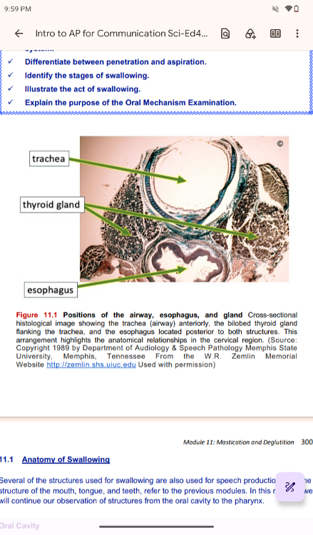
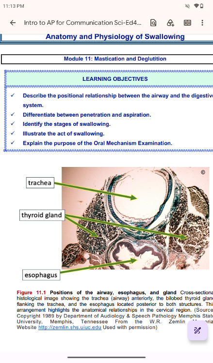
Positions of the airway, esophagus, and gland Cross-sectional
histological image showing the trachea (airway) anteriorly, the
bilobed thyroid gland
flanking the trachea, and the esophagus
located posterior to both structures. This
arrangement
highlights the anatomical relationships in the cervical region.
(Source:
Copyright 1989 by Department of Audiology & Speech
Pathology Memphis State
University, Memphis, Tennessee From the
W.R. Zemlin Memorial
Website http://zemlin.shs.uiuc.edu Used
with permission)
..
Anatomy of Swallowing
Several of the structures used for swallowing are also used for
speech production. For the
structure of the mouth, tongue, and
teeth, refer to the previous modules. In this
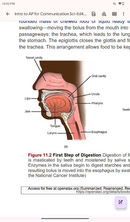
First Step of Digestion Digestion of food begins in the (a) oral
cavity. Food
is masticated by teeth and moistened by saliva
secreted from the (b) salivary glands.
Enzymes in the saliva
begin to digest starches and fats. With the help of the tongue, the
resulting bolus is moved into the esophagus by swallowing.
(credit: modification of work by
the National Cancer Institute)
Oral Cavity
The oral cavity, or mouth, is the point of entry of
food into the digestive system, illustrated
in Figure 11.2. The
food consumed is broken into smaller particles by
mastication.
Mastication is the act of chewing of the teeth.
The extensive chemical process of digestion begins in the mouth.
As food is being chewed,
saliva, produced by the salivary
glands, mixes with the food. Saliva is a watery substance
produced in the mouths of many animals. The chewing and wetting
action provided by the
teeth and saliva prepare the food into a
mass called the bolus for swallowing. A bolus is a
rounded mass
of chewed food or liquid ready to be swallowed. The tongue helps in
swallowing—moving the bolus from the mouth into the pharynx. The
pharynx opens to two
passageways: the trachea, which leads to
the lungs, and the esophagus, which leads to
the stomach. The
epiglottis closes the glottis and food passes into the esophagus and
not
the trachea. This arrangement allows food to be kept out of
the trachea.
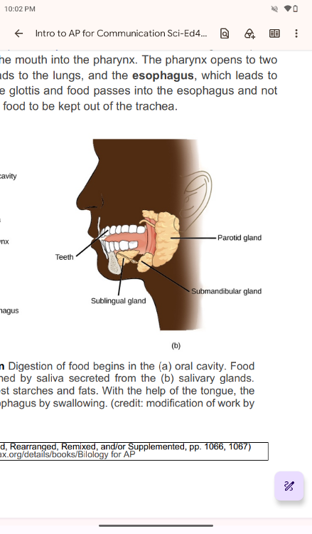
First Step of Digestion Digestion of food begins in the (a) oral
cavity. Food
is masticated by teeth and moistened by saliva
secreted from the (b) salivary glands.
Enzymes in the saliva
begin to digest starches and fats. With the help of the tongue, the
resulting bolus is moved into the esophagus by swallowing.
(credit: modification of work by
the National Cancer Institute)
The Salivary Glands
The Salivary Glands
Salivary glands are housed within the mucous
membranes of the mouth and tongue. These
glands are constantly
secreting saliva, either directly into the oral cavity or indirectly
through
ducts, even while you sleep. In fact, an average of 1 to
1.5 liters of saliva is secreted each
day. Usually, just enough
saliva is present to moisten the mouth and teeth. Secretion
increases when you eat, because saliva is essential to moisten
food and initiate the chemical
breakdown of carbohydrates. Small
amounts of saliva are also secreted by the labial glands
in the
lips. In addition, the buccal glands in the cheeks, palatal glands in
the palate, and
lingual glands in the tongue help ensure that
all areas of the mouth are supplied with
adequate saliva.
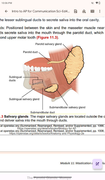
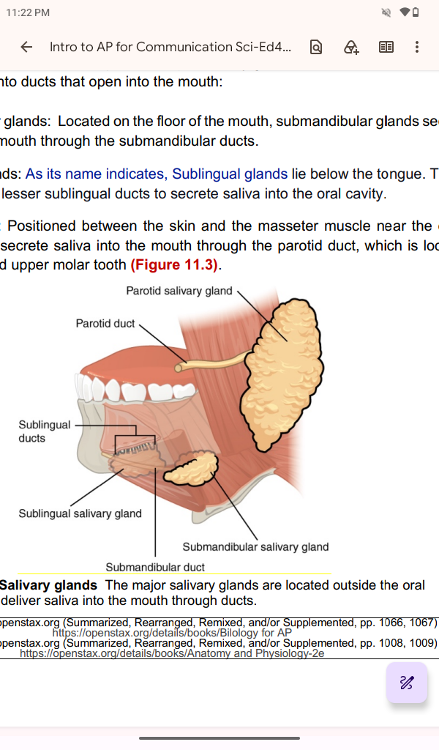
Outside the oral mucosa are three pairs of major salivary glands,
which secrete the
majority of saliva into ducts that open into
the mouth:
• Submandibular glands: Located on the floor of the
mouth, submandibular glands secrete
saliva into the mouth
through the submandibular ducts.
• Sublingual glands: As its name
indicates, Sublingual glands lie below the tongue. These
glands
use the lesser sublingual ducts to secrete saliva into the oral
cavity.
• Parotid glands: Positioned between the skin and the
masseter muscle near the ears,
parotid glands secrete saliva
into the mouth through the parotid duct, which is located
near
the second upper molar tooth
Salivary glands The major salivary glands are located outside the
oral
mucosa and deliver saliva into the mouth through ducts.
The Parotid Glands: Mumps
The Parotid Glands: Mumps
Infections of the nasal passages and
pharynx can attack any salivary gland. The parotid glands
are
the usual site of infection with the virus that causes mumps
(paramyxovirus). Mumps
manifests by enlargement and inflammation
of the parotid glands, causing a characteristic
swelling between
the ears and the jaw. Symptoms include fever and throat pain, which
can be
severe when swallowing acidic substances such as orange
juice.
In about one-third of men who are past puberty, mumps also
causes testicular inflammation,
typically affecting only one
testis and rarely resulting in sterility. With the increasing use and
effectiveness of mumps vaccines, the incidence of mumps has
decreased dramatically.
According to the U.S. Centers for
Disease Control and Prevention (CDC), the number of
mumps cases
dropped from more than 150,000 in 1968 to fewer than 1700 in 1993 to
only 11
reported cases in 2011.
Saliva
Saliva
Saliva is essentially (95.5 percent) water. The remaining
4.5 percent is a complex mixture
of ions, glycoproteins,
enzymes, growth factors, and waste products. Perhaps the most
important ingredient in saliva from the perspective of digestion
is the enzyme called salivary
amylase. Salivary amylase
initiates the breakdown of carbohydrates. Food does not spend
enough time in the mouth to allow all the carbohydrates to break
down. Salivary amylase
continues acting until it is inactivated
by stomach acids. Bicarbonate and phosphate ions
function as
chemical buffers, maintaining saliva at a power of the hydrogen ion
(pH) level
between 6.35 and 6.85. Salivary mucus helps lubricate
food, facilitating movement in the
mouth, bolus formation, and
swallowing. Saliva contains immunoglobulin A, which prevents
microbes from penetrating the epithelium, and lysozyme, which
makes saliva antimicrobial.
Saliva also contains epidermal
growth factor, which might have given rise to the adage “a
mother’s kiss can heal a wound.”
Each of the major
salivary glands secretes a unique formulation of saliva according to
its
cellular makeup. For example, the parotid glands secrete a
watery solution that contains
salivary amylase. The
submandibular glands have cells similar to those of the parotid
glands,
as well as mucus-secreting cells. Therefore, saliva
secreted by the submandibular glands
also contains amylase. The
sublingual glands contain mostly mucous cells, and they secrete
the thickest saliva with the least amount of salivary amylase.
Regulation of Salivation
Regulation of Salivation
The autonomic nervous system regulates
salivation (the secretion of saliva). In the
absence of food,
parasympathetic stimulation keeps saliva flowing at just the right
level for
comfort as you speak, swallow, sleep, and generally go
about life.
Over-salivation can occur, for example, if you are stimulated by the
smell of food, but that
food is not available for you to eat.
Drooling is an extreme instance of the overproduction of
saliva.
During times of stress, such as before speaking in public, sympathetic
stimulation
takes over, reducing salivation and producing the
symptom of dry mouth often associated
with anxiety. When you are
dehydrated, salivation is reduced, causing the mouth to feel dry
and prompting you to take action to quench your thirst.
Salivation can be stimulated by the
sight, smell, and taste of
food. It can even be stimulated by thinking about food. You might
notice whether reading about food and salivation right now has
had any effect on your
production of saliva.
How does the
salivation process work while you are eating? Food contains chemicals
that
stimulate taste receptors on the tongue, which send
impulses to the superior and inferior
salivatory nuclei in the
brain stem. These two nuclei then send back parasympathetic
impulses through fibers in the glossopharyngeal and facial
nerves, which stimulate salivation.
Even after you swallow food,
salivation is increased to cleanse the mouth and water down
and
neutralize any irritating chemical remnants, such as that hot sauce in
your burrito. Most
saliva is swallowed along with food and is
reabsorbed so that fluid is not lost.
..
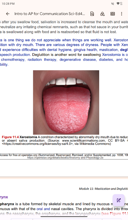
Saliva is one thing we do not appreciate when things are working
well. Xerostomia is a
condition with dry mouth. There are
various degrees of dryness. People with Xerostomia
could
experience difficulties with dental hygiene, gingiva health,
mastication, deglutition,
and speech production. Deglutition is
another word for swallowing Xerostomia is common
with
chemotherapy, radiation therapy, degenerative disease, diabetes, and
hormonal
instability.
Xerostomia A condition characterized by abnormally dry mouth due to
reduced
or absent saliva production. (Source:
www.scientificanimations.com, CC BY-SA 4.0
<https://creativecommons.org/licenses/by-sa/4.0>, via
Wikimedia Commons
Pharynx
Pharynx
The pharynx is a tube formed by skeletal muscle and
lined by mucous membrane that is
continuous with that of the
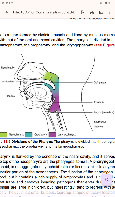
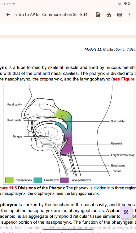
oral and nasal cavities. The pharynx is divided into three major
regions: the nasopharynx, the oropharynx, and the laryngopharynx
(see Figure 11.5)
Divisions of the Pharynx The pharynx is divided into three regions:
the nasopharynx, the oropharynx, and the laryngopharynx.
The nasopharynx is flanked by the conchae of the nasal cavity, and it
serves only as an
airway. At the top of the nasopharynx are the
pharyngeal tonsils. A pharyngeal tonsil, also
called an adenoid,
is an aggregate of lymphoid reticular tissue similar to a lymph node
that
lies at the superior portion of the nasopharynx. The
function of the pharyngeal tonsil is not
well understood, but it
contains a rich supply of lymphocytes and is covered with ciliated
epithelium that traps and destroys invading pathogens that enter
during inhalation. The
pharyngeal tonsils are large in children,
but interestingly, tend to regress with age and may
even
disappear. The uvula is a small bulbous, teardrop-shaped structure
located at the apex
of the soft palate. Both the uvula and soft
palate move like a pendulum during swallowing,
swinging upward
to close off the nasopharynx to prevent ingested materials from
entering
the nasal cavity. In addition, the auditory
(Eustachian) tube connects to each middle ear
cavity and opens
into the nasopharynx. This connection is why colds often lead to
middle
ear infections and why individuals with a cold may
complain of a full feeling in their ears. ,
the nasopharynx, is
involved only in breathing and speech.
..
the oropharynx and the laryngopharynx are used for both breathing and
digestion. The
oropharynx begins inferior to the nasopharynx and
is continuous below with the
laryngopharynx (Figure 11.6, p.
306). The inferior border of the laryngopharynx connects
to the
esophagus, whereas the anterior portion connects to the larynx,
allowing air to flow
into the bronchial tree.
..
The oropharynx is a passageway for both air and food. The oropharynx
is bordered
superiorly by the nasopharynx and anteriorly by the
oral cavity. The fauces are the opening
at the connection
between the oral cavity and the oropharynx. As the nasopharynx becomes
the oropharynx, the epithelium changes from pseudostratified
ciliated columnar epithelium
to stratified squamous epithelium.
Both epithelium cells have multiple layers. Because the
nasopharynx passes only air through the nasal cavity, the
covering is rather simple. In
contrast, the oropharynx could
interact with incoming food (liquid) and air. Therefore, its
covering is the stratified squamous epithelium, which has
protective lining structures. The
oropharynx contains two
distinct sets of tonsils, the palatine and lingual tonsils. A palatine
tonsil is one of a pair of structures located laterally in the
oropharynx in the area of the
fauces. The lingual tonsil is
located at the base of the tongue. Similar to the pharyngeal
tonsil, the palatine and lingual tonsils are composed of
lymphoid tissue, and trap and destroy
pathogens entering the
body through the oral or nasal cavities.
..
The laryngopharynx is inferior to the oropharynx and posterior to the
larynx. It continues the
route for ingested material and air
until its inferior end, where the digestive and respiratory
systems diverge. The stratified squamous epithelium of the
oropharynx is continuous with
the laryngopharynx. Anteriorly,
the laryngopharynx opens into the larynx, whereas
posteriorly,
it enters the esophagus.
..
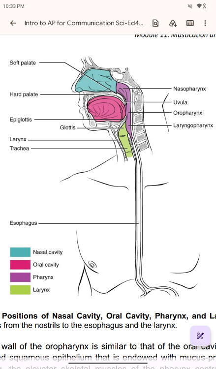
The pharynx is involved in both digestion and respiration. It
receives food and air from the
mouth, and air from the nasal
cavities. When food enters the pharynx, involuntary muscle
contractions close off the air passageways. The pharynx, a short
tube of skeletal muscle
lined with a mucous membrane, runs from
the posterior oral and nasal cavities to the
opening of the
esophagus and larynx.
Positions of Nasal Cavity, Oral Cavity, Pharynx, and Larynx The
pharynx runs from the nostrils to the esophagus and the larynx.
..
Histologically, the wall of the oropharynx is similar to that of the
oral cavity. The mucosa
includes a stratified squamous
epithelium that is endowed with mucus-producing glands.
During
swallowing, the elevator skeletal muscles of the pharynx contract,
raising and
expanding the pharynx to receive the bolus of food.
Once received, these muscles relax,
and the constrictor muscles
of the pharynx contract. The contraction forces the bolus into
the esophagus and initiating peristalsis.
Usually, during
swallowing, the soft palate and uvula rise reflexively to close off
the entrance
to the nasopharynx. At the same time, the larynx is
pulled superiorly. Epiglottic inversion
happens to cover the
glottis (the opening to the larynx); this process effectively blocks
access to the trachea and bronchi. When the food “goes down the
wrong way,” it goes into
the trachea. When food enters the
trachea, we cough and force the food up and out of the
trachea.
Further, the food will move back into the pharynx. The cough is the
body’s
mechanism to clear the airway from unwanted matter, such
as food, saliva, or other foreign
matters.
..
Muscles of the Anterior Neck
Muscles of the Anterior Neck
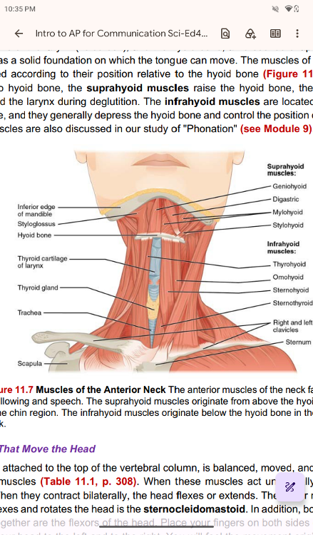
The muscles of the anterior neck
assist in deglutition (swallowing) and speech by controlling
the
positions of the larynx (voice box), and the hyoid bone, a
horseshoe-shaped bone that
functions as a solid foundation on
which the tongue can move. The muscles of the neck are
categorized according to their position relative to the hyoid
bone (Figure 11.7). Located
superior to hyoid bone, the
suprahyoid muscles raise the hyoid bone, the floor of the
mouth,
and the larynx during deglutition. The infrahyoid muscles are located
inferiorly to
hyoid bone, and they generally depress the hyoid
bone and control the position of the larynx.
These muscles are
also discussed in our study of "Phonation" (see Module 9).
Muscles of the Anterior Neck The anterior muscles of the neck
facilitate
swallowing and speech. The suprahyoid muscles
originate from above the hyoid bone
in the chin region. The
infrahyoid muscles originate below the hyoid bone in the lower
neck.
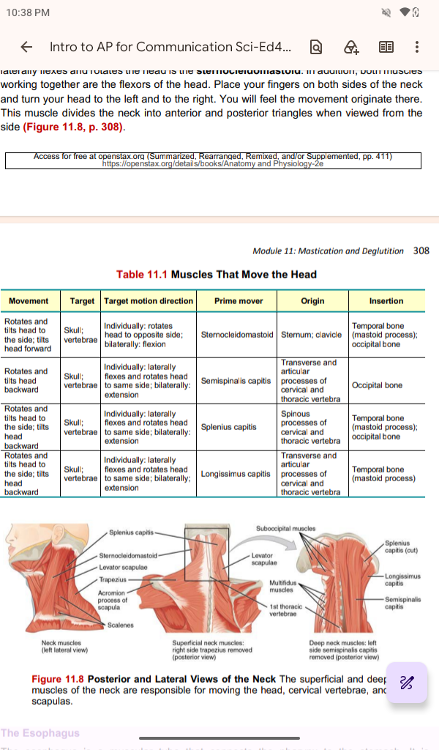
Muscles That Move the Head
Muscles That Move the Head
The head, attached to the top of the
vertebral column, is balanced, moved, and rotated by
the neck
muscles (Table 11.1, p. 308). When these muscles act unilaterally, the
head
rotates. When they contract bilaterally, the head flexes or
extends. The major muscle that
laterally flexes and rotates the
head is the sternocleidomastoid. In addition, both muscles
working together are the flexors of the head. Place your fingers
on both sides of the neck
and turn your head to the left and to
the right. You will feel the movement originate there.
This
muscle divides the neck into anterior and posterior triangles when
viewed from the
side (Figure 11.8, p. 308).
..
..
Posterior and Lateral Views of the Neck The superficial and deep
muscles of the neck are responsible for moving the head,
cervical vertebrae, and
scapulas.
..
Esophagus
The Esophagus
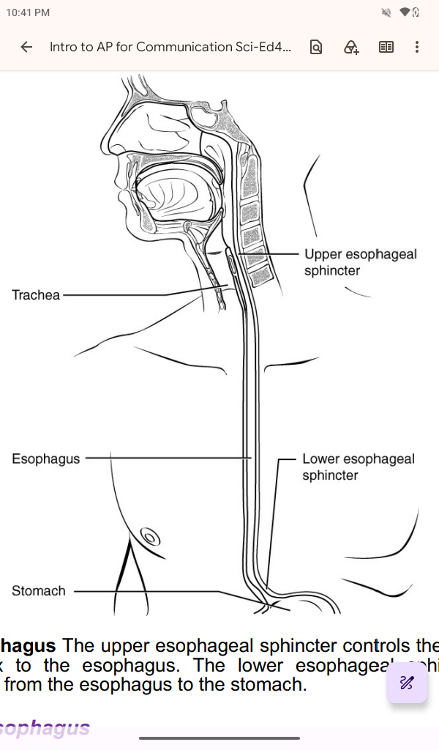
The esophagus is a muscular tube that connects the
pharynx to the stomach. It is
approximately 25.4 cm (10 in) in
length, located posterior to the trachea. It remains in a
collapsed form when not engaged in swallowing. As you can see in
Figure 11.9, p. 309, the
esophagus runs a mainly straight route
through the mediastinum of the thorax. To enter the
abdomen, the
esophagus penetrates the diaphragm through an opening called the
esophageal hiatus
Esophagus The upper esophageal sphincter controls the movement of
food
from the pharynx to the esophagus. The lower esophageal
sphincter controls the
movement of food from the esophagus to
the stomach.
Histology of the Esophagus
Histology of the Esophagus
The mucosa of the esophagus is made
up of an epithelial lining that contains non-
keratinized,
stratified squamous epithelium, with a layer of basal and parabasal
cells. This
epithelium protects against erosion from food
particles. Under the epithelium, the esophagus
has lamina
propria, which is a layer of connective tissue. The mucosa’s lamina
propria
contains mucus-secreting glands. The muscularis layer
changes according to location: In
the upper third of the
esophagus, the muscularis is skeletal muscle. In the middle third, it
is
both skeletal and smooth muscle. In the lower third, it is
smooth muscle. As mentioned
previously, the most superficial
layer of the esophagus is called the adventitia, not the
serosa.
In contrast to the stomach and intestines, the loose connective tissue
of the
adventitia is not covered by a fold of the visceral peritoneum.
Passage of Food through the Esophagus
Passage of Food through the Esophagus
The upper esophageal
sphincter (UES), which is continuous with the inferior pharyngeal
constrictor, controls the movement of food from the pharynx into
the esophagus. The upper
two-thirds of the esophagus consists of
both smooth and skeletal muscle fibers, with the
latter fading
out in the bottom third of the esophagus
Rhythmic waves of peristalsis (see Figure 11.10), which begin in the
upper esophagus,
propel the bolus of food toward the stomach.
Peristalsis is the involuntary, wave-like muscle
contraction
that moves food and liquids through the digestive tract. Meanwhile,
secretions
from the esophageal mucosa lubricate the esophagus
and food. Food passes from the
esophagus into the stomach at the
lower esophageal sphincter (LES) (also called the
gastroesophageal or cardiac sphincter). Sphincters are muscles
surrounding tubes and
serve as valves, closing the tube when the
sphincters contract and opening it when they
relax. The lower
esophageal sphincter relaxes to let food pass into the stomach, and
then
contracts to prevent stomach acids from backing up into the
esophagus. Surrounding this
sphincter is the muscular diaphragm,
which helps close off the sphincter when food is not
being swallowed.
..
When the lower esophageal sphincter does not completely close, the
stomach’s contents
can reflux (that is, back up into the
esophagus), causing heartburn or gastroesophageal
reflux disease
(GERD). When the upper esophageal sphincter does not completely close,
the stomach acid can flow out of the esophagus, results in
laryngopharyngeal reflux
disease (LPRD). Unlike GERD, it often
does not cause heartburn. Because the digestive
way and airway
are next to each other, the stomach acid often leaks to the airway,
causing
edema (swelling) of the posterior part of vocal folds.
LPRD often causes voice disorders
without other digestive
symptoms. For this reason, LPRD is called “silent reflux.” Individuals
with LPRD experience a hoarse voice in the morning, and the
voice improves later in the
day. The management of GERD and LPRD
includes the pharmacological approach,
behavioral modifications,
and surgical treatment, depending on the degree of the problem
Peristalsis The esophagus transfers food from the mouth to the
stomach
through peristaltic movements. Peristalsis creates
involuntary wave-like motions of
continuous partial constriction
and relaxation of the esophagus to propel the food into
the stomach.
Aging and the...
Digestive System: From Appetite Suppression to
Constipation
Age-related changes in the digestive system begin in
the mouth and can affect virtually
every aspect of the digestive
system. Taste buds become less sensitive, so food isn’t as
appetizing as it once was. A slice of pizza is a challenge, not
a treat, when you have lost
teeth, your gums are diseased, and
your salivary glands aren’t producing enough saliva.
Swallowing
can be difficult, and ingested food moves slowly through the
alimentary
canal because of reduced strength and tone of muscular
tissue. Neurosensory feedback
is also dampened, slowing the
transmission of messages that stimulate the release of
enzymes
and hormones. Pathologies that affect the digestive organs—such as
hiatal
hernia, gastritis, and peptic ulcer disease—can occur at
greater frequencies as you age.
Problems in the small intestine
may include duodenal ulcers, maldigestion, and
malabsorption.
Problems in the large intestine include hemorrhoids, diverticular
disease,
and constipation. Conditions that affect the function
of accessory organs—and their
abilities to deliver pancreatic
enzymes and bile to the small intestine—include jaundice,
acute
pancreatitis, cirrhosis, and gallstones.
..
Physiology of Swallowing
Physiology of Swallowing
When we orally intake solid or liquid,
we arrange the material in a manageable way for our
oral
structure. For example, when we have a cookie, we put a bite in our
mouth and chew.
The food forms bolus. After mastication, we
swallow. The act of swallowing is called
deglutition.
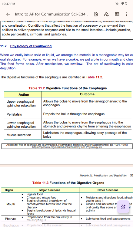
The
digestive functions of the esophagus are identified in
..
..
Deglutition
Deglutition
Deglutition is another word for swallowing—the
movement of food from the mouth to the
stomach. The entire
process takes about 4 to 8 seconds for solid or semisolid food, and
about 1 second for very soft food and liquids. Although this
sounds quick and effortless,
deglutition is, in fact, a complex
process that involves both the skeletal muscle of the tongue
and
the muscles of the pharynx and esophagus.
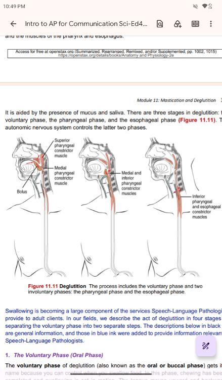
It is aided by the presence of mucus and saliva. There are three
stages in deglutition: the
voluntary phase, the pharyngeal
phase, and the esophageal phase (Figure 11.11). The
autonomic
nervous system controls the latter two phases.
Deglutition The process includes the voluntary phase and two
involuntary phases: the pharyngeal phase and the esophageal phase.
Swallowing is becoming a large component of the services
Speech-Language Pathologists
provide to adult clients. In our
fields, we describe the act of deglutition in four stages by
separating the voluntary phase into two separate steps. The
descriptions below in black ink
are general information, and
those in blue ink were added to provide information relevant to
Speech-Language Pathologists.
..
The Voluntary Phase (Oral Phase)
The Voluntary Phase (Oral Phase)
The voluntary phase of
deglutition (also known as the oral or buccal phase) gets its
name because you can control when you swallow food. In this
phase, chewing has been
completed and swallowing is set in
motion. The tongue moves upward and backward
against the palate,
pushing the bolus to the back of the oral cavity and into the
oropharynx.
Other muscles keep the mouth closed and prevent food
from falling out. At this point, the
two involuntary phases of
swallowingbegin. This phase can be classified into two separate
stages.
1a. Oral Preparatory phase
1a. Oral Preparatory phase
• The food is masticated, mixed with
saliva, and formed into a cohesive bolus held
against the hard
palate.
• The duration of this phase is variable by the type of
material to swallow, individual
difference in oral motor
capacity and preference.
• We can control this phase, and
therefore, it is voluntary.
1b. Oral phase
1b. Oral phase
• The oral phase begins when the lips seal and
the tongue begins moving the bolus
posteriorly.
• The
tongue forms a central groove so that the food can move following to
the ramp.
• The oral phase is considered voluntary.
• This
phase typically takes less than one second.
The Pharyngeal Phase
The Pharyngeal Phase
In the pharyngeal phase, stimulation of
receptors in the oropharynx sends impulses to the
deglutition
center (a collection of neurons that controls swallowing) in the
medulla oblongata.
Impulses are then sent back to the uvula and
soft palate, causing them to move upward and
close off the
nasopharynx. The laryngeal muscles also constrict to prevent
aspiration of food
into the trachea. At this point, deglutition
apnea takes place, which means that breathing
ceases for a very
brief time.
Contractions of the pharyngeal constrictor muscles
move the bolus through the oropharynx
and laryngopharynx.
Relaxation of the upper esophageal sphincter then allows food to enter
the esophagus.
The pharyngeal phase takes one second or less.
• This phase
begins with the triggering of the swallow response or pharyngeal
response at the anterior facial pillars.
• The triggering
of the swallow causes several physiologic activities to occur in the
pharynx simultaneously:
o Protection of nasal passage:
velopharyngeal closure
o Protection of the airway: laryngeal
elevation (superior and posterior or up and
back); inversion of
the epiglottis; closure of aryepiglottic folds, false vocal folds,
and true vocal folds
o Facilitation of swallowing:
Initiation of pharyngeal peristalsis to help bolus
moving from
pharynx to esophagus.
...
Protection of nasal passage
Protection of nasal passage: velopharyngeal closure
Protection of the airway:
Protection of the airway: laryngeal elevation (superior and posterior
or up and
back); inversion of the epiglottis; closure of
aryepiglottic folds, false vocal folds,
and true vocal folds
Facilitation of swallowing: Initiation of pharyngeal peristalsis to
help bolus
moving from pharynx to esophagus. •
Access for
free at openstax.org (Summarized, Rearranged, R
Facilitation of swallowing
The Esophageal Phase
The entry of food into the esophagus marks
the beginning of the esophageal phase of
deglutition and the
initiation of peristalsis. As in the previous phase, the complex
neuromuscular actions are controlled by the medulla oblongata.
Peristalsis propels the bolus
through the esophagus and toward
the stomach. The circular muscle layer of the muscularis
contracts, pinching the esophageal wall and forcing the bolus
forward. At the same time, the
longitudinal muscle layer of the
muscularis also contracts, shortening this area and pushing
out
its walls to receive the bolus. In this way, a series of contractions
keep moving food
toward the stomach. When the bolus nears the
stomach, distention of the esophagus
initiates a short reflex
relaxation of the lower esophageal sphincter that allows the bolus to
pass into the stomach. During the esophageal phase, esophageal
glands secrete mucus
that lubricates the bolus and minimizes
friction. It is important to note that esophageal
dysphagia is
not in the scope of practice of SLPs. In the event that esophageal
dysphagia
is suspected, the evaluating SLP is expected to make a
referral to a physician who
specializes in Gastrointestinal (GI) processes
..
The esophageal phase begins as the bolus enters the esophagus, and
the bolus is
passed through into the stomach. Normal esophageal
transit time is 8 to 20 seconds.
• When the bolus goes into the
airway instead of the digestive way and stops at above the
vocal
folds level, we call it penetration. When it passes the vocal folds
and enters into
the lower airway, we call it aspiration.
Aspiration pneumonia occurs when food or
liquid passes into the
lungs and causes an infection.
..
Role of Speech-Language Pathologists in Swallowing
Role of Speech-Language Pathologists in Swallowing
Swallowing
evaluation and treatment are within the scope of the practice of
Speech-
Language Pathologists (SLP). SLPs assess and evaluate
swallowing disorders by observing
in person or using various
instruments, such as a Modified Barium Swallow Study (MBSS).
For
swallowing, SLPs work in a team of professionals, including
physicians, nurses,
Gastroenterologists, and other allied health
professionals.
Swallowing is personal, and therefore,
individuals with swallowing disorders may have
desire not to
follow swallowing advice presented by SLPs. It is important for SLPs
to present
factual and evidence-based information to individuals
with swallowing disorders so that they can make informed decisions to
address their situations. Because eating is a large part of
the
quality of life and social activities, it is often difficult for
individuals with dysphagia, or a
swallowing disorder, to adhere
to lifestyle changes that may be necessary to manage their
disorder.
Oral Mechanism Examination
An oral mechanism examination (OME)
is also referred to as "oral peripheral examination.
OME is
one of the first and essential parts of a speech and language and/or
swallowing
function evaluation following the chart or case
history review. It allows the SLP to assess
the structures and
functions of a client’s anatomy to determine if and why a disorder is
present. Ideally, your client or patient will be sitting up
during this assessment, but in cases
where patients are
medically fragile, this may not be the case. Let’s study how to
administer
an OME for a patient during your initial
evaluation.
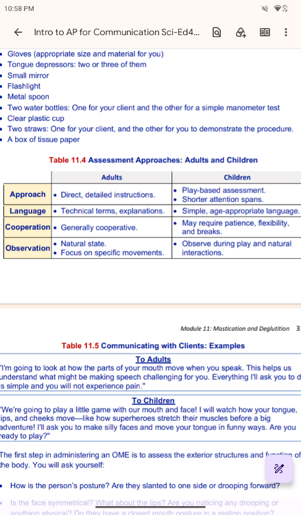
In general, SLPs use the following materials for
OME.
• Your OME procedure form: SLPs use various forms that are
customized for their needs.
In our diagnostic lab today, we will
use a published screening tool, Oral Speech
Mechanism Screening
Examination (OSMSE)-3.
• Writing materials
• Hand
sanitizer
• Gloves (appropriate size and material for you)
•
Tongue depressors: two or three of them
• Small mirror
•
Flashlight
• Metal spoon
• Two water bottles: One for your
client and the other for a simple manometer test
• Clear plastic
cup
• Two straws: One for your client, and the other for you to
demonstrate the procedure.
• A box of tissue paper
..
..
..
Communicating with Clients: Examples
To Adults
"I'm
going to look at how the parts of your mouth move when you speak. This
helps us
understand what might be making speech challenging for
you. Everything I'll ask you to do
is simple and you will not
experience pain."
To Children
"We're going to play
a little game with our mouth and face! I will watch how your tongue,
lips, and cheeks move—like how superheroes stretch their muscles
before a big
adventure! I'll ask you to make silly faces and
move your tongue in funny ways. Are you
ready to play?"
The first step in administering an OME is to assess the exterior
structures and function of
the body. You will ask
yourself:
• How is the person’s posture? Are they slanted to one
side or drooping forward?
• Is the face symmetrical? What about
the lips? Are you noticing any drooping or
anything atypical? Do
they have a closed mouth posture in a resting position?
• Is the
person short of breath? Do they appear to be chunking their words into
phrases
or single word utterances, or is their speech smooth and natural?
After you note the function of the exterior, you may proceed to the
next step of assessing an
individual’s function:
• Ask the
individual to pucker and then smile. You will check for coordination
during the
movement and the fluidity of the movement.
•
Afterward, you will ask the client to open their mouth. You will check
their dentition or
teeth to note any unusual structures or
formations, the size of the tongue, and their uvula.
For some
people, you may observe a bi-fid uvula, which is where they have a
split uvula.
Be sure to note the palatal arch and if there is
enlargement of the tonsils.
• Next, you will check the tongue.
Ask your client to stick their tongue straight out. Look for
anything atypical on the tongue (color, shape, or tremors in a
resting state).
• Then, ask your client to move their tongue
side-to-side and up-and-down. Here, you are
checking the range
of motion and coordination. A typical individual will be able to
complete these tasks with ease.
• After checking exterior
coordination, you will want to check how the tongue can handle
repetitive movements for speech. This step is completed using
the
speech diadochokinetic test or the ability to coordinate
lingual motion to complete
speech-like tasks. First, you will
have your client repeat saying /p/ as fast and accurately
as
possible until you stop. Next, elicit the same repetitive productions
of /t/ and /k/. After
checking the lingual movement in each
sound separately, you will combine the
movement to see how the tongue can coordinate the movements from the
anterior to
posterior directions. This trial can be completed by
having the client repeat “puh tuh kuh”
as fast and accurately as
possible until you stop. Check for fluidity in the production and
ability to move front-to-back with ease. Keep in mind that if
your client is a child with an
articulation disorder, speech
diadochokinetic test may be difficult, as they may not have
certain sounds in their inventory yet. In this case, the result
may not reflect the difficulties
in coordination but rather an
issue with producing the speech sound.
In a medical setting, you
may proceed to check the swallowing function or complete a
cognitive-linguistic evaluation to assess cognition and language
deficits. For our purposes,
it is important to check from the
top down for any sort of explanation to the deficits. As you
develop your clinical skills, you will develop a procedure for
completing an Oral Mechanism
Evaluation. There are some
screening tool procedures available, such as the Mann
Assessment
of Swallowing Ability, but often this procedure is based on clinical
judgment and
is used to get an initial impression of the
patient’s structures, function, and potential
explanations for deficits.
..
laryngopharynx
(p. 305) portion of the pharynx bordered by the oropharynx
superiorly and esophagus and trachea inferiorly; serves as a
route for both air
and food
aspiration Pneumonia
(p. 315) when food or liquid that passes into the lungs
causing
an infection, which results in pneumonia
deglutition
(p. 312) three-stage process of swallowing
dysphagia
(p. 316) a swallowing disorder
esophagus
(p. 300) muscular tube that runs from the pharynx to the stomach
esophageal phase
(p. 315) the entry of the food into the esophagus as it travels
into the stomach
gastroesophageal reflux disease (GERD)
(p. 310) condition with which the
stomach acid comes out of the
lower sphincter and re-enter the esophagus
laryngopharyngeal reflux disease (LPRD)
(p. 310) condition with which the
stomach acid comes out of the
upper sphincter and often leaks to the airway,
causing edema
(swelling) of the posterior part of vocal folds.
laryngopharynx
(p. 305) portion of the pharynx bordered by the oropharynx
superiorly and esophagus and trachea inferiorly; serves as a
route for both air
and food
oropharynx
(p. 305) part of the pharynx continuous with the oral cavity that
functions in respiration and digestion
oral mechanism examination (OME)
(p. 316) a preliminary step in assessment
for speech
pathologist to observe the structure and function of oral and
pharyngeal structures informally
penetration
(p. 315) when food or liquid passes to or just above the vocal folds
peristalsis
(p. 310) muscular contractions and relaxations that propel food
through
the GI tract
pharynx
(p. 304) throat. the canal that extends from the nasal and oral
cavities to
the larynx and esophagus
pharyngeal phase
(p. 314) the second phase of the swallowing process in which
the bolus travels through the oropharynx and laryngopharynx
upper esophageal sphincter
(p. 309) skeletal muscle sphincter that regulates
food movement
from the pharynx to the esophagus
voluntary phase
(p. 313) initial phase of deglutition, in which the bolus moves from
the mouth to the oropharynx
..
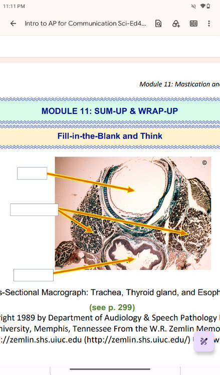
trachea,
thyroid gland,
esophagus
..
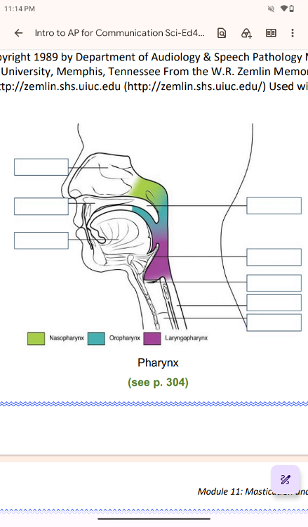
nasal cavity,
hard palate,
tongue,
soft palate,
epiglottis,
larynx (voice box),
esophagus,
traches
..
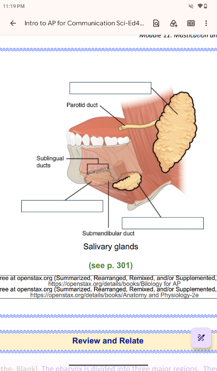
parotid salivary gland,
parotid duct,
sublingual ducts,
sublingual salivary gland,
submandibular duct,
Submandibular salivary gland
The pharynx is divided into three major regions. They are
_______________, ____________, and _____________
nasopharynx, oropharynx, laryngopharynx
2. [True/False] The pharynx is involved in both digestion and
respiration. It receives food
and air from the mouth, and air
from the nasal cavities. When food enters the pharynx,
involuntary muscle contractions close off the air passageways.
True
] ____________ creates involuntary wave-like motions of continuous
partial constriction and relaxation of the esophagus to propel
the food to the stomach.
Peristalsis
The act of chewing is called
mastication
] The act of swallowing is called
deglutition
] When we swallow, we briefly cease breathing. It is called
deglutition apnea