An Anterior View
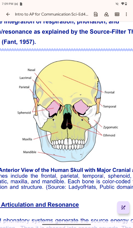
An Anterior View of the Human Skull with Major Cranial and Facial
ones
The labeled bones include the frontal, parietal, temporal,
sphenoid, ethmoid, nasal,
lacrimal, zygomatic, maxilla, and
mandible. Each bone is color-coded to distinguish its
anatomical
position and structure. (Source: LadyofHats, Public domain, via
Wikimedia
Commons)
Anatomy of Articulation and Resonance
Anatomy of Articulation and Resonance
The respiratory and
phonatory systems generate the source energy of speech production
for verbal communication. Then it is shaped into speech sounds.
The three subsystems of
speech production are respiration,
phonation, and articulation/resonance. They work in a
coordinated fashion to make verbal communication functional. The
complicated
synchronization of these subsystems produces speech
sounds sequentially to produce
meaningful utterances With oral
phonemes, the air escapes from the oral cavity, and with nasal
phonemes, it
escapes from the nasal cavity. We open and close
the velopharyngeal port to use nasal
or oral tract, respectively.
The smallest sound unit that makes meaning is called
"phoneme." In English, there are 44
basic phonemes,
and the strings of these form our verbal communication. An example of
a
phoneme is /p/. Phonemes are language-specific, which means
that different languages
have their own sets of
phonemes.
The voice generated at the level of vocal folds travels
through the upside-down L-shaped
cavity formed by the pharynx
and oral or nasal cavity. These passages from the vocal folds
to
the point that air escapes are called vocal tracts, and we have two of
them. The two
vocal tracts are the oral tract and nasal tract.
We use one of the two vocal tracts to produce
oral or nasal phonemes.
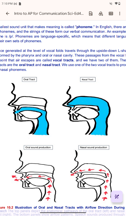
Illustration of Oral and Nasal Tracts with Airflow Direction
During
Speech The top panels depict the anatomical pathways of
the oral tract (left) and nasal
tract (right). The bottom
panels show airflow patterns. Speech sounds produced through
either pathway are shaped by resonance characteristics and
reflect specific places and
manners of articulation.
Velopharyngeal Port Movements During Speech
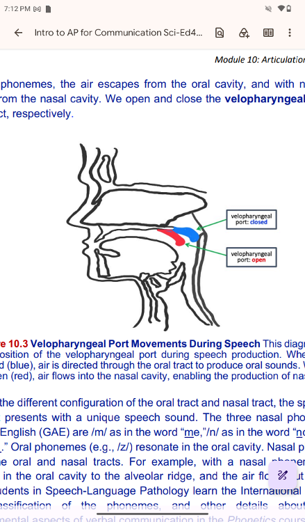
Velopharyngeal Port Movements During Speech This diagram illustrates
the position of the velopharyngeal port during speech
production. When the port is
closed (blue), air is directed
through the oral tract to produce oral sounds. When the port
is
open (red), air flows into the nasal cavity, enabling the production
of nasal sounds.
Based on the different configuration of the
oral tract and nasal tract, the speech sounds from
each tract
presents with a unique speech sound. The three nasal phonemes in
General
American English (GAE) are /m/ as in the word “me,”/n/
as in the word “no,” and /ŋ/ as in the
word “sing.” Oral
phonemes (e.g., /z/) resonate in the oral cavity. Nasal phonemes
resonate
in both the oral and nasal tracts. For example, with a
nasal phoneme /n/, the sound
resonates in the oral cavity to the
alveolar ridge, and the air flows out through the nasal
cavity.
Students in Speech-Language Pathology learn the International Phonetic
Alphabet
(IPA), classification of the phonemes, and other
details about segmental and
suprasegmental aspects of verbal
communication in the Phonetics course.
Articulation is the
contact, approximation, or separation of speech organs for speech
production. There are fixed articulators and movable
articulators. Fixed articulators do not
move independently, such
as teeth and hard palate. Movable articulators move freely, such
as lips, tongue, jaws, and soft palate. Next, we will study the
structures related to
articulation/resonance.
The Mouth and Surrounding Structures
The cheeks, tongue, and
palate frame the mouth, which is also called the oral cavity (or
buccal cavity). The structures of the mouth are illustrated in
At the entrance to the mouth are the lips, or labia (or labium if
talking about one lip). Their
outer covering is skin, which
transitions to a mucous membrane in the mouth. Lips are
very
vascular with a thin layer of keratin; hence, they are
"red." They have a substantial
representation on the
cerebral cortex. The lips cover the orbicularis oris muscle, which
surrounds the mouth and regulates what comes in and goes out of
the mouth.
..
labial frenulum
The labial frenulum is a midline fold of mucous membrane that
attaches the inner surface
of each lip to the gum. The cheeks
make up the oral cavity’s sidewalls. While their outer
covering
is skin, their inner covering is mucous membrane. This membrane
consists of non-
keratinized, stratified squamous epithelium,
featuring layers of cells that secrete mucus or
saliva to serve
as a protective barrier between the oral cavity and internal organs.
The
function of stratified squamous epithelium is to protect
structures against abrasion. Between
the skin and mucous
membranes are connective tissue and buccinator (the cheek)
muscles.
The next time you eat some food, notice how the
buccinator muscles in your cheeks and the
orbicularis oris muscle
around your lips contract, helping you keep the food from falling out
of your mouth. Additionally, notice how these muscles work when
you are speaking.
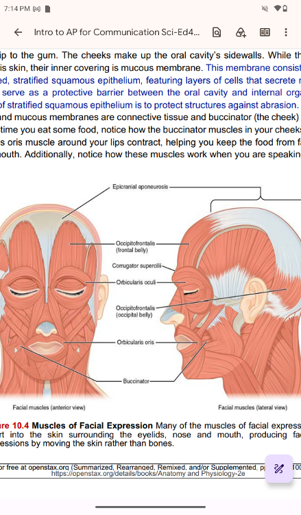
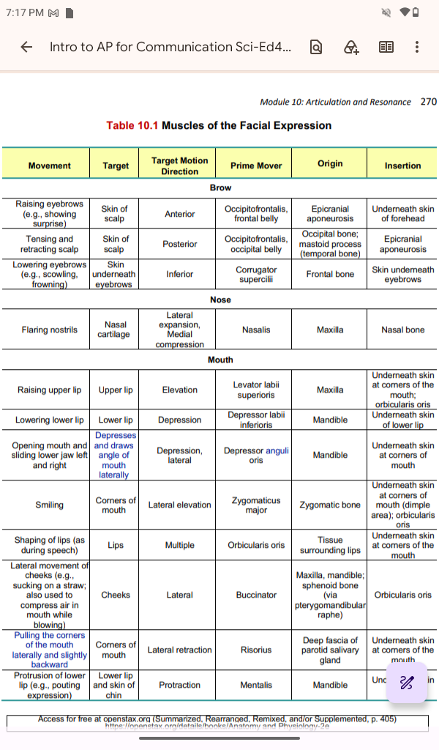
Muscles of Facial Expression Many of the muscles of facial
expression
insert into the skin surrounding the eyelids, nose
and mouth, producing facial
expressions by moving the skin
rather than bones.
orbicularis oris
The orbicularis oris is a circular muscle that moves the lips. The
orbicularis oculi is a
circular muscle that closes the eye. The
occipitofrontalis muscle moves up the scalp and
eyebrows. The
muscle has a frontal belly and an occipital (near the occipital bone
on the
posterior part of the skull) belly. In other words, there
is a muscle on the forehead (frontalis)
and one on the back of
the head (occipitalis), but there is no muscle across the top of the
head. Instead, the two bellies are connected by a broad tendon
called epicranial
aponeurosis, or galea aponeurosis (galea =
“apple”) (Figure 10.4, p. 268). The physicians
originally
studying human anatomy thought the skull looked like an apple.
A large portion of the face is composed of the buccinator muscle,
which compresses the
cheek. This muscle allows you to whistle,
blow, and suck; and it contributes to the action of
chewing.
There are several small facial muscles, one of which is the corrugator
supercilii,
which is the prime mover of the eyebrows. Place your
finger on your eyebrows at the point
of the bridge of the nose.
Raise your eyebrows as if you were surprised and lower your
eyebrows as if you were frowning. With these movements, you can
feel the action of the
corrugator supercilii. Additional muscles
of facial expression are presented in
..
Knowledge about the muscles in facial expression is essential for
SLPs. Facial expression
impacts communication greatly and
transmits various non-linguistic information. Also,
damages to
certain muscles for facial expression could impact the articulator and
resonator
movements. For example, facial paralysis, secondly to
the nerve damages that affect the
lower part of the face may
cause difficulties in closing the lips completely. Individuals in such
situations could experience difficulty in producing bilabial
stop consonants.
We learned about Peripheral Nervous System
(PNS) in Module 4. Many of you remember
that Cranial Nerve (CN)
VII, facial nerve, innervates many muscles of facial expressions
(Module 4, p. 136). Also, Cranial Nerve (CN) V, trigeminal nerve
innervates muscles of the
lower jaw (Table 10.2, p. 273).
Dysfunctions of these muscles could be presented as the
loss of
intentional muscular activities (paralysis) or muscle weakness
(paresis). As with the
somatic regions, either paralysis or
paresis of these muscles of facial areas could be
secondary to
the deficits of the lower motor neurons or those of the upper motor neurons.
..
..
..
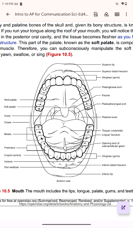
The pocket-like part of the mouth that is framed on the inside by the
gums and teeth, and
the outside by the cheeks and lips is called
the oral vestibule. Moving farther into the mouth,
the opening
between the oral cavity and throat (oropharynx) is called the fauces
(like the
kitchen "faucet"). The main open area of the
mouth, or oral cavity proper, runs from the
gums and teeth to
the fauces.
When you are chewing, you do not find it difficult to
breathe simultaneously. The next time
you have food in your
mouth, notice how the arched shape of the roof of your mouth allows
you to handle both digestion and respiration at the same time.
This arch is called the palate.
The anterior region of the
palate serves as a wall (or septum) between the oral and nasal
cavities. This region is a rigid shelf against which the tongue
can push food. It is created by
the maxillary and palatine bones
of the skull and, given its bony structure, is known as the
hard
palate. If you run your tongue along the roof of your mouth, you will
notice that the hard
palate ends in the posterior oral cavity,
and the tissue becomes fleshier as you feel the end
of the hard
structure. This part of the palate, known as the soft palate, is
composed mainly
of skeletal muscle. Therefore, you can
subconsciously manipulate the soft palate—for
instance, to yawn,
swallow, or sing
..
Mouth The mouth includes the lips, tongue, palate, gums, and teeth.
A fleshy bead of tissue called the uvula drops down from the center
of the posterior edge of
the soft palate. Although some have
suggested that the uvula is a vestigial organ, it serves
an
important purpose. A vestigial organ is a body structure that has lost
most or all of its
original function through evolution. When you
swallow, the soft palate and uvula move
upward, helping to keep
foods and liquid from entering the nasal cavity. Unfortunately, it can
also contribute to the sound produced by snoring.
Two
muscular folds extend downward from the soft palate on either side of
the uvula.
Toward the front, the palatoglossal arch lies next to
the base of the tongue; behind it, the
palatopharyngeal arch
forms the superior and lateral margins of the fauces. Between
these two arches are the palatine tonsils, clusters of lymphoid
tissue that protect the
pharynx. The lingual tonsils are located
at the base of the tongue.
..
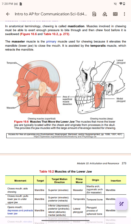
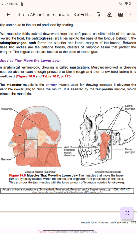
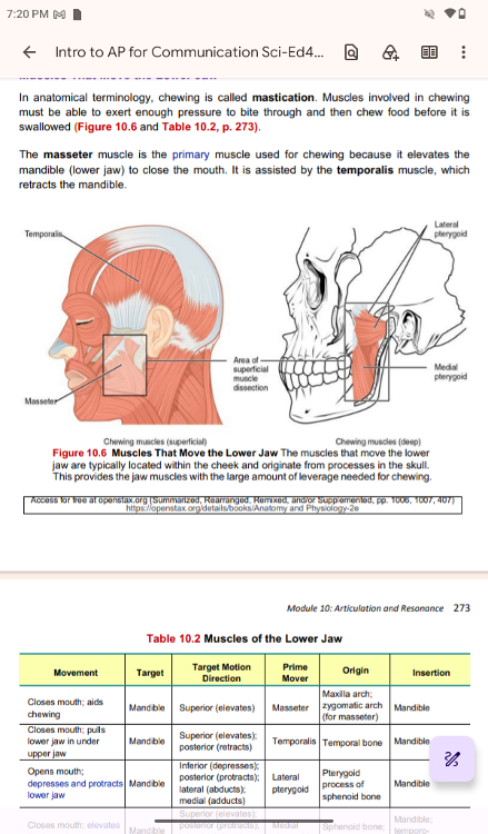
Muscles That Move the Lower Jaw
In anatomical terminology,
chewing is called mastication. Muscles involved in chewing
must
be able to exert enough pressure to bite through and then chew food
before it is
swallowed (Figure 10.6 and Table 10.2, p.
273).
The masseter muscle is the primary muscle used for chewing
because it elevates the
mandible (lower jaw) to close the mouth.
It is assisted by the temporalis muscle, which
retracts the mandible.
Muscles That Move the Lower Jaw The muscles that move the lower
jaw are typically located within the cheek and originate from
processes in the skull.
This provides the jaw muscles with the
large amount of leverage needed for chewing.
..
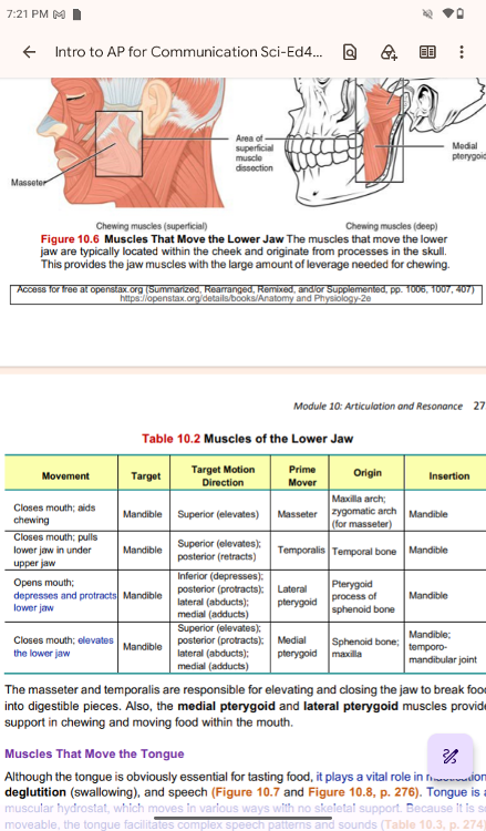
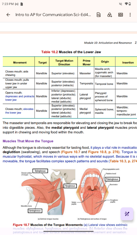
The masseter and temporalis are responsible for elevating and closing
the jaw to break food
into digestible pieces. Also, the medial
pterygoid and lateral pterygoid muscles provide
support in
chewing and moving food within the mouth.
..
Muscles That Move the Tongue
Muscles That Move the Tongue
Although the tongue is obviously
essential for tasting food, it plays a vital role in
mastication,
deglutition (swallowing), and speech (Figure 10.7
and Figure 10.8, p. 276). Tongue is a
muscular hydrostat, which
moves in various ways with no skeletal support. Because it is so
moveable, the tongue facilitates complex speech patterns and
sounds (Table 10.3, p. 274).
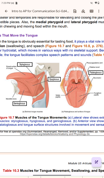
Figure 10.7 Muscles of the Tongue
Movements (a) Lateral view shows extrinsic
muscles:
styloglossus, hyoglossus, and genioglossus. (b) Anterior view shows
the
palatoglossus and tongue surface structures involved in
movement and sensatio
The masseter and temporalis are responsible for elevating and closing
the jaw to break food
into digestible pieces. Also, the medial
pterygoid and lateral pterygoid muscles provide
support in
chewing and moving food within the mouth.
..
Muscles That Move the Tongue
Muscles That Move the Tongue
Although the tongue is obviously
essential for tasting food, it plays a vital role in
mastication,
deglutition (swallowing), and speech (Figure 10.7
and Figure 10.8, p. 276). Tongue is a
muscular hydrostat, which
moves in various ways with no skeletal support. Because it is so
moveable, the tongue facilitates complex speech patterns and
sounds (Table 10.3, p. 274).
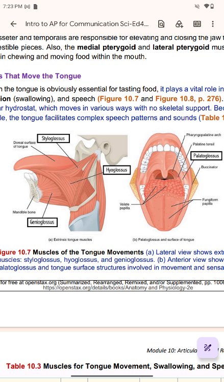
Figure 10.7 Muscles of the Tongue
Movements (a) Lateral view shows extrinsic
muscles:
styloglossus, hyoglossus, and genioglossus. (b) Anterior view shows
the
palatoglossus and tongue surface structures involved in
movement and sensation.
..
..
Tongue
The Tongue
Perhaps you have heard it said that the tongue is the
strongest muscle in the body. Those
who stake this claim cite
its strength proportionate to its size. Although it is difficult to
quantify
the relative strength of different muscles, it remains
indisputable that the tongue is a
workhorse, facilitating
ingestion, mechanical digestion, chemical digestion (lingual lipase),
sensation (of taste, texture, and temperature of food),
swallowing, and vocalization. The
process of mechanical
digestion breaks the food into small pieces using muscular activities,
such as chewing. Chemical digestion involves breaking the food
down to aid digestion by
using acids and enzymes in our body
fluid.
The tongue is attached to the mandible, the styloid
processes of the temporal bones, and
the hyoid bone. As we
discussed in Module 9 Phonation, the hyoid is unique in that it only
distantly/indirectly articulates with other bones. The tongue is
positioned over the floor of the
oral cavity. A medial septum
extends the entire length of the tongue, dividing it into
symmetrical halves.
Tongue muscles are classified into
extrinsic or intrinsic. Extrinsic tongue muscles insert into
the
tongue from outside origins. The intrinsic tongue muscles insert into
the tongue from
origins within it. The extrinsic muscles move
the whole tongue in different directions. The
intrinsic muscles
allow the tongue to change its shape (curling the tongue in a loop or
flattening it).
Beneath its mucous membrane covering, each half of the tongue is
composed of the same
number and type of intrinsic and extrinsic
skeletal muscles. The intrinsic muscles (those
within the
tongue) are the longitudinalis inferior, longitudinalis superior,
transversus linguae,
and verticalis linguae muscles. These
intrinsic and extrinsic tongue muscles allow you to
change the
size and shape of your tongue, and to stick it out, respectively.
Having such a
flexible tongue facilitates both swallowing and speech.
..
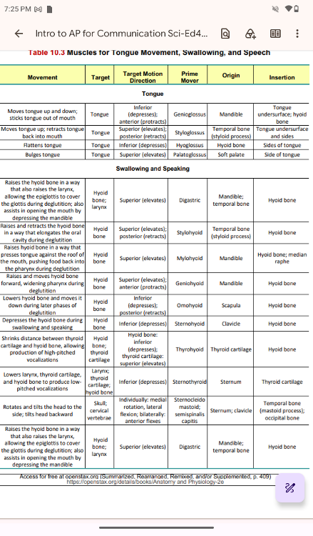
The extrinsic muscles (Figure 10.7, p. 273) of the tongue are the
palatoglossus
(glossopalatine), hyoglossus, styloglossus, and
genioglossus muscles. These muscles
originate outside the tongue
and insert into connective tissues within the tongue. The
extrinsic muscles all include the word root glossa (glossa =
"tongue"). The muscle names
are derived from where the
muscle originates. The genioglossus (genio = "chin")
originates
on the mandible and allows the tongue to move
downward and forward.
The styloglossus originates on the styloid
bone and allows upward and backward motion.
The palatoglossus
originates on the soft palate to elevate the back of the tongue, and
the hyoglossus originates on the hyoid bone to move the tongue
downward and flatten it.
These muscles perform three essential
digestive functions in the mouth: (1) position food
for optimal
chewing, (2) gather food into a bolus (rounded mass of food we form
for
swallowing), and (3) position food so it can be swallowed.
..
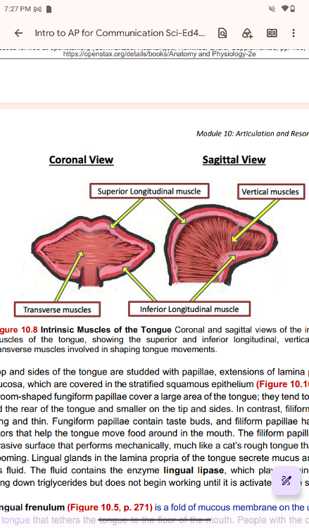
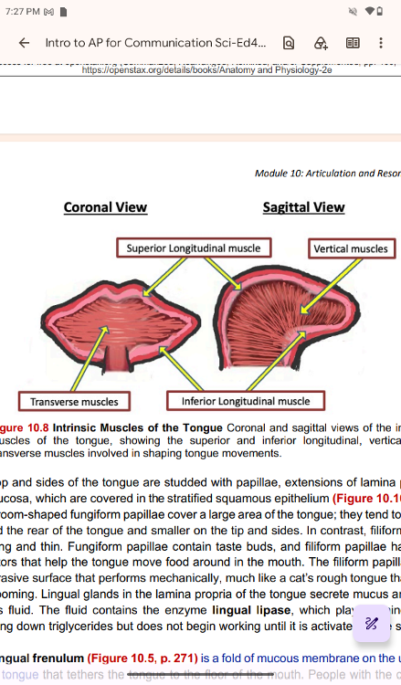
Intrinsic Muscles of the Tongue Coronal and sagittal views of the
intrinsic
muscles of the tongue, showing the superior and
inferior longitudinal, vertical, and
transverse muscles involved
in shaping tongue movements.
The top and sides of the tongue are
studded with papillae, extensions of lamina propria of
the
mucosa, which are covered in the stratified squamous epithelium
(Figure 10.10, p.277).
Mushroom-shaped fungiform papillae cover a
large area of the tongue; they tend to be larger
toward the rear
of the tongue and smaller on the tip and sides. In contrast, filiform
papillae
are long and thin. Fungiform papillae contain taste
buds, and filiform papillae have touch
receptors that help the
tongue move food around in the mouth. The filiform papillae create
an abrasive surface that performs mechanically, much like a
cat’s rough tongue that is used
for grooming. Lingual glands in
the lamina propria of the tongue secrete mucus and watery
serous
fluid. The fluid contains the enzyme lingual lipase, which plays a
minor role in
breaking down triglycerides but does not begin
working until it is activated in the stomach.
..
The lingual frenulum
The lingual frenulum (Figure 10.5, p. 271) is a fold of mucous
membrane on the underside
of the tongue that tethers the tongue
to the floor of the mouth. People with the congenital
anomaly
ankyloglossia, also known by the non-medical term “tongue-tie,” have a
lingual
frenulum that is too short or otherwise malformed.
Severe ankyloglossia could impair
feeding or cause oral-motor
deficiency. Many of you may think the short lingual frenulum
could affect speech. Although it could depend on the degree of
the impact on tongue mobility,
ankyloglossia seldom causes
problems with speech in English speakers (Kummer, 2020).
Except
for feeding difficulties, surgical management (frenulectomy) is not
indicated for
speech purposes alone. Although SLPs may be the
first to identify a reduced range of motion
(ROM) of the tongue,
it is not in our scope of practice to diagnose ankyloglossia. When an
SLP observes potential problems with ankyloglossia, he or she
may refer the client to a
physician for further
examination.
Access for free at openstax.org (Sum
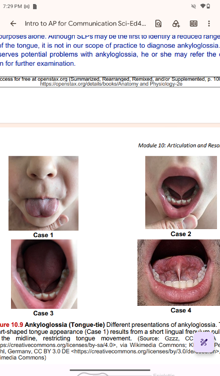
Ankyloglossia (Tongue-tie) Different presentations of ankyloglossia.
The
heart-shaped tongue appearance (Case 1) results from a short
lingual frenulum pulling
at the midline, restricting tongue
movement. (Source: Gzzz, CC BY-SA 4.0
<https://creativecommons.org/licenses/by-sa/4.0>, via
Wikimedia Commons; Klaus D. Peter,
Wiehl, Germany, CC BY 3.0 DE
<https://creativecommons.org/licenses/by/3.0/de/deed.en>, via
Wikimedia Commons)
..
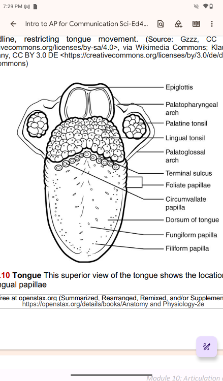
Tongue This superior view of the tongue shows the locations and
types of lingual papillae
..
Anesthesia and the Tongue Muscles
Anesthesia and the Tongue Muscles
Before surgery, general
anesthesia may be used. General anesthesia is a medically
induced state of unconsciousness with loss of sensation,
awareness, and reflexes used
during surgical procedures. The
normal homeostatic controls of the body are put “on
hold” so
that the patient can be prepped for surgery. Control of respiration
must be
switched from the patient’s homeostatic control to the
control of the anesthesiologist.
The drugs used for anesthesia
relax a majority of the body’s muscles.
Among the muscles
affected during general anesthesia are those that are necessary
for breathing and moving the tongue. Under anesthesia, the
tongue can relax and
partially or fully block the airway, and
the muscles of respiration may not move the
diaphragm or chest
wall. To avoid possible complications, the safest procedure to use
on a patient is called endotracheal intubation. Placing a tube
into the trachea allows the
doctors to maintain a patient’s
(open) airway to the lungs and seal the airway off from
the
oropharynx. Post-surgery, the anesthesiologist gradually changes the
mixture of
the gases that keep the patient unconscious. When the
muscles of respiration begin to
function, the tube is removed.
It still takes about 30 minutes for a patient to wake up,
and
for breathing muscles to regain control of respiration. After surgery,
most people
have a sore or scratchy throat for a few days.
The teeth
The Teeth
The teeth, or dentes (dens when talking about one
tooth), are organs similar to bones that
you use to tear, grind,
and otherwise mechanically break down (chew) food.
Types of Teeth
Types of Teeth
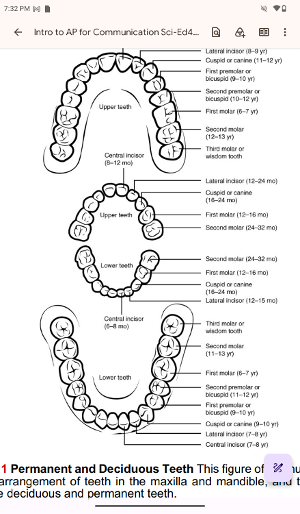
You have two sets of teeth during your lifetime (one set of teeth is called dentition). Your
20 deciduous teeth, or baby teeth, first begin to appear around 6 months of age. Between
approximately ages 6 and 12, these teeth are replaced by 32 permanent teeth. Moving
from the center of the mouth toward the side, these are as follows
Permanent and Deciduous Teeth This figure of two human dentitions
shows the arrangement of teeth in the maxilla and mandible, and
the relationship
between the deciduous and permanent teeth.
Permanent and Deciduous Teeth This figure of two human dentitions
shows the arrangement of teeth in the maxilla and mandible, and
the relationship
between the deciduous and permanent teeth.
Permanent and Deciduous Teeth This figure of two human dentitions
shows the arrangement of teeth in the maxilla and mandible, and
the relationship
between the deciduous and permanent teeth.
..
The eight incisors (four each on the top and the bottom) are the
sharp front teeth you
use for biting into food (used for cutting
or tearing).
• The four cuspids (or canines) flank the incisors
and have a pointed edge (cusp) to tear
up food. These fang-like
teeth are superb for piercing tough or fleshy foods.
• Posterior
to the cuspids are the eight premolars (or bicuspids), which have an
overall
flatter shape with two rounded cusps useful for mashing
foods.
• The most posterior and largest are the 12 molars, which
have several pointed cusps used
to crush food, preparing for
swallowing. The third members of each set of three molars,
top
and bottom, are commonly referred to as the wisdom teeth. Their
eruption is
commonly delayed until early adulthood. It is not
uncommon for wisdom teeth to fail to
erupt; that is, they remain
impacted (within the gums). In these cases, the teeth
are
typically removed by orthodontic surgery.
..
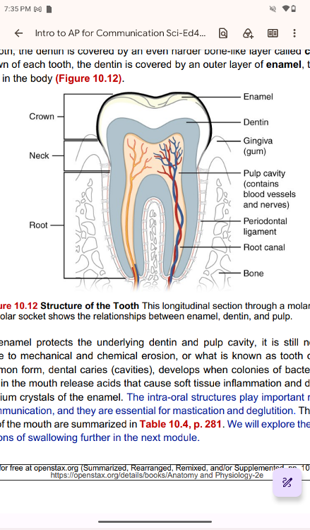
Anatomy of a Tooth
Anatomy of a Tooth
The teeth are secured in the alveolar
processes (sockets) of the maxilla (upper jaw) and
the mandible
(lower jaw). Gingivae (commonly called the gums) are soft tissues that
line
the alveolar processes and surround the necks of the teeth.
Teeth are also held in their
sockets by a connective tissue
called the periodontal ligament.
The two main parts of a tooth
are the crown and root. The crown is the portion projecting
above the gum line, and the root is embedded within the maxilla
and mandible. Both parts
contain an inner pulp cavity,
containing loose connective tissue through which run nerves
and
blood vessels. The region of the pulp cavity that runs through the
root of the tooth is
called the root canal. Surrounding the pulp
cavity is dentin, a bone-like tissue. In the root
of each tooth,
the dentin is covered by an even harder bone-like layer called
cementum.
In the crown of each tooth, the dentin is covered by
an outer layer of enamel, the hardest
substance in the body
Structure of the Tooth This longitudinal section through a molar in
its
alveolar socket shows the relationships between enamel,
dentin, and pulp.
Although enamel protects the underlying dentin
and pulp cavity, it is still nonetheless
susceptible to
mechanical and chemical erosion, or what is known as tooth decay. The
most common form, dental caries (cavities), develops when
colonies of bacteria feeding
on sugars in the mouth release
acids that cause soft tissue inflammation and degradation
of the
calcium crystals of the enamel. The intra-oral structures play
important roles in our
verbal communication, and they are
essential for mastication and deglutition. The digestive
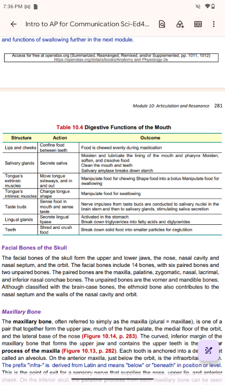
functions of the mouth are summarized in Table 10.4, p. 281. We
will explore the structures
and functions of swallowing further
in the next module.
..
..
..
Facial Bones of the Skull
Facial Bones of the Skull
The facial bones of the skull form the
upper and lower jaws, the nose, nasal cavity and
nasal septum,
and the orbit. The facial bones include 14 bones, with six paired
bones and
two unpaired bones. The paired bones are the maxilla,
palatine, zygomatic, nasal, lacrimal,
and inferior nasal conchae
bones. The unpaired bones are the vomer and mandible bones.
Although classified with the brain-case bones, the ethmoid bone
also contributes to the
nasal septum and the walls of the nasal
cavity and orbit.
Maxillary Bone
Maxillary Bone
The maxillary bone, often referred to simply as
the maxilla (plural = maxillae), is one of a
pair that together
form the upper jaw, much of the hard palate, the medial floor of the
orbit,
and the lateral base of the nose (Figure 10.14, p. 283).
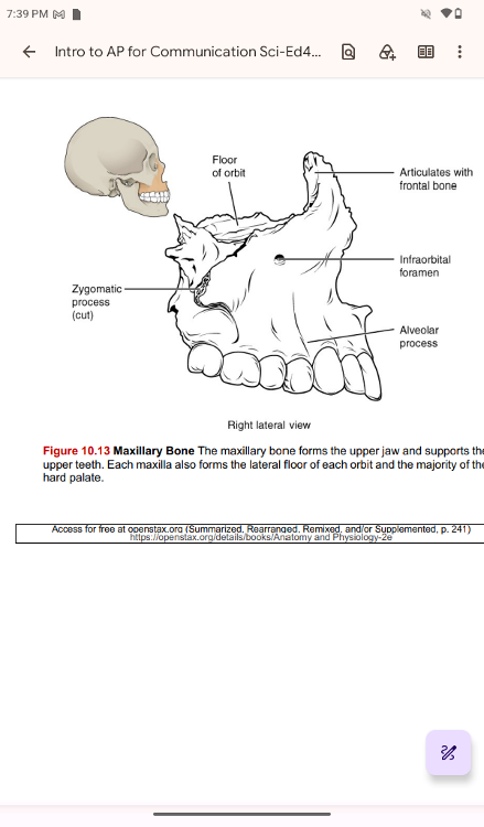
The curved, inferior margin of the
maxillary bone that forms the
upper jaw and contains the upper teeth is the alveolar
process
of the maxilla (Figure 10.13, p. 282). Each tooth is anchored into a
deep socket
called an alveolus. On the anterior maxilla, just
below the orbit, is the infraorbital foramen.
The prefix
"infra-" is derived from Latin and means "below"
or "beneath" in position or level.
This is the point
of exit for a sensory nerve that supplies the nose, upper lip, and
anterior
cheek. On the inferior skull, the palatine process from
each maxillary bone can be seen
joining together at the midline
to form the anterior three-quarters of the hard palate (Figure
10.16, p. 284). The hard palate is the bony plate that forms the
roof of the mouth and floor
of the nasal cavity, separating the
oral and nasal cavities.
Maxillary Bone The maxillary bone forms the upper jaw and supports
the
upper teeth. Each maxilla also forms the lateral floor of
each orbit and the majority of the
hard palate.
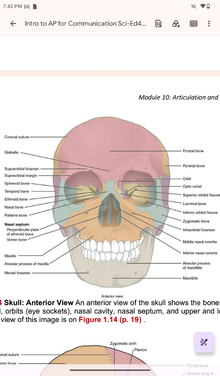
Skull: Anterior View An anterior view of the skull shows the bones
that form
the forehead, orbits (eye sockets), nasal cavity,
nasal septum, and upper and lower jaws.
An enlarged view of this
image is on Fi
..
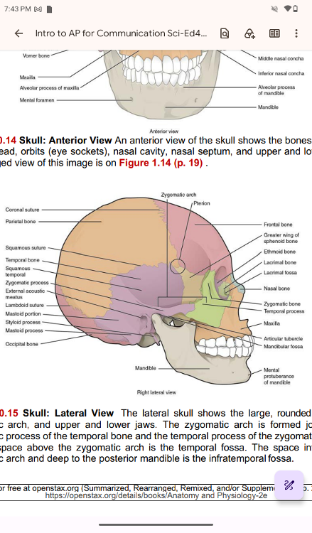
Skull: Lateral View The lateral skull shows the large, rounded brain
case,
zygomatic arch, and upper and lower jaws. The zygomatic
arch is formed jointly by the
zygomatic process of the temporal
bone and the temporal process of the zygomatic bone. The
shallow
space above the zygomatic arch is the temporal fossa. The space
inferior to the
zygomatic arch and deep to the posterior
mandible is the infratemporal fossa.
..
..
..
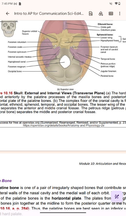
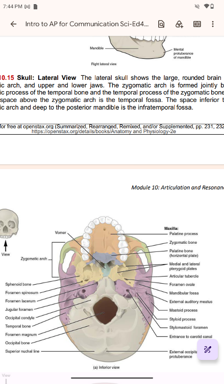
Skull: External and Internal Views (Transverse Plane) (a) The hard
palate is
formed anteriorly by the palatine processes of the
maxilla bones and posteriorly by the
horizontal plate of the
palatine bones. (b) The complex floor of the cranial cavity is formed
by
the frontal, ethmoid, sphenoid, temporal, and occipital
bones. The lesser wing of the sphenoid
bone separates the
anterior and middle cranial fossae. The petrous ridge (petrous portion
of
temporal bone) separates the middle and posterior cranial fossae
..
Palatine Bone
Palatine Bone
The palatine bone is one of a pair of irregularly
shaped bones that contribute small areas
to the lateral walls of
the nasal cavity and the medial wall of each orbit. The largest region
of each of the palatine bones is the horizontal plate. The
plates from the right and left
palatine bones join together at
the midline to form the posterior quarter of the hard
palate
(Figure 10.16, a, p. 284). Thus, the palatine bones are
best seen in an inferior view of the
skull and hard palate.
Zygomatic Bone
Zygomatic Bone
The zygomatic bone is also known as the
cheekbone. Each of the paired zygomatic bones
forms much of the
lateral wall of the orbit and the lateral-inferior margins of the
anterior
orbital opening (Figure 10.14, p. 283). The short
temporal process of the zygomatic bone
projects posteriorly,
where it forms the anterior portion of the zygomatic arch (Figure
10.15,
p. 283)
Nasal bone
Nasal Bone
The nasal bone is one of two small bones that
articulate (join) with each other to form the
bony base (bridge)
of the nose. They also support the cartilages that form the lateral
walls
of the nose (Figure 10.17). These are the bones that are
damaged when the nose is broken.
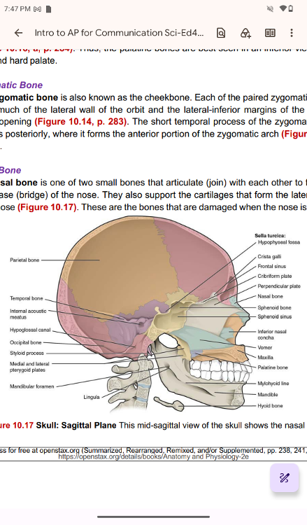
Skull: Sagittal Plane This mid-sagittal view of the skull shows the nasal septum
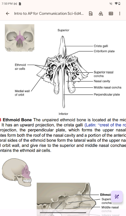
Ethmoid Bone
Ethmoid Bone The unpaired ethmoid bone is located at the midline
within the
central skull. It has an upward projection, the
crista galli (Latin: “crest of the rooster”), and a
downward
projection, the perpendicular plate, which forms the upper nasal
septum. The
cribriform plates form both the roof of the nasal
cavity and a portion of the anterior cranial fossa
floor. The
lateral sides of the ethmoid bone form the lateral walls of the upper
nasal cavity, part
of the medial orbit wall, and give rise to
the superior and middle nasal conchae. The ethmoid
bone also
contains the ethmoid air cells.
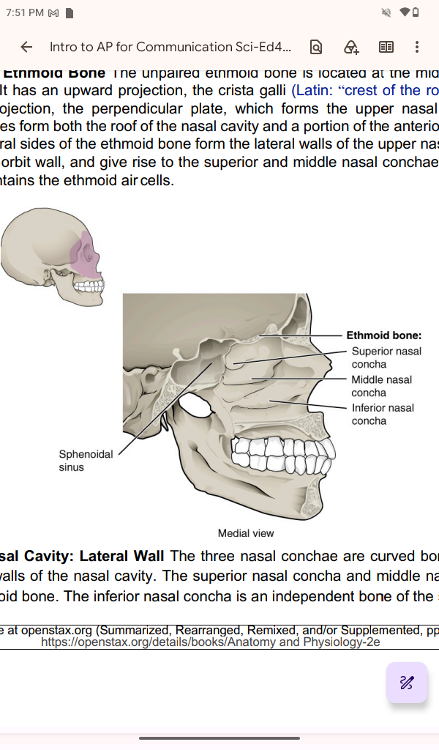
Nasal Cavity
Nasal Cavity: Lateral Wall The three nasal conchae are curved bones
that project
from the lateral walls of the nasal cavity. The
superior nasal concha and middle nasal concha are
parts of the
ethmoid bone. The inferior nasal concha is an independent bone of the skull.
Lacrimal Bone
Lacrimal Bone
Each lacrimal bone is a small, rectangular bone
that forms the anterior, medial wall of the
orbit (see Figure
10.14 and Figure 10.15, p. 283). The anterior portion of the lacrimal
bone
forms a shallow depression called the lacrimal fossa.
Extending inferiorly from lacrimal
fossa is the nasolacrimal
canal. The lacrimal fluid (tears of the eye), which serves to
maintain the moist surface of the eye, drains at the medial
corner of the eye into the
nasolacrimal canal. This duct then
extends downward to open into the nasal cavity, behind
the
inferior nasal concha. In the nasal cavity, the lacrimal fluid
normally drains posteriorly,
but with an increased flow of tears
due to crying or eye irritation, some fluid will also
drain
anteriorly, thus causing a runny nose
Inferior Nasal Conchae
Inferior Nasal Conchae
The right and left inferior areas of
nasal conchae form a curved bony plate that projects into
the
nasal cavity space from the lower lateral wall (see Figure 10.19, p.
286). The inferior
concha is the largest of the nasal conchae.
It can easily be seen when looking into the
anterior opening of
the nasal cavity.
Vomer Bone
Vomer Bone
The unpaired vomer bone often referred to simply as
the vomer, is triangular-shaped and
forms the posterior-inferior
part of the nasal septum (see Figure 10.17, p. 285). The vomer
is best seen when looking from behind into the posterior
openings of the nasal cavity (see
Figure 10.16a, p. 284). In this
view, the vomer is seen to form the entire height of the
nasal
septum. A much smaller portion of the vomer can also be
seen when looking into the anterior
opening of the nasal cavity.
Mandible
Mandible
The mandible forms the lower jaw and is the only
moveable bone of the skull. At the time of
birth, the mandible
consists of paired right and left bones, but these bones fuse
together
during the first year to form the single U-shaped
mandible of the adult skull. Each side of the
mandible consists
of a horizontal body, and posteriorly, a vertically oriented ramus of
the
mandible (ramus = “branch”). The outside margin of the
mandible, where the body and
ramus come together, is called the
angle of the mandible (
The ramus on each side of the mandible has two upward-going bony
projections. The more
anterior projection is the flattened
coronoid process of the mandible, which provides
attachment for
one of the biting muscles. The posterior projection is the condylar
process
of the mandible, which is topped by the oval-shaped
condyle. The condyle of the mandible
articulates (joins) with
the mandibular fossa and articular tubercle of the temporal bone.
Together, these articulations form the temporomandibular joint,
allowing the opening and
closing of the mouth (Figure 10.15, p.
283). The broad U-shaped curve located between the coronoid and
condylar processes is the
mandibular notch. Important landmarks
for the mandible include the following.
..
Alveolar process of the mandible
Alveolar process of the mandible—This is the upper border of the
mandibular body
and serves to anchor the lower teeth.
• Mental protuberance
• Mental protuberance—The forward projection from the inferior margin
of the anterior
mandible that forms the chin (mental = “chin”).
Mental foramen
Mental foramen—The opening is located on each side of the
anterior-lateral mandible,
the exit site for a sensory nerve that
supplies the chin.
Mylohyoid line
Mylohyoid line—This bony ridge extends along the inner aspect of the
mandibular
body (see Figure 10.17, p. 285). The muscle that
forms the floor of the oral cavity
attaches to the mylohyoid
lines on both sides of the mandible.
Mandibular foramen
Mandibular foramen—This opening is located on the medial side of the
ramus of the
mandible. The opening leads into a tunnel that runs
down the length of the mandibular
body. The sensory nerve and
blood vessels that supply the lower teeth enter the
mandibular
foramen and then follow this tunnel. Thus, to numb the lower teeth
prior to
dental work, the dentist must inject anesthesia into
the lateral wall of the oral cavity at a
point where this
sensory nerve enters the mandibular foramen.
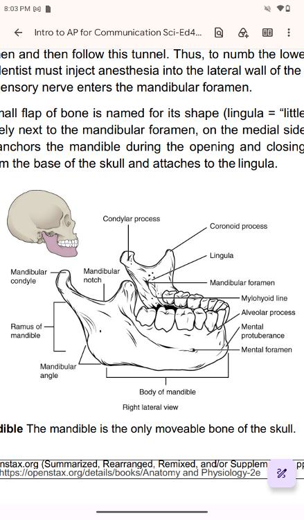
Lingula
Lingula—This small flap of bone is named for its shape (lingula =
“little tongue”). It is
located immediately next to the
mandibular foramen, on the medial side of the ramus.
A ligament
that anchors the mandible during the opening and closing of the mouth
extends down from the base of the skull and attaches to the lingula.
Mandible The mandible is the only moveable bone of the skull.
Lateral View of the Human Facial Skeleton with a horizontal reference
line drawn from the lower border of the orbit to the upper
margin of the ear canal. (Source:
Copyright 1989 by Department
of Audiology & Speech Pathology Memphis State
University,
Memphis, Tennessee From the W.R. Zemlin Memorial
Website
http://zemlin.shs.uiuc.edu Used with permission)
The lateral view
of the human facial skeleton above shows an example of the normal
dentition and occlusion of the upper and lower teeth. There are
several different
classifications of occlusion. Some of them
could cause various challenges in verbal
communication (“Angle’s
classification, 1899,” explained in Zemlin, 1998). The following
three classes are common.
• Class I normal occlusion
•
Class II malocclusion: Overjet. Upper teeth are located ahead of the
mandible, often
presenting the appearance of a receding chin.
• Class III malocclusion: lower teeth are located ahead of the
maxillary incisors,
presenting the appearance of a prognathic jaw.
..
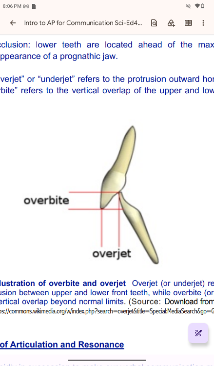
NOTE: The term “overjet” or “underjet” refers to the protrusion
outward horizontally, while
“overbite” or “underbite” refers to
the vertical overlap of the upper and lower teeth out of
normal
limits.
Figure 10.22 Illustration of overbite and overjet
Overjet (or underjet) refers to the
horizontal protrusion
between upper and lower front teeth, while overbite (or underbite)
refers to their vertical overlap beyond normal limits. (Source:
Download from Wikimedia
Commons: at https://commons.wikimedia.org/w/index.php?search=overjet&title=Special:MediaSearch&go=Go&type=image)
..
Physiology of Articulation and Resonance
Physiology of Articulation and Resonance
Articulators work
rapidly in succession to make our verbal communication meaningful.
Let’s
consider the articulators and vocal tracts contribution in
vowels and consonants production
separately. Vowels of English
are all produced with the oral tract open. The levator veli
palatini and muscular uvulae are primary muscles for closing the
nasal tract for oral sound
production.
If the nasal tract
does not close sufficiently, hypernasality in speech occurs.
Individuals with
cleft palate often experience incomplete
closure of the nasal tract, result in hypernasality in
speech.
Hypernasality often interferes with the intelligibility of speech. In
contrast,
individuals who cannot open their nasal tract,
potentially due to allergies or a head cold, may
sound
hyponasal. For vowel production, we move speech articulators
horizontally and
vertically to make distinctions in vowels.
Five factors generally classify consonants. The main three factors of
the five are voicing
features (voiced or unvoiced), place of
articulation, and manner of articulations (Remember:
VPM – V for
"Voice," P for "Place," and M for
"Manner" of articulation). We vibrate our vocal
folds
for voiced sounds. If you touch your neck with your hand and say
“bin,” you will feel
the vibration on your hand at the beginning
of the word. If you say “pin” in the same way,
you do not feel
the vibration on your hand at the beginning of your production because
the
/p/ in the word “pin” is a voiceless phoneme.
Place of
articulation refers to the location of the speech articulators when
each phoneme is
produced. In English, the places of articulation
are bilabial (both lips), labio-dental (upper
teeth and lower
lip – think lip and teeth), inter-dental (upper and lower teeth –
between the
teeth), alveolar (alveolar ridge), post- or
palate-alveolar (behind the alveolar ridge), velar
(posterior
part of the soft palate), and glottis (vocal-folds level). Manner of
articulation refers
to the way with which each phoneme is
produced.
The workings of the articulation and resonance organs
involve coordination and timing with
two other subsystems of
speech production, including respiration and phonation.
..
Role of Articulation and Resonance in Communication
Role of Articulation and Resonance in Communication
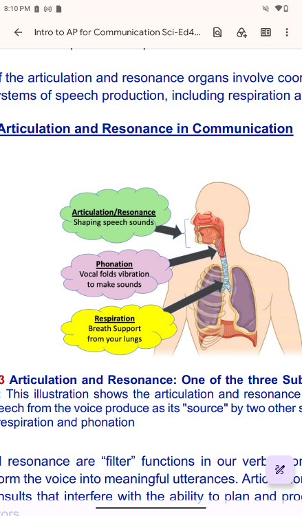
Figure 10.23
Articulation and Resonance: One of the three Subsystems of Speech
Production: This illustration shows the articulation and
resonance as the subsystem to
shape up speech from the voice
produce as its "source" by two other subsystems of speech
production, respiration and phonation
Articulation and
resonance are “filter” functions in our verbal communication. The
filter
functions transform the voice into meaningful utterances.
Articulation could be disrupted due
to neurogenic insults that
interfere with the ability to plan and program the movements of
speech articulators
In some cases, children do not develop proper movements of speech
articulators because
of various causes, such as Cerebral Palsy,
Childhood Apraxia of Speech (CAS), Autism
Spectrum Disorders
(ASD), and Chromosome disorders, such as Down Syndrome.
The
effects of early intervention are widely reported. Speech-Language
Pathologists (SLPs)
work with young children to guide them
through speech sound development to improve
communicative
competence. We acquire speech sounds in sequence. Utilizing the
published developmental normative information, SLPs facilitate
movements of speech
articulators for young clients to develop
intelligible speech.
Resonance disorders present with
hypernasality, hypo nasality, cul-de-sac resonance, or a
combination of two or more of those. The structural causes of
resonance disorders are
generally due to cleft lips, cleft
palate, both cleft lips and palate, or other craniofacial
disorders. These structural differences could be attributable to
genetic factors or secondary
to other conditions. In these
cases, SLPs work in inter-professional teams to guide clients
with proper behavioral management for verbal communication
before or after the care of
other professionals, such as
cleft-repair surgeries.
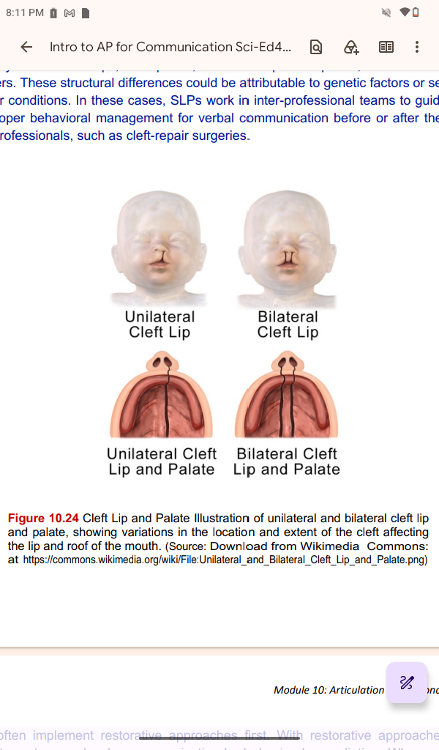
Cleft Lip and Palate Illustration of unilateral and bilateral cleft
lip
and palate, showing variations in the location and extent of
the cleft affecting
the lip and roof of the mouth. (So
SLPs often implement restorative approaches first. With restorative
approaches, SLPs
intend to restore or develop communication by
behavioral remediation. When restorative
approaches are not
applicable, SLPs take substitutive approaches. In some cases, SLPs
may support a combination of oral communication and the use of
an Alternative
Augmentative Communication (AAC) device to
support functional communication.
Resonance disorders could occur
without structural deviation due to velopharyngeal
mislearning
of the sound production presented for certain phonemes or in certain
situations
of phoneme production. If this is the case, SLPs
provide behavioral guidance to facilitate
proper re-learning of
the phoneme production. Velopharyngeal mislearning is confirmed by
assessing for anatomical differences through an endoscopy of
speech structures that
examines structure and function and
objective examination of produced speech.
..
Cleft Lip and Cleft Palate
Cleft Lip and Cleft Palate
During embryonic development, the
right and left maxilla bones come together at the
midline to
form the upper jaw. At the same time, the muscle and skin overlying
these bones
join together to form the upper lip. Inside the
mouth, the palatine processes of the maxilla
bones, along with
the horizontal plates of the right and left palatine bones, join
together to
form the hard palate. If an error occurs during
these developmental processes, atypical
development, such as a
cleft lip or cleft palate, may result.
Cleft lip is a common
developmental condition that affects approximately 1 in 1,000 births,
with a higher incidence in males. This condition involves a
partial or complete failure of
the right and left portions of
the upper lip to fuse, leaving a cleft (gap).
A more severe
developmental defect is cleft palate, which affects the hard palate.
The
hard palate is the bony structure that separates the nasal
cavity from the oral cavity. It is
formed during embryonic
development by the midline fusion of the horizontal plates from
the right and left palatine bones and the palatine processes of
the maxilla bones. Cleft
palate affects approximately 1 in 2,500
births and is more common in females. It results
from a failure
of the two halves of the hard palate to completely come together and
fuse
at the midline, thus leaving a gap between them. This gap
allows for communication
between the nasal and oral cavities. In
severe cases, the bony gap extends into the
anterior upper jaw,
where the alveolar processes of the maxilla bones also fail to join
together above the front teeth properly. If this occurs, a cleft
lip will also be seen. Because
of the communication between the
oral and nasal cavities, a cleft palate makes it very
difficult
for an infant to generate the suckling needed for nursing, thus
leaving the infant at
risk for malnutrition. Surgical repair is
required to correct cleft palate defects.
Bolus
bolus (p. 275) mass of chewed food
Crown
crown (p. 280) portion of tooth visible superior to the gum line
Cuspid
(p. 279) (also, canine) pointed tooth used for tearing and shredding food
deciduous teeth
(p. 278) “baby teeth” (20 in total)
Dentin
(p. 280) bone-like tissue immediately deep to the enamel of the crown
or cementum
of the root of a tooth
Dentition
(p. 278) set of teeth
enamel
p. 280) covering of the dentin of the crown of a tooth
Fauces
(p. 271) opening between the oral cavity and the oropharynx
gingivae
(p. 280) gums.
Labia
(p. 268) lips. singular = labium
lingual frenulum
(p. 276) mucous membrane fold that attaches the bottom of the tongue
to the floor of the mouth
lingual lipase
(p. 276) digestive enzyme from glands in the tongue that acts on triglycerides
molar
(p. 279) tooth used for crushing and grinding food
oral cavity
(p. 267) (also, buccal cavity) mouth
oral vestibule
(p. 271) part of the mouth bounded externally by the cheeks and lips,
and
internally by the gums and teeth
palatoglossal arch
(p. 272) muscular fold that extends from the lateral side of the soft
palate to the base of the tongue
palatopharyngeal arch
(p. 272) muscular fold that extends from the lateral side of the soft
palate to the side of the pharynx
permanent teeth
(p. 278) adult teeth (32 in total)
premolar
(p. 279) (also, bicuspid) transitional tooth used for mastication,
crushing, and
grinding food
pulp cavity
(p. 280) deepest portion of a tooth, containing nerve endings and blood vessels
Root
(p. 280) portion of a tooth embedded in the alveolar processes beneath the gum line
soft palate
(p. 271) posterior region of the bottom portion of the nasal cavity
that consists
of skeletal muscle
tongue
(p. 275) accessory digestive organ of the mouth, the bulk of which is
composed of
skeletal muscle
..
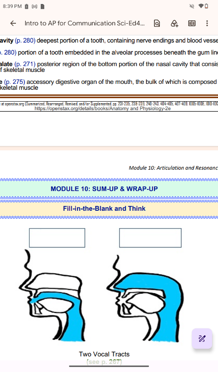
oral tract,
nasal tract
..
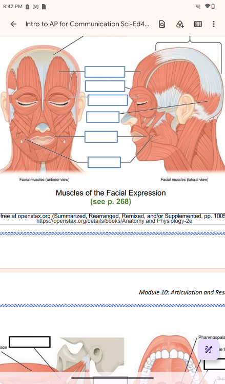
epicranial aponeurosis,
occipitofrontalis (frontal belly),
corrugator supercilio,
orbicularis oculi, o
ccipitofrontalis (occipital belly),
orbicularies oris, buccinator
..
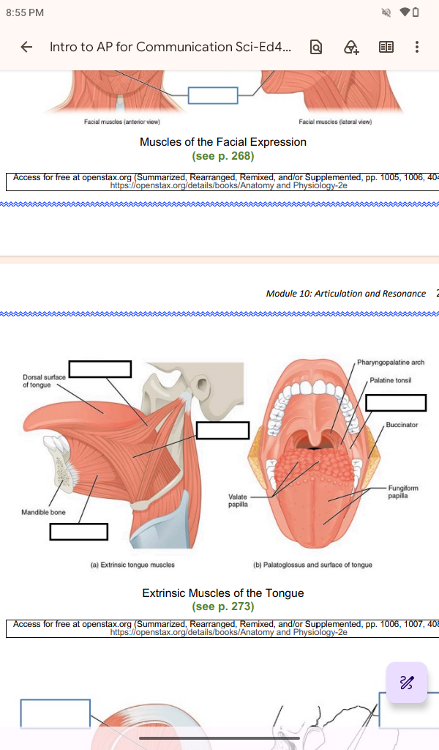
styloglossus,
genioglossus,
hyoglossus,
palatoglossus
..
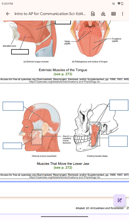
temporalis,
masseter,
area of superficial muscle dissection,
lateral pterygoid,
medial pterygoid
..
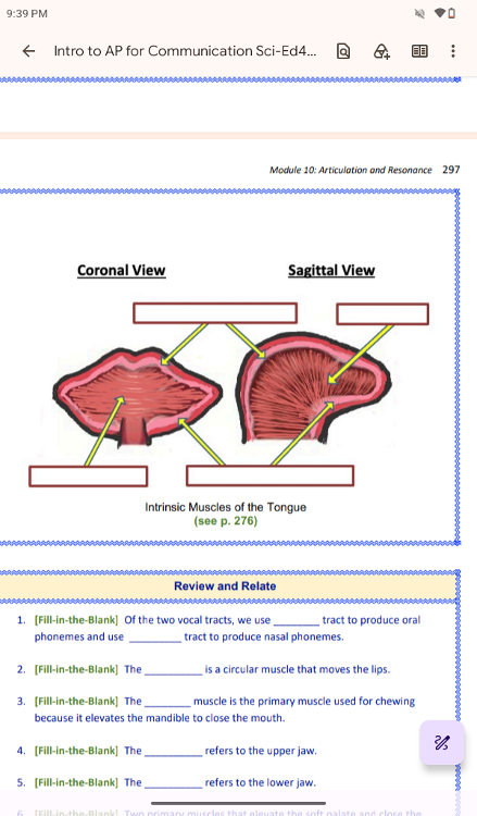
superior longitudinal muscle,
verrical muscles,
transverse muscles,
inferior longitudinal muscle
] Of the two vocal tracts, we use ________ tract to produce oral
phonemes and use _________ tract to produce nasal phonemes.
oral, nasal
The __________ is a circular muscle that moves the lips.
orbicularis oris
The ________ muscle is the primary muscle used for chewing
because it elevates the mandible to close the mouth.
masseter
The __________ refers to the upper jaw.
maxilla
The __________ refers to the lower jaw.
mandible
Two primary muscles that elevate the soft palate and close the
velopharyngeal port are ________________ and _______________.
levator veli palatini; muscular uvulae