Human lungs Anterior view of the human lungs. The lungs are paired
organs of the respiratory system located in the thoracic cavity. The
right lung consists of three lobes, while the left lung has two lobes
to accommodate the heart. Together, they facilitate gas exchange,
delivering oxygen to the bloodstream and removing carbon dioxide from
the body. (Source: Copyright 1989 by Department of Audiology
& Speech
Pathology Memphis State University, Memphis,
Tennessee From the W.R. Zemlin
Memorial Website
http://zemlin.shs.uiuc.edu Used with permission)
..
Anatomy of Respiration
Anatomy of Respiration
As we discussed in Module 6, the function
of respiration for speech production is to provide
the source
energy for our speech production. In a larger sense, respiration
covers a
biological process of gas exchange at the cellular
level by means of the act of breathing. An
important part of
breathing is ventilation, which involves moving air into and out of
the lower
airway. We use the following basic terms in breathing
in and out.
• Inspiration (inhalation) The movement of air into
our upper and lower airways
• Expiration (exhalation) The
movement of air out of the lower and upper airways.
We often
hear the term “upper airway infection” as related to head cold. The
upper airway
refers to the structures above the vocal folds. The
lower airway refers to those below the
vocal folds used for
breathing. Breathing is essential for our survival, and large skeletal
structures contain important organs for breathing.
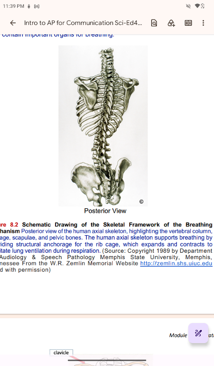
Schematic Drawing of the Skeletal Framework of the Breathing
Mechanism Posterior view of the human axial skeleton,
highlighting the vertebral column,
rib cage, scapulae, and
pelvic bones. The human axial skeleton supports breathing by
providing structural anchorage for the rib cage, which expands
and contracts to
facilitate lung ventilation during respiration.
(Source: Copyright 1989 by Department
of Audiology & Speech
Pathology Memphis State University, Memphis,
Tennessee From the
W.R. Zemlin Memorial Website http://zemlin.shs.uiuc.edu
Used with permission)
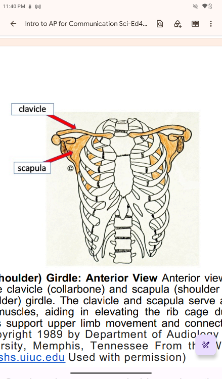
Figure 8.3 Pectoral (Shoulder) Girdle: Anterior View Anterior view of
the upper thoracic
skeleton highlighting the clavicle
(collarbone) and scapula (shoulder blade), which together
form
the pectoral (shoulder) girdle. The clavicle and scapula serve as
attachment sites for
accessory respiratory muscles, aiding in
elevating the rib cage during deep or labored
breathing. These
bones support upper limb movement and connect the arms to the axial
skeleton. (Source: Copyright 1989 by Department of Audiology
& Speech Pathology
Memphis State University, Memphis,
Tennessee From the W.R. Zemlin Memorial
Website
http://zemlin.shs.uiuc.edu Used with permission)
..
Hold your breath. Really! See how long you can hold your breath as
you continue reading.
How long can you do it? Chances are you
are feeling uncomfortable already. A typical
human cannot
survive without breathing for more than 3 minutes, and even if you
wanted to
hold your breath longer, your autonomic nervous system
would take control. This is because
every cell in the body needs
to run the oxidative stages of cellular respiration, the process
by which energy is produced in the form of adenosine
triphosphate (ATP). For oxidative
phosphorylation to occur,
oxygen is used as a reactant and carbon dioxide is released as a
waste product.
..
Although oxygen is a critical need for cells, it is actually the
accumulation of carbon dioxide
that primarily drives your new
inhalation (breathing in). Carbon dioxide is exhaled, and
oxygen
is inhaled through the respiratory system, which includes muscles to
move air into
and out of the lungs, passageways through which
air moves, and microscopic gas exchange
surfaces covered by
capillaries. The circulatory system transports gases from the lungs to
tissues throughout the body and vice versa. A variety of
diseases can affect the respiratory
system, such as asthma,
emphysema, chronic obstructive pulmonary disease (COPD), and
lung cancer. All of these conditions affect the gas exchange
process and result in labored
breathing and other difficulties.
The major organs of the respiratory system function primarily to
provide oxygen to body
tissues for cellular respiration, remove
the waste product carbon dioxide, and help to
maintain acid-base
balance. Portions of the respiratory system are also used for
non-vital
functions, such as sensing odors, speech production,
and for straining, such as during
childbirth or coughing (Figure
8.4, p. 202).
..
The major organs of the respiratory system function primarily to
provide oxygen to body
tissues for cellular respiration, remove
the waste product carbon dioxide, and help to
maintain acid-base
balance. Portions of the respiratory system are also used for
non-vital
functions, such as sensing odors, speech production,
and for straining, such as during
childbirth or coughing (Figure
8.4, p. 202).
..
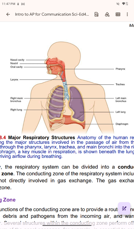
Major Respiratory Structures Anatomy of the human respiratory system,
illustrating the major structures involved in the passage of air
from the nasal and oral
cavities through the pharynx, larynx,
trachea, and main bronchi into the right and left lungs.
The
diaphragm, a key muscle in respiration, is shown beneath the lungs,
highlighting its
role in driving airflow during breathing.
Functionally, the respiratory system can be divided into a
conducting zone and a
respiratory zone. The conducting zone of
the respiratory system includes the organs and
structures not
directly involved in gas exchange. The gas exchange occurs in the
respiratory zone.
..
Conducting Zone
Conducting Zone
The major functions of the conducting zone are
to provide a route for incoming and outgoing
air, remove debris
and pathogens from the incoming air, and warm and humidify the
incoming air. Several structures within the conducting zone
perform other functions as well.
The epithelium of the nasal
passages, for example, is essential to sensing odors, and the
bronchial epithelium that lines the lungs can metabolize some
airborne carcinogens
The Nose and its Adjacent Structures
The major path for the respiratory system is through the nose. When
discussing the nose, it
is helpful to divide it into two major
sections: the external nose, and the nasal cavity or
internal nose.
external nose
The external nose consists of the surface and skeletal structures
that result in the
outward appearance of the nose and contribute
to its numerous functions (Figure 8.5, p.
203). The root is the
region of the nose located between the eyebrows. The bridge is the
part of the nose that connects the root to the rest of the nose.
The dorsum nasi is the
length of the nose.
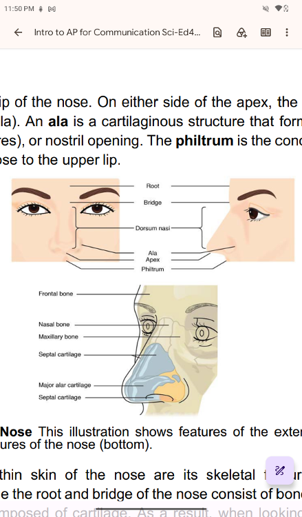
Apex
The apex is the tip of the nose. On either side of the apex, the
nostrils are formed by the
alae (singular = ala). An ala is a
cartilaginous structure that forms the lateral side of each
naris (plural = nares), or nostril opening. The philtrum is the
concave surface that connects
the apex of the nose to the upper lip
Nose This illustration shows features of the external nose (top) and
skeletal features of the nose (bottom
Positions of Ethmoid Bone and Vomer Bone
Underneath the thin skin of the nose are its skeletal features
(Figure 8.6, lower
illustration). While the root and bridge of
the nose consist upof bone, the protruding portion
of the nose
is composed of cartilage. As a result, when looking at a skull, the
nose is
missing. The nasal bone is one of a pair of bones that
lies under the root and bridge of
the nose. The nasal bone
articulates superiorly with the frontal bone and laterally with the
maxillary bones. Septal cartilage is flexible hyaline cartilage
connected to the nasal bone,
forming the dorsum nasi. The alar
cartilage consists of the apex of the nose; it surrounds
the naris.
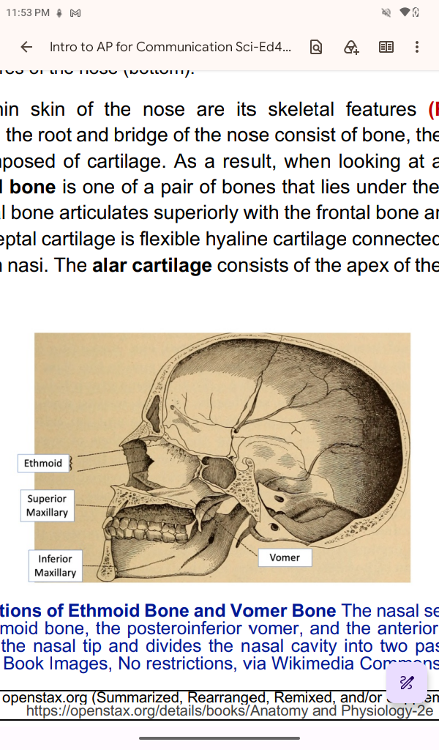
Positions of Ethmoid Bone and Vomer Bone The nasal septum consists
of
the superior ethmoid bone, the posteroinferior vomer, and the
anterior septal cartilage,
which supports the nasal tip and
divides the nasal cavity into two passages. (Source:
Internet
Archive Book Images, No restrictions, via Wikimedia Commons