Describe the structure of a DNA molecule:
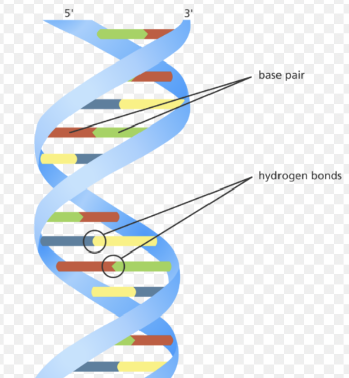
- two strands coiled together to form a double helix
- each strand contains chemicals called bases
- bonds between pairs of bases hold the strands together
- the bases always pair up in the same way: A with T, and C with G
Describe a catalyst
a substance that increases the rate of a chemical reaction and is not changed by the reaction.
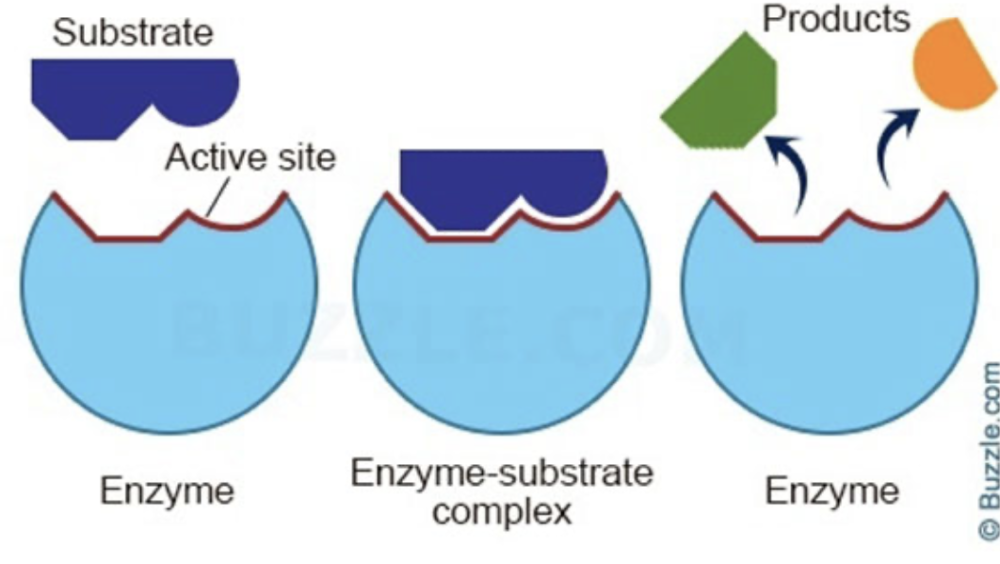
Describe enzymes
Proteins that are involved in all metabolic reactions, where they function as biological catalysts
Enzymes reaction parts
Describe the effect of changes in temperature and pH on enzyme activity
Each enzyme has an optimum temperature and pH. Deviations from these optima can lead to reduced enzyme activity or even denaturation, where the enzyme loses its functional shape and becomes inactive.
Describe photosynthesis
The process by which plants synthesise carbohydrates from raw materials using energy from light.
carbon dioxide + water → glucose + oxygen ---- in the presence of light and chlorophyll
6CO2 + 6H2O → C6H12O6 + 6O2
Outline the use and storage of the carbohydrates made in photosynthesis.
- Converted into starch molecules which act as an effective energy store
- Converted into cellulose to build cell walls
- Glucose can be used in respiration to provide energy
- Converted to sucrose for transport in the phloem
- As nectar to attract insects for pollination
Why plants need magnesium ions and nitrate ions
- Magnesium ions are a central component of the chlorophyll molecule, which is vital for photosynthesis. Chlorophyll absorbs light energy and uses it to convert carbon dioxide and water into glucose. A deficiency in magnesium leads to a lack of chlorophyll, severely impacting the plant's ability to photosynthesize.
- Nitrate ions are essential for plants to synthesize amino acids, which are the building blocks of proteins. These proteins are crucial for various cellular functions, including growth, repair, and enzyme production. Without sufficient nitrate ions, plants cannot produce enough amino acids, leading to stunted growth and yellowing leaves.
Experiment to test what affects the rate of photosynthesis.
- Group 1 (Control): A healthy plant with normal exposure to light and carbon dioxide.
- Group 2: A plant with no chlorophyll (e.g., a variegated leaf or a plant treated with a chlorophyll inhibitor) exposed to light and carbon dioxide.
- Group 3: A plant kept in the dark but with access to carbon dioxide.
- Group 4: A plant exposed to light but with no carbon dioxide.
Chlorophyll is essential. Light is essential. Carbon dioxide is essential.
- Increased light intensity generally leads to increased photosynthesis:
- Increased carbon dioxide concentration generally leads to increased photosynthesis:
- Increased temperature generally leads to increased photosynthesis
Investigate and describe the effect of light and dark conditions on gas exchange in an aquatic plant
Place an aquatic plant (like Elodea) in a container with hydrogencarbonate indicator solution (which changes colour depending on the pH). Expose one container to light and the other to dark conditions. Observe the color change in each container.
- In light conditions:The plant will produce oxygen through photosynthesis, raising the pH and changing the indicator solution to a more alkaline color.
- In dark conditions:The plant will undergo respiration, consuming oxygen and lowering the pH, changing the indicator solution to a more acidic color.
What is needed for photosynthesis
- Plants need several factors for photosynthesis to occur:
- The presence of chlorophyll
- A supply of carbon dioxide
- A supply of water
- Light energy
- A suitable temperature
- The main external
factors that affect the rate of photosynthesis are:
- light intensity: The greater the light intensity, the more energy is supplied to the plant and therefore the faster the light-dependent stage of photosynthesis can occur.
- carbon dioxide concentration
- temperature: As temperature increases the rate of photosynthesis increases as the reaction is controlled by enzymes However, as the reaction is controlled by enzymes, this trend only continues up to a certain temperature beyond which the enzymes begin to denature and the rate of reaction decreases
Leaf Adaptations and parts for Photosynthesis:
- Large Surface Area: Maximises the area exposed to sunlight for capturing light energy, a key component of photosynthesis.
- Thin Structure: Minimises the distance carbon dioxide needs to diffuse to reach the cells where photosynthesis occurs, ensuring efficient gas exchange.
Plant parts for Photosynthesis:
- Chloroplasts: Contain chlorophyll, the pigment that absorbs light energy for photosynthesis.
- Cuticle: A waxy, waterproof layer on the leaf surface that reduces water loss.
- Guard Cells: Specialized cells that surround stomata and control their opening and closing, regulating gas exchange and water loss.
- Stomata: Pores on the leaf surface that allow for gas exchange.
- Upper and Lower Epidermis: Protective layers on the top and bottom of the leaf. Upper is a transparent layer that allows light to reach the palisade mesophyll. The lower epidermis is a single layer of cells on the underside of a leaf, containing stomata and guard cells. It plays a crucial role in regulating gas exchange and water loss through transpiration.
- Palisade Mesophyll: A layer of tightly packed, elongated cells rich in chloroplasts, located near the top of the leaf to maximise light absorption.
- Spongy Mesophyll: A layer of loosely arranged cells with air spaces that facilitate gas exchange.
- Air Spaces: Gaps between spongy mesophyll cells that allow for the diffusion of carbon dioxide and oxygen.
- Vascular Bundles: Bundles of xylem and phloem that transport water and nutrients throughout the leaf.
- Xylem: Transports water to the leaf.
- Phloem: Transports sugars produced during photosynthesis to other parts of the plant.
Describe what is meant by a balanced diet
A balanced diet is a diet consisting of the right proportions of every type of nutrient in suitably sized portions for the right amount of energy.
State the principal dietary sources and describe the importance of:
(a) carbohydrates - found in almost any food, but are present in large quantities in staple foods such as rice, potatoes, wheat, cereal, bread, etc.
They are broken down to release energy in respiration.
(b) fats and oils - found in oil, butter, margarine, the white stuff on animal meat, etc.
Insulate the body, helping reduce fluctuations in our body temperature. They are also a good store of energy – fats have a higher chemical potential energy per gram than carbohydrates, so they can store more energy in the same space.
(c) proteins - found in meats, such as chicken, beef, fish, etc. It is also found in vegetables such as lentils and beans.
Proteins form our muscles, enzymes, skin etc, need for growth
(d) vitamin C - found in many fresh fruits, especially citrus fruits. It is also found in dark leafy greens.
- Required for the development and maintenance of scar tissue, blood vessels and cartilage.
- It’s needed to make ATP (your source of energy), too.
- Vitamin C contributes to healthy teeth and gums as well. It regulates the flow of calcium into the bloodstream by promoting the absorption of calcium from food.
(e) vitamin D - formed under our skin as a reaction to sunlight. Food sources include oily fish, eggs, fortified fat spreads, fortified breakfast cereals, and some powdered milks.
Build bones and keep them healthy. The body can absorb calcium only if it has enough vitamin D.
(f) mineral ion, calcium - found in so many foods! Dairy foods such as milk, cheese and yoghurt contain it; greens like kale, broccoli, most grainy food, like bread or rice; etc.
- Almost all calcium is stored in bones and teeth, where it supports their structure and hardness.
- Calcium is required for muscles to move and for nerves to carry messages between the brain and every body part.
- It’s used to help blood vessels move blood through the body and to help release hormones and enzymes (almost every function in the body is regulated by hormones and enzymes).
(g) mineral ion, iron - found in liver, meat, beans, nuts, dried fruit, whole grains, fortified breakfast cereals, clams, oysters, shrimps and dark green leafy vegetables.
Iron is primarily needed to form the haemoglobin in RBCs.
(g) fibre - Insoluble fibre sources include wholemeal bread, bran, cereals, nuts and seeds.
Insoluble fibre keeps your bowels healthy and helps prevent digestive problems.
(h) water - Every cell in our body is made of and surrounded by water (unless they’re dead. Then maybe not.) – and as a whole, we are approximately 70% water.
- Every reaction in our body (respiration, digestion, growth, etc.) occurs in water.
- Hormones and other substances are dissolved in water;
- most of your blood is made of plasma (which is 92% water),
- gas exchange is possible because the gas exchange surface area moistened using water
State the cause of rickets
Caused by a deficiency in Vitamin D and/or calcium, leading to soft and weakened bones, causing deformities and pain,
State the cause of scurvy
- A lack of vitamin C in the diet. This is often due to insufficient intake of fresh fruits and vegetables, as these are primary sources of vitamin C.
- Effect:Scurvy can lead to bleeding under the skin and around the gums, premature stopping of bone growth in children (stunted growth), and dry skin and hair.
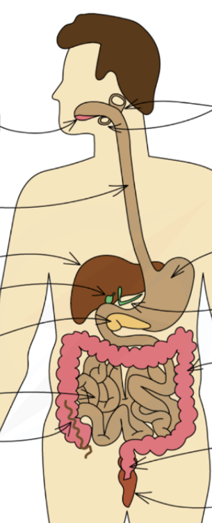
Identify in diagrams and images the main organs of the digestive system
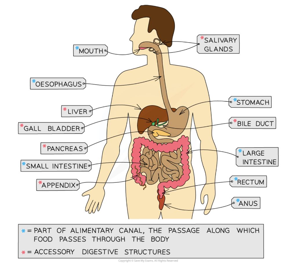
Give functions of mouth, salivary gland, oesophagus, stomach, liver, gall bladder, pancreas, duodenum, ileum, colon, rectum, anus.
- Mouth- Food is ingested here and the teeth break it down into smaller pieces during mechanical digestion
- Salivary glands- Saliva is secreted into the mouth. The enzyme amylase in saliva begins to digest starch into maltose. Saliva lubricates the food for easy swallowing
- Oesophagus- This tube connects the mouth to the stomach. Contractions of the walls of the oesophagus force the food downwards; this is peristalsis.
- Stomach- Churning of the muscular stomach walls continues the process of mechanical digestion. Protease enzymes begin protein digestion. Hydrochloric acid provides a suitable pH for the enzymes and also destroys any pathogens in food.
- Liver- Bile is produced here. Bile aids the digestion of fats, as well as neutralising stomach acid as it exits the stomach.
- Gall bladder- Bile is stored here before being released into the duodenum via the bile duct.
- Pancreas- Amylase, protease and lipase enzymes are produced here before being released into the duodenum.
- Small intestine: duodenum- Food enters the small intestine from the stomach here. The acidic stomach contents are neutralised by bile and become slightly alkaline. Enzymes complete chemical digestion here.
- Small intestine: ileum- Food and water are absorbed into the blood via villi in the lining of the ileum.
- Large intestine: colon- Remaining water is absorbed from food into the blood, and the solid waste left behind in the colon forms faeces.
- Large intestine: rectum - Faeces are stored here prior to egestion
- Large intestine: anus - Faeces leave the body via the anus; this is egestion
Definition of ingestion, digestion, absorption, assimilation, egestion
(a) ingestion – the taking of substances, e.g. food and drink, into the body
(b) digestion – the breakdown of food
(c) absorption – the movement of nutrients from the intestines into the blood
(d) assimilation – uptake and use of nutrients by cells
(e) egestion – the removal of undigested food from the body as faeces
Describe physical digestion and what it does
The breakdown of food into smaller pieces without chemical change to the food molecules.
*It increases the surface area of food for the action of enzymes in chemical digestion.
Describe the structure of human teeth
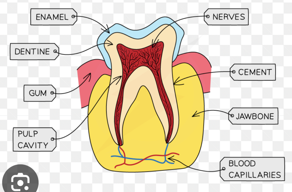
The outermost layer is the enamel, a hard, protective layer covering the crown of the tooth.Beneath the enamel is dentine, a bonelike substance that is less dense than enamel. The pulp, located in the center of the tooth, contains nerves and blood vessels. The nerves provide sensation to the tooth, while blood vessels supply it with nutrients. The tooth's root is covered by cement, a bonelike substance that anchors the tooth to the jawbone. Teeth are embedded in the jawbone and surrounded by the gums.
Describe chemical digestion
The break down of large insoluble molecules into small soluble molecules.
State functions of 3 enzymes. where secreted and act
(a) amylase breaks down starch to simple reducing sugars. Secreted by the salivary gland and pancreas, acts in mouth and duodenum.
(b lipase breaks down fats and oils to fatty acids and glycerol. Secreted by the pancreas and walls of stomach, acts in duodenum and stomach.
(c) proteases break down protein to amino acids. Secreted by the pancreas, acts in duodenum.
Describe the functions of hydrochloric acid in gastric juice
killing harmful microorganisms in food and providing an acidic pH for optimum enzyme activity
Describe the digestion of starch in the digestive system
(a) amylase breaks down starch to maltose
(b) maltase breaks down maltose to glucose on the membranes of the epithelium lining the small intestine
Describe the digestion of protein
(a) pepsin breaks down protein in the acidic conditions of the stomach
(b) trypsin breaks down protein in the alkaline conditions of the small intestine
Bile: where produced and stored. what it does
- Cells in the liver produce bile which is then stored in the gallbladder.
- Bile is an alkaline mixture that neutralises the acidic mixture of food and gastric juices entering the duodenum from the stomach to provide a suitable pH for enzyme action.
- It breaks down large drops of fat into smaller ones. This is known as emulsification. The larger surface area allows lipase to chemically break down the lipid into glycerol and fatty acids faster.
Describe structure of villus
- A large surface area
- Microvilli on the surface of the villus further increase the surface available for absorption
- A short diffusion distance
- The wall of a villus is only one cell thick
- A steep
concentration gradient
- The villi are well supplied with a network of blood capillaries that transport glucose and amino acids away from the small intestine in the blood
- A lacteal (lymph vessel) runs through the centre of the villus to transport fatty acids and glycerol away from the small intestine in the lymph
- Enzymes produced in the walls of the villi assist with chemical digestion
- The movement of villi helps to move food along and mix it with the enzymes present
State the functions of xylem and phloem

(a) xylem – transport of water and mineral ions, and supports plant
(b) phloem – transport of sucrose and amino acids
On diagram red is xylem and blue is phloem
Relate the structure of xylem vessels to their function, 3
- Thick walls with lignin: Lignin is a strong, polymer that reinforces the cell walls of xylem vessels, giving them rigidity and preventing them from collapsing under pressure. This is crucial for withstanding the water pressure during transport.
- No cell contents: Mature xylem vessels are dead cells with no cytoplasm or organelles inside. This allows for the free passage of water through the vessel without obstruction.
- Cells joined end to end with no cross walls: Forms a long, continuous tube. This uninterrupted structure facilitates efficient water flow.
Outline the pathway taken by water
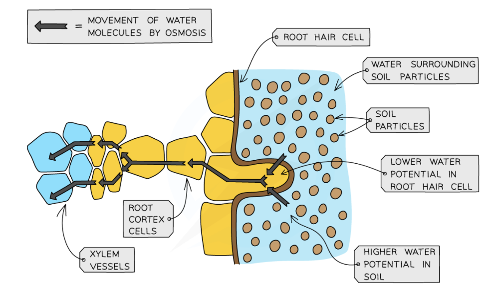
root hair cell → root cortex cells → xylem → leaf mesophyll cells
Diagram and adaptations and functions of root hairs
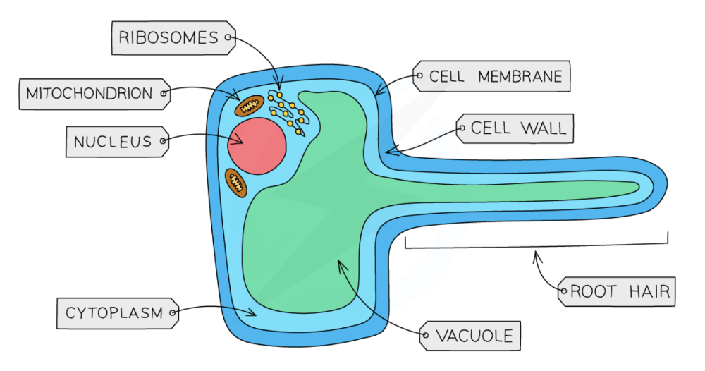
-
- They contain mitochondria which release energy for active transport
- Root hairs increase the surface area of plant roots, increasing the rate at which water and minerals can be taken up
- The water concentration of the cell cytoplasm is reduced due to the presence of mineral ions
Experiment to witness xylem
1. Prepare the plant and stain:Cut a celery stem under water to prevent air from entering the xylem. Prepare a solution of red food colouring in water.
2. Place the stem in the stain.
3. Observe the stained areas over set time intervals: Allow the plant to sit for a few hours. You will observe the stain traveling up the stem and into the leaves.
4. Analyze the stained tissue:Cut the stem at intervals and examine the stained areas. This will reveal the location of the xylem vessels, which are responsible for water transport in the plant.
Describe and define transpiration, and effects of outside world on it.
The loss of water vapour from leaves.
State that water evaporates from the surfaces of the mesophyll cells into the air spaces and then diffuses out of the leaves through the stomata as water vapour.
More wind makes it faster: Moving air carries away water vapor from the leaf's surface, preventing it from saturating the surrounding air. This maintains a concentration gradient, allowing more water to evaporate and diffuse out of the leaf.
Higher temp makes it faster: Higher temperatures provide water molecules with more kinetic energy, accelerating evaporation from the leaf surface.
High humidity makes it slower; High humidity means the air is already saturated with water vapor, reducing the concentration gradient between the leaf and the air.
Explain the mechanism by which water moves upwards in the xylem
- As the water moves into a root hair, across to xylem vessels, up to leaves and then out into air, it is moving down a water potential gradient..
- The low water potential in leaves is caused by a loss of water vapour from leaves by transpiration.
- This produces a transpiration pull from above, drawing a column of water molecules held together by forces of attraction between water molecules.
Define translocation
The movement of sucrose and amino acids in phloem from sources to sinks.
(a) sources as the parts of plants that release sucrose or amino acids
(b) sinks as the parts of plants that use or store sucrose or amino acids
Explain why some parts of a plant may act as a source and a sink at different times.
Dissolved food is always transported from source(where it’s made) to sink(where it’s stored or used):
- During winter, when many plants have no leaves, the phloem tubes may transport dissolved sucrose and amino acids from the storage organs to other parts of the plant so that respiration can continue.
- During a growth period (eg during the spring), the storage organs (eg roots) would be the source and the many growing areas of the plant would be the sinks.
- After the plant has grown(usually during the summer), the leaves are photosynthesizing and producing large quantities of sugars; so they become the source and the roots become the sinks – storing sucrose as starch until it is needed again.
Define the circulatory system
A system of blood vessels with a pump and valves to ensure one-way flow of blood.
Describe the single circulation of a fish
- Blood flows from the heart to the gills for oxygenation.
- Oxygenated blood then travels to the rest of the body.
- Deoxygenated blood returns directly to the heart.
- The heart in fish has two chambers: one atrium and one ventricle.
- This single circuit is less efficient for oxygen delivery, as the blood loses pressure in the capillaries of the gills.
Describe the double circulation of a mammal
- Blood travels through two separate circuits: the pulmonary and systemic circuits.
- Pulmonary circulation: Deoxygenated blood is pumped from the heart to the lungs. Oxygenated blood returns from the lungs to the heart.
- Systemic circulation: Oxygenated blood is pumped from the heart to the rest of the body. Deoxygenated blood returns to the heart.
The heart in mammals has four chambers: two atria and two ventricles.
This separation of circuits allows for higher blood pressure and more efficient delivery of oxygen and nutrients to body tissues.
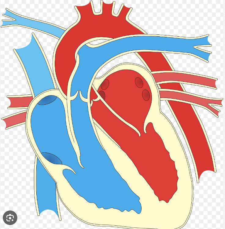
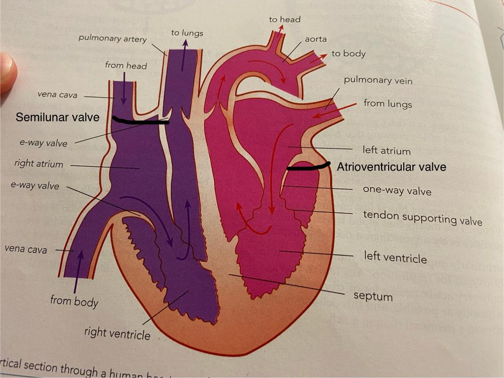
Identify in diagrams and images the structures of the mammalian heart, limited to: muscular wall, septum, left and right ventricles, left and right atria, one-way valves and coronary arteries, atrioventricular and semilunar valves.
Describe the functioning of the beating heart
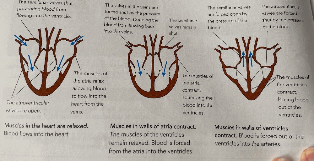
Heart muscles contract and relax.
- Muscles in the heart are relaxed allowing blood to flow into the atrium. Valves in the veins are open to let it in and the atrioventricular valves are open, semilunar valves are shut to stop blood leaving heart.
- Muscles on walls of atria contract and ventricles remain relaxed pushing blood into the ventricles. Valves in veins and semilunar valves are shut to trap blood inside the heart. The atrioventricular valves between atria and ventricles remains open.
- Walls of ventricles contract pushing blood out of ventricles into the arteries. The semilunar valves are opened by the pressure and atrioventricular valves close.
Explain the importance of the septum, wall thickness, arteries and veins.
The septum in separating oxygenated and deoxygenated blood.
Atria only supply blood to the ventricles so have thinnest walls. Ventricles supply to whole body so are thinner. The left ventricle has thicker wall than the right as the left pumps blood to the entire body and the right only to the lungs.
Blood is pumped away from the heart in arteries and returns to the heart in veins.
State that the activity of the heart may be monitored by:
Monitoring Heart Activity:
- ECG: An electrocardiogram records the electrical activity of the heart, which correlates with the contractions of the atria and ventricles.
- Pulse Rate:The pulse rate, typically measured at the wrist or neck, reflects the number of heartbeats per minute as blood is pumped through the arteries.
- Heart Sounds:A stethoscope can be used to listen to the sounds of the heart valves closing.
Describe coronary heart disease
CHD occurs when the coronary arteries, which supply blood to the heart muscle, become narrowed or blocked. This blockage restricts blood flow, potentially leading to angina (chest pain), heart attack, or other complications.
Possible risk factors including:
- Diet: A diet high in saturated and trans fats, cholesterol, and sodium can contribute to plaque buildup in the arteries.
- Lack of Exercise:A sedentary lifestyle increases the risk of obesity, high blood pressure, and high cholesterol, all of which are risk factors for CHD.
- Stress: Chronic stress can elevate blood pressure and heart rate, potentially damaging the arteries over time.
- Smoking: Smoking damages blood vessels, increases blood clotting, and elevates blood pressure, all of which contribute to CHD.
- Genetic Predisposition: Family history of early heart disease (especially before age 55 for men and 65 for women) increases the risk.
- Age: The risk of CHD increases with age, particularly after 45.
- Sex: Men generally have a higher risk of CHD compared to women, although the risk increases for women after menopause.
Describe the structure of arteries, veins and capillaries, reasons and functions of capillaries.
- Arteries: Carry blood away from the heart. They have thick, muscular walls with elastic fibers to withstand the high pressure of blood pumped from the heart. The lumen (internal space) is relatively narrow compared to veins. The elastic fibers allow the artery to stretch and recoil, helping to maintain a continuous flow of blood.
- Veins: Carry blood back to the heart. They have thinner walls and a larger lumen compared to arteries. They also contain valves that prevent backflow of blood, especially in the limbs, where blood must travel against gravity.
- Capillaries: They have extremely thin walls, often just one cell thick. The single-cell thickness of the capillary wall facilitates the exchange of substances (oxygen, nutrients, waste) between the blood and surrounding tissues. The extensive network of capillaries provides a large surface area for efficient diffusion of substances.
- Nutrient and waste exchange: Capillaries are the primary sites for the exchange of nutrients, oxygen, and waste products between the blood and body tissues.
- Blood pressure regulation: By adjusting their diameter, capillaries play a role in regulating blood pressure and blood flow.
- Formation of the blood-brain barrier: In the brain, capillaries form a barrier that regulates the passage of substances into the brain tissue, protecting it from harmful substances.
- Filtering blood in the kidneys: Specialized capillaries in the kidneys filter blood to produce urine.
- Hormone delivery: Capillaries deliver hormones to specific target organs.
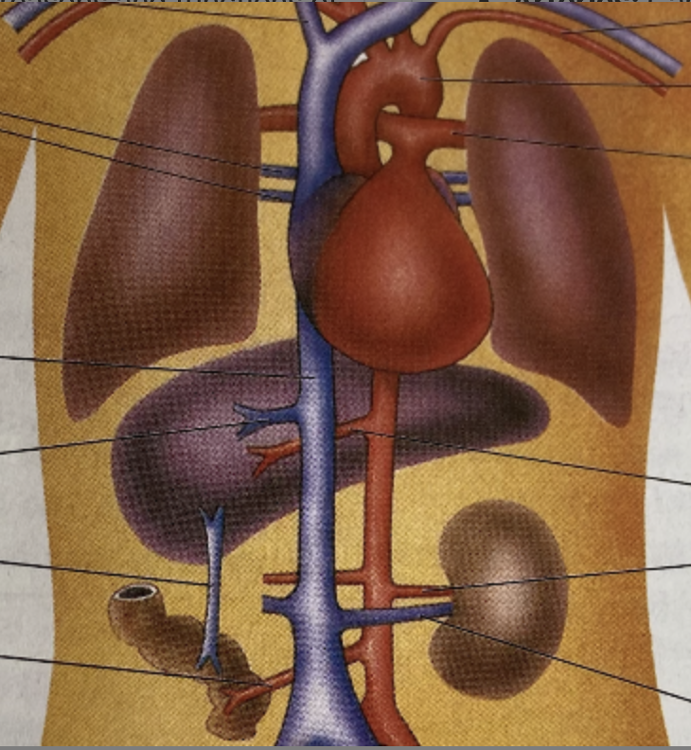
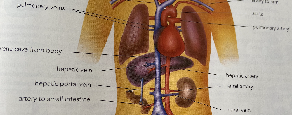
3 Identify in diagrams and images the main blood vessels to and from the:
(a) heart, limited to: vena cava, aorta, pulmonary artery and pulmonary vein
(b) lungs, limited to: pulmonary artery and pulmonary vein
(c) kidney, limited to: renal artery and renal vein
(d) liver : hepatic artery, hepatic veins and hepatic portal vein