Physiology
The study of how the body and its parts function. It focuses on how organs, tissues, cells, and molecules work together to keep the body alive and healthy.
Example Topics in Physiology:
- How the heart pumps blood
- How lungs exchange oxygen and carbon dioxide
- How muscles contract
- How hormones regulate body functions
Homeostasis
The body’s ability to maintain a stable internal environment, even when the external environment changes.
The body constantly adjusts things like: Temperature, pH, Blood sugar, Water balance, and Blood pressure
Goal: Keep conditions within a narrow, healthy range so the body functions properly.
Ex: Negative Feedback
When you get too hot:
- You sweat to cool down
When your blood sugar drops: - Your body releases glucagon to raise it
The key features of the scientific method
a systematic, logical process used by scientists to ask questions, gather data, and test explanations about the natural world.
Observation: Notice something in the natural world.
- Ask: What’s happening? Why does this occur?
Question: Turn your observation into a clear, focused question.
- Example: Why do plants grow faster in sunlight?
Hypothesis: Make an educated guess or prediction that explains your observation. Must be testable and falsifiable.
- Example: If a plant gets more sunlight, then it will grow taller.
Experiment: Design a controlled test to check your hypothesis.
Use variables:
- Independent variable = what you change (e.g., amount of sunlight)
- Dependent variable = what you measure (e.g., plant height)
- Controlled variables = kept constant
Data Collection: Record measurements and observations carefully.
Analysis: Look at your data to find patterns or trends. Use charts, graphs, or statistics if needed.
Conclusion: Decide whether the data supports or rejects your hypothesis. Repeat experiments to be sure of results.
Communication: Share your findings through reports, papers, or presentations.
Steps in Drug development
Discovery and Preclinical Research
Goal: Find a possible drug and test if it’s safe in the lab.
Scientists: Identify a target (like a protein involved in a disease), Design or find molecules that might affect it and Test these in cells and animals to check for: Safety, Effectiveness and How the drug is absorbed, used, and removed
Investigational New Drug (IND) Application
Before testing in humans, the company must apply to the FDA (or other regulatory agency) with: Preclinical results, Manufacturing info, Proposed clinical trial plan and If approved, they can begin clinical trials.
Who is considered the father of modern physiology?
Claude Bernard
Introduced the concept of the “internal environment”, which laid the foundation for our modern understanding of homeostasis—the body's ability to maintain a stable internal state.
Conducted pioneering experiments in: Digestion, Blood sugar regulation, Nervous system function, Liver metabolism and Strongly emphasized the importance of using the scientific method and controlled experiments in biological research.
Who coined the term “homeostasis?”
Walter Cannon (1871–1945)
An American physiologist, Built on Claude Bernard’s idea of the "internal environment", Coined “homeostasis” from Greek words: "homeo" = similar and "stasis" = standing still
He described it as the body's ability to maintain a stable internal state despite changes in the external environment.
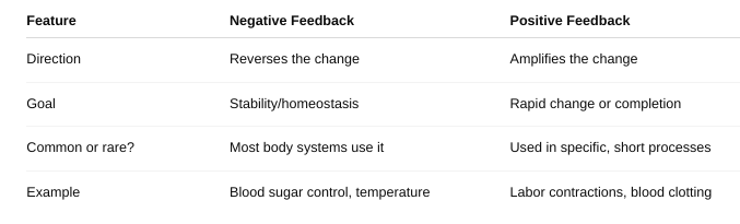
What are the four main features of a negative feedback loop? Give some examples.
A negative feedback loop is a biological control system that helps the body maintain homeostasis by reversing a change to return to a set point (like a thermostat turning off the heat when it gets too warm).
1. Stimulus: A change or disruption in the environment.
Ex: Body temperature rises above normal.
2. Sensor (Receptor): Detects the change.
Ex: Temperature sensors in the skin and brain detect the heat.
3. Control Center: Processes the information and decides how to respond. Usually the brain or endocrine glands.
Ex: The hypothalamus in the brain processes the temperature change.
4. Effector: Carries out the response to reverse the change.
Ex: Sweat glands release sweat; blood vessels widen to cool the body.
Explain the regulatory mechanisms involved in maintaining homeostasis.
Negative Feedback Loops: Reverse a change to bring the body back to its set point.
Components: Stimulus – A change occurs (e.g., increase in body temperature). Sensor (Receptor) – Detects the change. Control Center – Usually the brain or endocrine system (e.g., hypothalamus); processes info. Effector – Responds to correct the imbalance (e.g., sweat glands cool the body).
Ex: Thermoregulation (body temperature), Blood glucose regulation (via insulin and glucagon), Blood pressure regulation
Positive Feedback Loops: Amplify a change until a specific outcome is reached. Not homeostatic by themselves but help the body complete critical processes.
✅ Examples: Childbirth: Oxytocin increases contractions, which cause more oxytocin release. Blood clotting: Clotting factors accelerate clot formation until bleeding stops.
Neural Regulation: The nervous system sends quick electrical signals to respond to immediate changes.
Example: Baroreceptors detect blood pressure → brain signals heart to adjust rate.
Hormonal (Endocrine) Regulation: The endocrine system uses hormones (chemical messengers) for slower, longer-lasting responses.
Example: Low calcium → parathyroid hormone is released to raise calcium levels.
What is negative feedback inhibition?
a specific type of negative feedback where the end product of a process inhibits an earlier step in that same process — slowing or stopping it to prevent overproduction.
How does positive feedback differ from negative feedback?
Describe the four general tissue types and explain how the structure of each tissue relates to its function
Epithelial Tissue: Covers body surfaces, lines hollow organs and cavities, and forms glands.
Structure: Closely packed cells with very little extracellular matrix. Forms continuous sheets that can be one layer (simple) or multiple layers (stratified). Cells often have special surface features like cilia or microvilli.
How structure relates to function: The tight packing creates effective barriers for protection, absorption, secretion, and filtration. Multiple layers protect against abrasion (like skin). Specialized surfaces (cilia) help move substances, and microvilli increase surface area for absorption (like in the intestines).
Connective Tissue: Supports, binds together, and protects tissues and organs; stores energy; transports substances.
Structure: Widely spaced cells embedded in a large amount of extracellular matrix made of fibers (collagen, elastin) and ground substance.
How structure relates to function: The abundant matrix provides strength and flexibility (e.g., bone is rigid, cartilage is flexible). Different types (loose, dense, bone, blood, cartilage, adipose) adapt to roles like cushioning, connecting muscles to bones, storing fat, or transporting nutrients and oxygen.
Muscle Tissue: Produces movement by contracting
Structure: Composed of elongated cells called muscle fibers, rich in contractile proteins (actin and myosin). There are three types: skeletal (striated, voluntary), cardiac (striated, involuntary, with intercalated discs), and smooth (non-striated, involuntary).
How structure relates to function: The arrangement of contractile proteins allows shortening of cells to generate force. Skeletal muscle’s striations allow rapid voluntary movements. Cardiac muscle’s intercalated discs enable synchronized heart contractions. Smooth muscle’s spindle shape and involuntary control regulate movements in organs (like blood vessels and intestines).
Nervous Tissue: Transmits electrical impulses to coordinate body activities.
Structure: Consists mainly of neurons (nerve cells) with long extensions (axons and dendrites) and supportive glial cells.
How structure relates to function: Neurons’ long processes allow rapid transmission of signals over distances. Dendrites receive signals; axons send them. Glial cells support and protect neurons, ensuring efficient communication.
Describe the major organ systems, the organs involved, and the function(s) of the system.
1. Integumentary System
Organs: Skin, hair, nails, sweat glands, oil glands
Functions: Protects the body from injury, infection, and dehydration, Regulates body temperature, Provides sensory information, and Synthesizes vitamin D
2. Skeletal System
Organs: Bones, cartilage, ligaments, joints
Functions: Provides structural support and shape, Protects internal organs (e.g., skull protects brain), Enables movement by serving as attachment points for muscles, Produces blood cells (in bone marrow) and Stores minerals like calcium and phosphorus
3. Muscular System
Organs: Skeletal muscles, tendons
Functions: Produces voluntary movement, Maintains posture and body position, and Generates heat during activity
4. Nervous System
Organs: Brain, spinal cord, peripheral nerves, sensory organs (eyes, ears, etc.)
Functions: Detects and processes sensory information, Controls voluntary and involuntary actions, Coordinates body functions through electrical signals
5. Endocrine System
Organs: Glands such as the pituitary, thyroid, adrenal, pancreas, ovaries/testes
Functions: Produces hormones that regulate metabolism, growth, reproduction, and homeostasis and Controls long-term body processes
6. Cardiovascular System
Organs: Heart, blood vessels (arteries, veins, capillaries), blood
Functions: Pumps and transports blood, carrying oxygen, nutrients, hormones, and waste products and Helps regulate body temperature and pH balance
7. Lymphatic (Immune) System
Organs: Lymph nodes, lymphatic vessels, spleen, thymus, tonsils
Functions: Returns excess tissue fluid to the bloodstream and Filters pathogens and produces immune cells to defend against infection
8. Respiratory System
Organs: Nose, pharynx, larynx, trachea, bronchi, lungs
Functions: Facilitates gas exchange: takes in oxygen and removes carbon dioxide, Helps regulate blood pH, and Produces vocal sounds
9. Digestive System
Organs: Mouth, esophagus, stomach, intestines (small and large), liver, pancreas, gallbladder
Functions: Breaks down food into nutrients, Absorbs nutrients into the blood and Eliminates solid waste
10. Urinary System
Organs: Kidneys, ureters, bladder, urethra
Functions: Removes metabolic waste from blood, Regulates water, electrolyte balance, and blood pressure and Maintains acid-base balance
11. Reproductive System
Male Organs: Testes, penis, seminal vesicles, prostate gland, vas deferens
Female Organs: Ovaries, fallopian tubes, uterus, vagina, mammary glands
Functions: Produces sex cells (sperm and eggs), Supports fertilization and fetal development (female), Produces sex hormones, Enables childbirth and lactation (female)
Use the lock-and-key model to explain how enzymes function as catalysts.
- Enzymes are biological catalysts that speed up chemical reactions without being consumed.
- The lock-and-key model compares an enzyme to a lock and the substrate (the molecule it acts on) to a key.
- Each enzyme has a specific active site — a uniquely shaped region that fits only a particular substrate, just like a key fits a specific lock.
- When the substrate binds perfectly into the enzyme’s active site, an enzyme-substrate complex forms.
- This binding lowers the activation energy needed for the reaction to proceed, making it easier and faster for the substrate to be converted into the product.
- After the reaction, the product is released, and the enzyme remains unchanged, ready to catalyze another reaction.
Explain how enzymes are named
- Most enzymes are named based on the type of reaction they catalyze.
- The names often end in the suffix “-ase.”
- Typically, the name is formed by taking the substrate’s name or
the reaction type and adding “-ase.”
- Example:
- Lactase breaks down lactose.
- DNA polymerase helps synthesize DNA.
- Lipase breaks down lipids.
- Example:
- Sometimes, names include additional descriptors for specificity (e.g., “alcohol dehydrogenase” catalyzes the removal of hydrogen from alcohol).
The nature of isoenzymes.
- Isoenzymes are different forms of an enzyme that catalyze the
same chemical reaction but differ in:
- Their amino acid sequences
- Their kinetic properties
- Their regulation
- Their tissue distribution
- They allow the same reaction to be finely tuned in different tissues or under different conditions.
- Example:
- Lactate dehydrogenase (LDH) has multiple isoenzymes found in muscle, heart, liver, and other tissues.
- Measuring isoenzyme levels can help diagnose tissue-specific damage (e.g., heart attack).
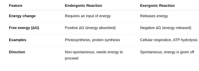
Distinguish between endergonic and exergonic reactions
Explain how ATP functions as a universal energy carrier.
- ATP (adenosine triphosphate) stores energy in its high-energy phosphate bonds, especially the bond between the 2nd and 3rd phosphate groups.
- When ATP is hydrolyzed (broken down) into ADP (adenosine diphosphate) + Pi (inorganic phosphate), energy is released.
- This released energy is used to drive endergonic (energy-requiring) reactions in cells.
- Because ATP links exergonic and endergonic processes, it acts as a universal energy currency, providing energy where and when cells need it.
- Cells constantly regenerate ATP from ADP and Pi using energy from food molecules during cellular respiration.
Distinguish between oxidation and reductions reactions
- Oxidation:
- Involves loss of electrons from a molecule.
- Often involves losing hydrogen atoms or gaining oxygen atoms.
- The molecule that loses electrons is said to be oxidized.
- Reduction:
- Involves gain of electrons by a molecule.
- Often involves gaining hydrogen atoms or losing oxygen atoms.
- The molecule that gains electrons is reduced.
- These reactions always occur together — when one molecule is oxidized, another is reduced. This is called a redox reaction.
Explain the functions of NAD and FAD.
- NAD (Nicotinamide Adenine Dinucleotide) and FAD (Flavin Adenine Dinucleotide) are important coenzymes that function as electron carriers in cellular metabolism.
- During metabolic reactions
(like cellular respiration), NAD and FAD:
- Accept electrons (and hydrogen atoms) from molecules being oxidized.
- Become reduced to NADH and FADH₂, respectively.
- Carry these high-energy electrons to the electron transport chain in mitochondria.
- In the electron transport chain, NADH and FADH₂ donate electrons, which helps generate ATP through oxidative phosphorylation.