viral detection + diagnosis
2/19/24
diagnosis of viral infections
- patient history + syptoms
- confirmation by lab
techniques
- antigen/nucleic acid detection
- serology
- viral culture/cell culture
- viral
replication
- cytopathic effects (CPE)
- hem adsorption/hemagglutination
- electron microscopy/light microscopy
specimens for viral diagnosis
quality of diagnosis depends on specimen
- timing of specimens in relation to patient's illness
- type of specimen (site of disease)
- quality and amount of specimen
- time and conditions of transport to the lab
specimens for viral diagnosis (lab diagnosis for viral disease)
common pathogenic viruses
resp tract
- adenovirus, flu, enterovirus (picorna), rhinovirus, paramyxovirus, rubella, HSV
GI tract
- reovirus, rotavirus, adenovirus, Norwalk virus, calcivirus
CNS
- enterovirus (picorna), arbovirus (togavirus, bunyavirus), rabies virus
- HSV, CMV, mumps, measles
blood
- HIV, human T cell leukemia virus, Hep B, C, D viruses
viral culture
Viral disease diagnosis traditionally relied on the isolation of viral pathogens in cell cultures.
Approach is often slow and requires considerable technical expertise, yet has been regarded for decades as the “ gold standard” for the laboratory diagnosis of viral disease.
With the development of nonculture methods for the rapid detection of viral antigens and/or nucleic acids, the usefulness of viral culture has been questioned.
However, virus diagnosis via viral cell culture, with classical and new technological advances such as stem cell lines and others, remains a useful approach:
•when viral tritation is needed,•when a viable isolate is needed, •if viable and nonviable virus must be differentiated, •when infection is not characteristic of any single virus (i.e., when testing for only one virus is not sufficient), •when available culture-based methods can provide a result in a more timely fashion than molecular methods.
systems for propagation of viruses
- animals
- embryonated eggs
- tissue culture
- primary
- secondary
- cell lines
*cell culture is most common method for propagation of viruses
steps in viral culture in cells
- processing of specimen and inoculation in cell culture
- maintenance of the inoculated cell culture
- detection of viral growth
detection of viral replication
cytopathic effects (CPE)
cytopathic effects can be defined as structural changes in host cell
- specifically in tissue culture resulting from viral infection
- generally, microscopically visible changes to infected cell
- however some viruses grow to high titers and do not produce visible CPE (rubella)
cytopathic effects (CPE) of viral cells
- change in cell morphology
- cell death
- apoptosis, necrosis
- cell fusion (syncytial)
- histologic changes (inclusion bodies)
- cell surface changes
- hemadsoprtion/hemagglutination
- change in growth/lifespan
CPE may be used in some cases to identify a virus unequivocally
in other cases, other detection methods used to confirm presence of suspected virus
syncytium formation
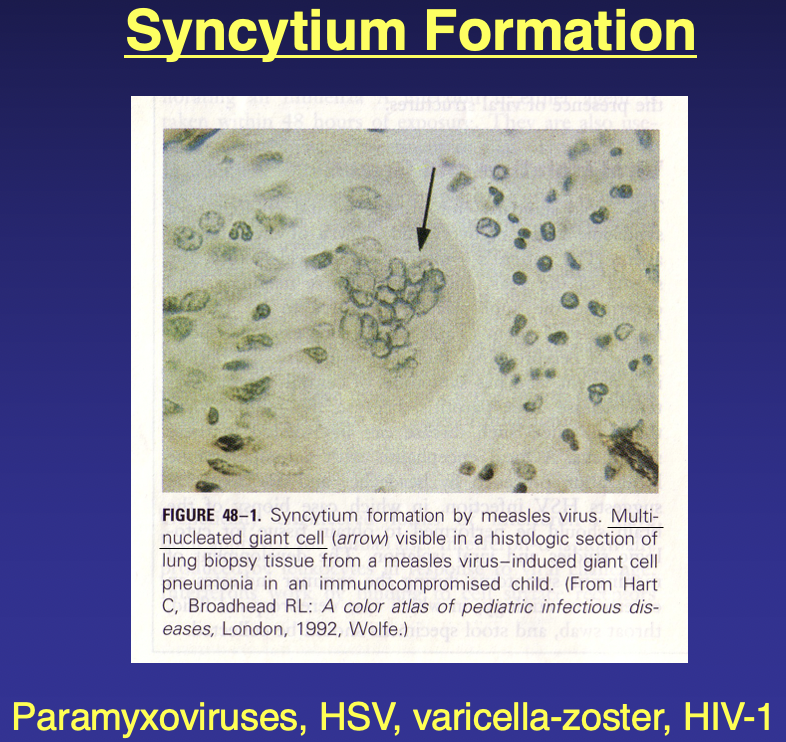
paramyxoviruses, HSM, varicella-zoster, HIV-1
hemadsorption/hemagglutination
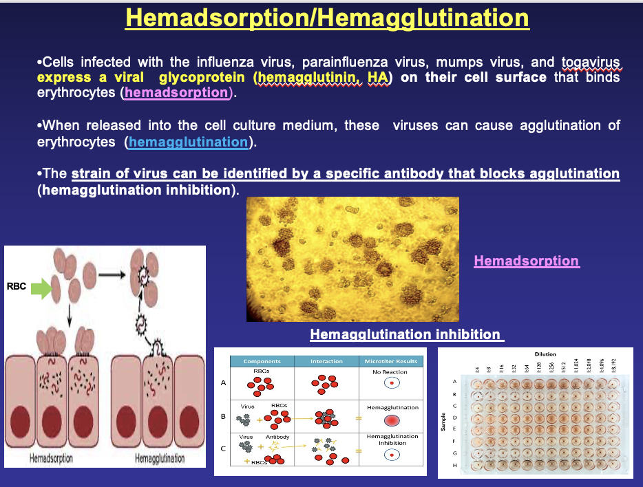
cells infected w/ flu virus, parainfluenza virus, mumps virus, and togavirus express a viral glycoprotein (hemagglutinin, HA) on their cell surface that binds erythrocytes (hemadsorption)
when released into a cell culture medium, these viruses cause agglutination of erythrocytes (hemagglutination)
the strain of virus can be identified by a specific antibody that blocks agglutination (hemagglutination inhibition)
virus titration (quantification)
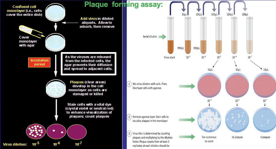
- cytopathic effects in a local area of cells may be observed as a plaque or focus of infection
- plaque assays count infectious particles of cytolytic viruses
- plaque assay is limite to only a subset of animal viruses that can lead to cell lysis, forming plaques on the monolayer of cells in a cell culture plate
PLAQUE ASSAY
the gold standard method to determine viral titer as plaque forming units (PFU)
quantitative measure of infectious virus: