Appendicular skeleton
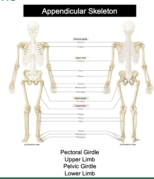
pectoral Girdle
Upper Limb
Pelvic Girdle
Lower Limb
The Pectoral Girdle - clavicle
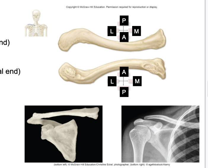
s-shaped
clavicle medial half
attached to the sternum and forms Sternoclavicular join
clavicle lateral half
attached to the acromoin and forms the Acromioclavicular join
The Pectoral Girdle - the scapula
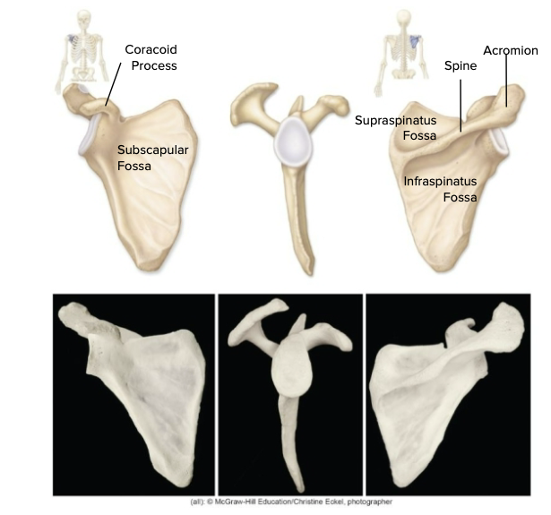
large flat irregular
Glenoid cavity – articulation with humerus
• Borders
•
Superior, medial, & Lateral
• Anterior surface: Subscapular
fossa
• Posterior surface
• Spine of scapula (ends at the
acromion)
• Fossa above: Supraspinatus fossa
• Fossa below:
Infraspinatus fossa
• Anterior surface: Subscapular fossa
•
Acromion process (posterior)
• Coracoid process (anterior)
Upper limb - Humerus
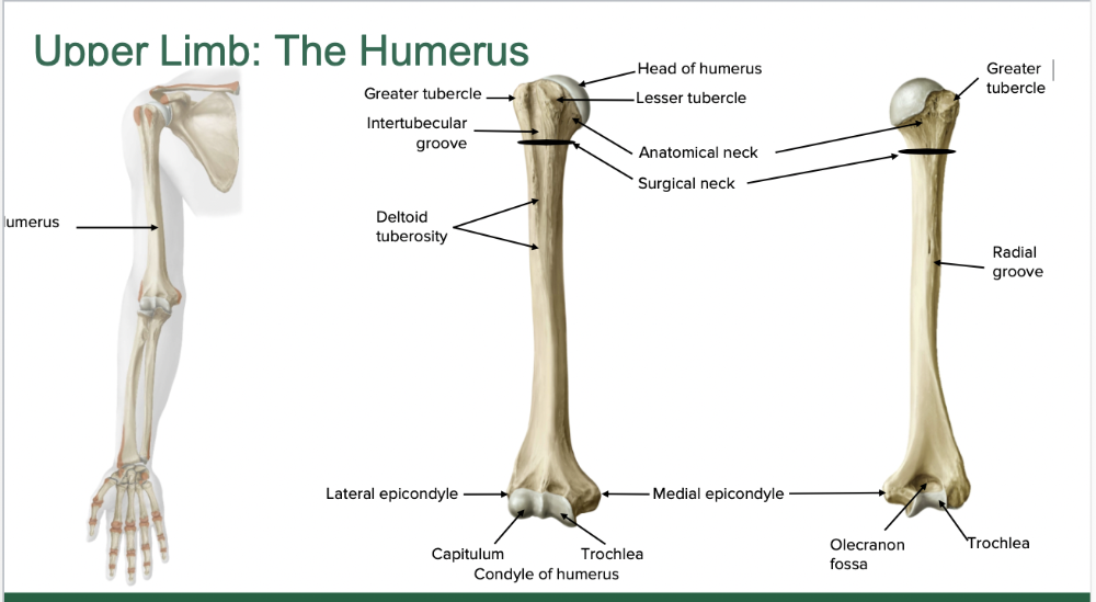
2 bones in the body
Upper limb - radius
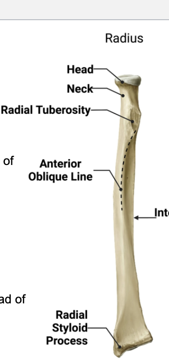
• Proximal features:
• Head: Articulates with capitulum
of
humerus
• Neck: Narrowest region
• Radial
tuberosity: For biceps
brachii muscle attachment
• Distal
features:
• Styloid process: Lateral “wrist
bump”
•
Ulnar notch: Medial dent for head of
ulna
Upper limb - ulna
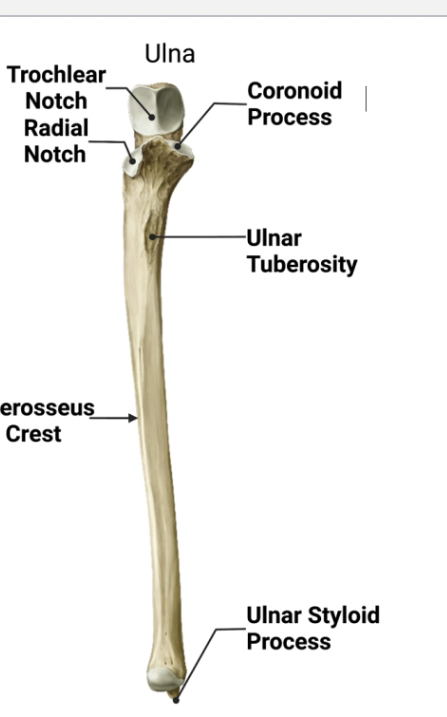
• Proximal features:
• Trochlear notch:
Accommodates
trochlea of humerus
• Olecranon: Projection
that forms the
posterior “bump” of the elbow;
attachment
site for triceps brachii
• Coronoid process: Inferior lip
of
trochlear notch
• Radial notch:
Lateral;
accommodates head of radius
• Distal
features:
• Styloid process: Posteromedial
“wrist bump
Upper limb - carpals
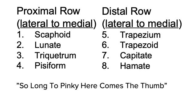
8 wrist bones
2 rows of four
Upper limb - metacarpals
five in the palm (I-V)
• I is the base of the thumb, V is the
base of the little
finger
Upper limb - phalanges
14 per hand • Three phalanges per finger
• Proximal, middle, and
distal phalanges
• But only two in the thumb (pollex)
•
Proximal and distal phalanges (no middle phalanx)
the pelvic girdle
The pelvic girdle consists of the right and
left ossa
coxae
• Commonly called “hip bone”
• os coxa =
singular
• With sacrum and coccyx = the bony pelvis
• Fusion
of ilium, ischium, and pubis between 13
and 15 years of age
the pelvic girdle -Articulations:
1. Anteriorly with other os coxae (2 os coxae =
pelvic
girdle)
2. Posteriorly with the sacrum
3. Laterally with
femur at acetabulum
• All three bones of the os coxa contribute
to the
acetabulum (socket)
the pelvic girdle - ilium
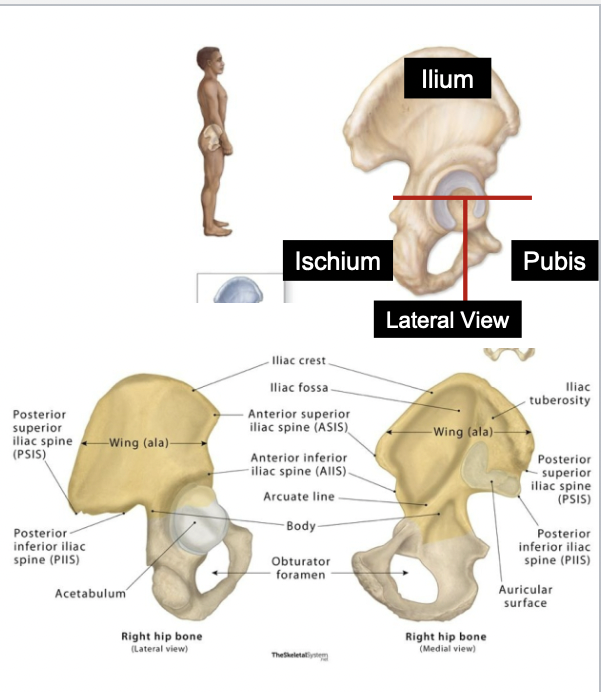
• Largest of the three fused coxal bones
• Makes up the superior
portions of os coxa
(and acetabulum)
• Features :
•
Ala
• Anterior superior iliac spine (ASIS)
• Iliac Crest
the pelvic girdle - the ischium
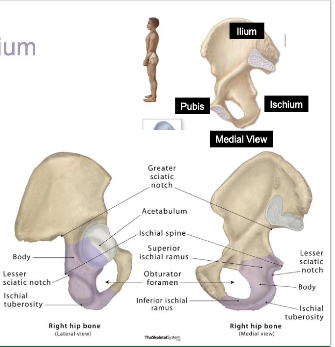
• Superior/posterior margin of os coxa
• Features:
•
Ischial spine: Prominent medial process
• Ischial tuberosity:
Rough inferior projection
that supports weight of body when seated
the pelvic girdle - pubis
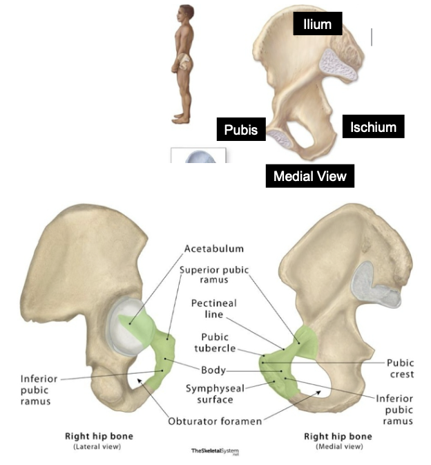
• Anterior region of os coxa
• Features:
• Pubic tubercle:
palpable point at anterior
border
• Obturator foramen: Large
space bordered
by pubic and ischial rami
the lower limb - the femur
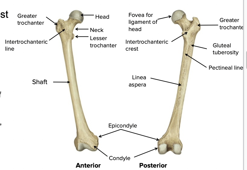
• Longest, strongest, and heaviest
bone in the
body
Important features:
• Head: Articulates with os coxa
at
acetabulum
• Ball-and-socket joint
• Greater and
lesser trochanters:
Massive processes for attachment
of
powerful hip and thigh muscles
• Shaft
• Medial and
lateral condyles: Smooth,
rounded articular surface
the lower limb - patella
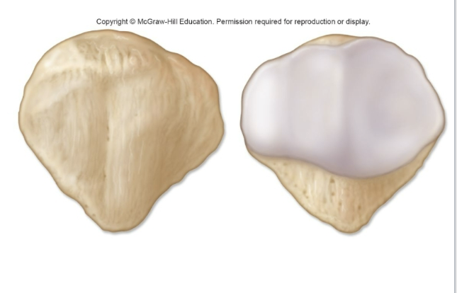
• “Kneecap” within tendon of quadriceps
femoris muscle
•
Triangular with broad superior base
and inferiorly pointed
apex
• Articular surface articulates with femur
the lower limb - tibia
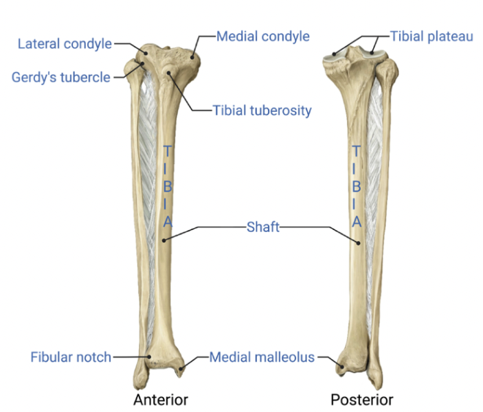
• Weight-bearing bone of lower leg
• Medial and lateral
condyles: Smooth
surfaces for articulation with femur
•
Tibial tuberosity: Rough anterior
projection inferior to
condyles
• Attachment site of patellar ligament
• Medial
malleolus: Inferior articular
surface: for the talus (a tarsal bone)
the lower limb - fibula
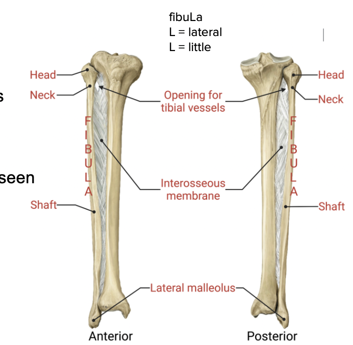
• No major weight-bearing properties
• Head
•
Neck/Shaft
• Lateral Malleolus: bony protrusion seen
on
outside of ankle
bones of ankles
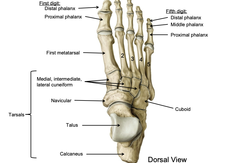
7 “ankle” bones called tarsals:
Tarsals:
1. Calcaneus
(forms the heel)
2. Talus, most superior,
weight-bearing,
articulates with the tibia
3.
Navicular
4-6. Cuneiform (wedges-medial,
intermediate,
lateral)
7. Cuboid
These seven bones are
collectively referred to
as “the tarsus
bones of the feet/toes

Five metatarsals are in the palm (I-V)
• I is the base of the
big toe, V is the base of the little
toe
Fourteen phalanges
per foot
• Three phalanges per toe
• Proximal, middle, and
distal phalanges
• But only two in the big toe (hallux)
•
Proximal and distal phalanges (no middle phal
Aging and the Appendicular Skeleton
• Skeletal mass and density decline with aging
• Potential
osteoporosis, susceptibility to fracture
• Begins in middle
age
• Osteoarthritis develops as articular surfaces
deteriorate
• Pubic symphysis changes with age
• Rough in
early adulthood
• Flattens in 20s
• Develops prominent rim
in 30s and 40s
• Develops concavities with arthritis in elderly