histology?
Study of the tissues of the body and how tissues are arranged into organs.
Tissues = cells + ECM
• Collected
• Fixed (formalin, paraformaldehyde, etc.)
•
Sectioned
• Stained (H&E, methylene blue, etc.)
• Imaged
(Brightfield, fluorescence)
H staining
Hematoxylin
basic stain that binds to acids
purple
ex. nucleus and RNA
E stain
Eosin
acidic stain that binds to basic
pink
ex. cytoplasm
what is bone?
skeletal bone are complex organs
primarily connective tissues
function of bones
• Support and protection of more delicate organs
• Movement –
attachment site for muscles
• Hematopoiesis – blood cell
production in red bone marrow
• Storage of mineral and energy reserves
long bones
greater length than width
ex. femur
short bones
nearly equal length and width
ex. tarsal bones
flat bones
thin surfaces
ex. frontal lobe
irregular bones
complex shapes
ex. vertebra
sesamoid bones
develop within tendons
diaphysis
elongated cylindrical shaft
epiphysis
• Knobby, enlarged regions at each end
• Strengthens
joints
• Attachment site for tendons and ligamen
metaphysis
• Region between diaphysis and epiphysis
• Contains epiphyseal
(growth) plat
articular cartilage
• Thin layer of hyaline cartilage covering the epiphysis
•
Reduces friction and absorbs shock in moveable join
medullary cavity
• Hollow, cylindrical space in diaphysis
• In adults, it
contains yellow bone marro
flat bones within the skull
Two layers of compact bone, with spongy bone (diploe) sandwiched between
Cartilage is a
strong, flexible connective
tissue that protects your joints and
bones
• Also supports soft tissues
compact bone
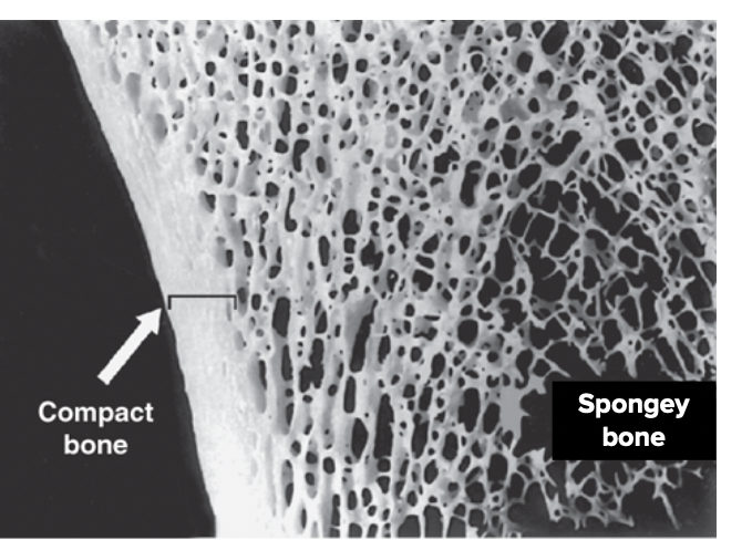
• Solid and relatively dense
• External surfaces of long and
flat bones
spongy bone
• Open lattice of narrow plates called trabeculae
• Internal
surface of bones
The basic unit of compact bone is
the osteon - Haversian system -small
central canal
Carries vessels and nerves in center of osteo
perforating canal
Run perpendicular to and help connect
multiple central canals;
Passageways for blood vessels and nerv
osteocytes
Housed in lacunae between lamellae
lacunae
spaces within bone which contain osteocytes
canaliculi
Tiny, interconnecting channels that extend between
lacunae and
allow osteocytes to connect and communica
concentric lamellae
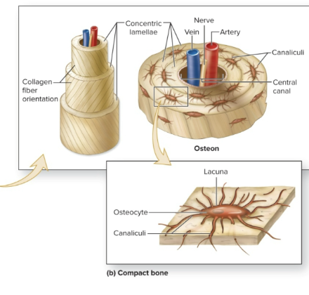
Rings of bone around central cana
The Histology of Bone
Abundant extracellular matrix:
• 15% water
• 30% collagen
fibers*
• 55% crystallized mineral salts
◦ Calcium
phosphate
◦ Calcium hydroxide
◦ Other:
• Calcium
carbonate
• Magnesium
• Fluoride
• Potassium
• Sulfate
osteoprogenitor cell
• Unspecialized bone stem cells from mesenchyme
• Only bone
cells to undergo cellular division
• Inner portions of
periosteum, endosteum and the
blood vessel canal
osteoblasts
bone builders
osteocytes
• Mature bone cells
• Maintains metabolism
osteoclasts
• HUGE cells which derived from up to 50 monocytes
• Within
endosteum
• Release lysozymes and acids to resorb bone
•
Bone Crushers
• Helps regulate serum (blood) calcium levels
red marrow bone
blood cell production
yellow marrow bone
blood vessels and adipocytes
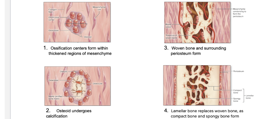
Intramembranous ossification
• Develops from mesenchyme
• Produces flat bones of the skull,
some facial bones, mandible, and central portion of clavicle
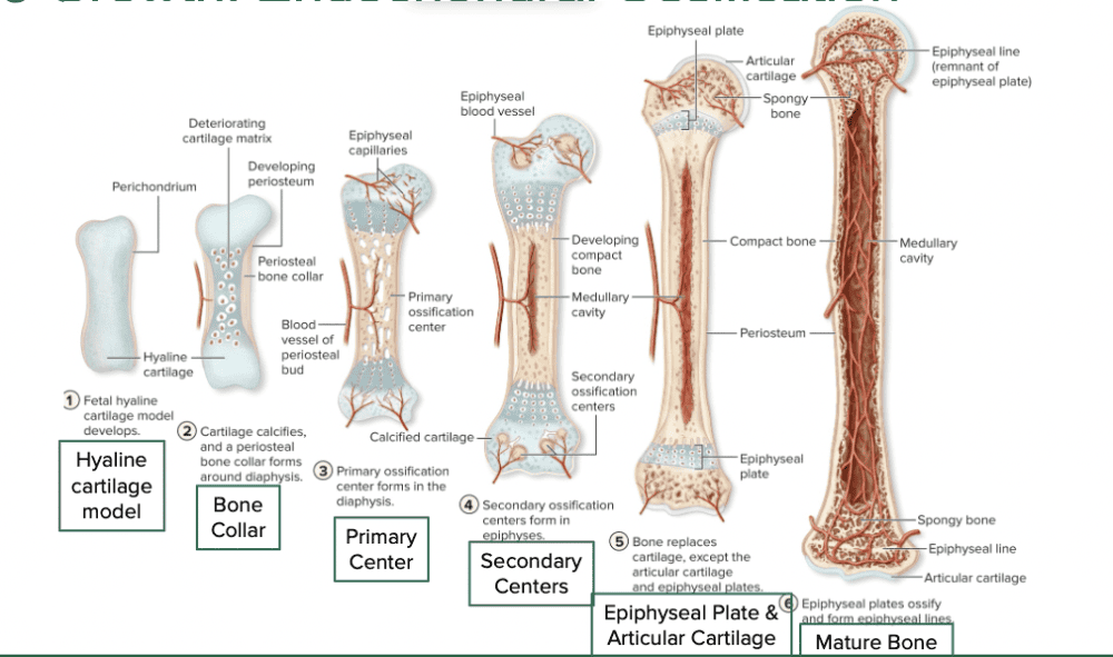
Endochondral ossification
• Begins with hyaline cartilage model
• Produces majority of
bones in the body
A long bone’s growth in length is referred to
as interstitial growth (occurs at the
epiphyseal plate)
Growth in a bone’s diameter is referred to
as appositional growth (occurs at the
periosteum)
bone growth: Intramembranous Ossification
bone growth: endochondral Ossification
zone 1 resting cartilage
Near the epiphysis; composed of small chondrocytes
zone 2 proliferating cartilage
Chondrocytes proliferate; align into stacks
zone 3 hypertrophic cartilage
Chondrocytes stop proliferating, but enlarge
(hypertrophy)
zone 4 calcified cartilage
Minerals are deposited, which kills the chondrocytes
zone 5 ossification
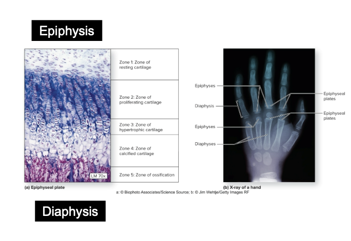
Matrix of bone is deposited on the remaining calcified
cartilage matrix
bone remodeling
bone is constantly renewing and get stronger with exercise
Involves bone resorption via osteoclast and new bone formation via osteoblast
1. Nutrient artery and vein:
Supply the diaphysis of a long
bone; usually just one nutrient
artery and vein per bone
2. Metaphyseal arteries and veins:
Supply the diaphyseal side
of the epiphyseal plate
3. Epiphyseal arteries and veins:
Supply the epiphyses
4. Periosteal arteries and veins:
Supply blood to the external
circumferential lamellae and
superficial osteons
Hormones control osteoblast/clast activity and calcium level
• Growth hormone stimulates cartilage growth at
epiphyseal
plate
• Thyroid hormone stimulates
osteoblasts
• Calcitonin (↓)/parathyroid (↑) hormone impact blood
calcium
Vitamins are needed for normal bone growth and maintenanc
• Vitamin A activates osteoblasts
• Vitamin C required for
collagen synthesis
• Vitamin D stimulates calcium absorption from
GI tract into
blood so that calcium is available for bold building
exercise
• Mechanical stress stimulates increase in bone density by increased
osteoblast activity
• Bones of athletes become thicker and
stronger as the result of repetitive and stressful exercise
•
Bones lose mass with age, but this can be slowed or reversed with
weight-bearing exercise
avulsion fracture
pulling a bone off from the rest of the bones
colles fracture
wrist fracture
comminuted fracture
bone is crushed into many pieces
compound fracture
Broken ends of the bone protrude through the skin
complete fracture
bone is broken into 2 or more pieces
displaced fracture
Fractured bone parts are out of anatomic alignment
greenstick fracture
Partial fracture; one side of bone breaks—the other side is bent
hairline fracture
Fine crack in which sections of bone remain aligned
linear fracture
fracture is parallel to the long axis of the bon
oblique fracture
fracture is at an angle
simple fracture
no break through skin
stress fracture
Thin fractures due to repeated, stressful impact such as running
closed fracture repair
• Manual realignment
• Casting/splinting
• Skin intact
open fracture repair
• Realignment with surgery
• Screws, plates, rods, pins,
wires
◦ Open reduction internal fixation
◦ Open reduction
external fixation
General Fracture Repair
BMD 310 - Clinical
Anatomy and Histology
Physiological Fracture Repair:
reactive phase
• Blood vessels crossing fracture line break
• Blood clots
resulting in a fracture hematoma
within 8 hrs after injury
•
Nearby bone cell death
• Swelling, inflammation
• Last up to
several week
Physiological Fracture Repair:
reparative
Formation of a fibrocartilaginous callus and then a bony callus (spongey bo
Physiological Fracture Repair:
bone remodeling
• Dead portion of original bone resorbed by osteoclasts
•
Compact bone will replace spongy bone surrounding the fracture site
osteoporosis disease
10m in the US
calcium and bone mass is lost
middle aged and older, women
treatment: diet, exercise, medication