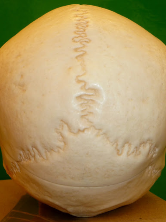
1
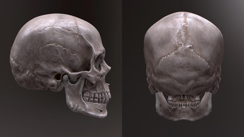
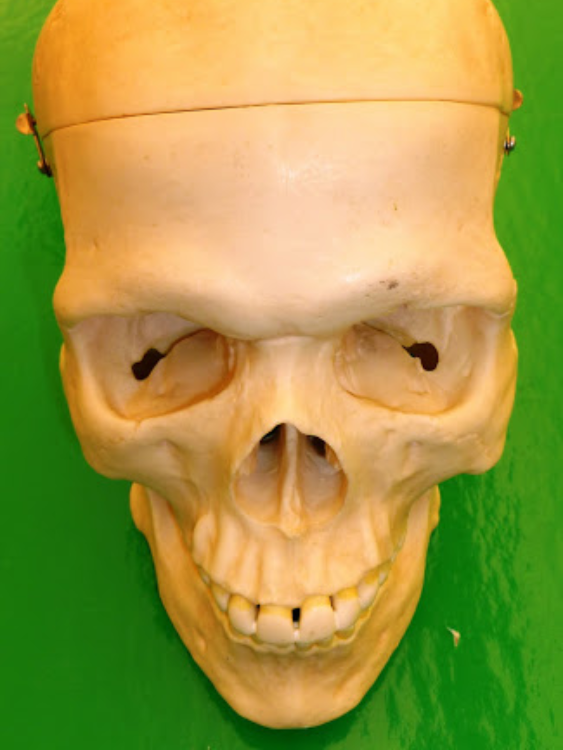
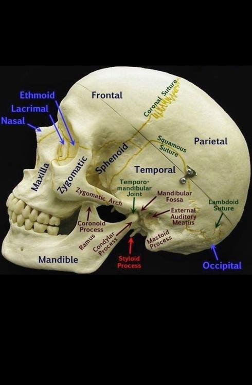
- Frontal Bone:The frontal bone is a bone in the human skull that forms the forehead, the upper part of the eye sockets (orbits), and the front part of the top of the skull. It is a single bone that contributes to the formation of the skullcap and the upper part of the eye sockets.
- Parietal Bones:The parietal bones are two large, flat bones that form the majority of the top and sides of the human skull.
- Sagittal Suture: The sagittal suture is situated along the sagittal plane, which divides the skull into left and right halves. It runs longitudinally from the anterior (front) to the posterior (back) part of the skull.
2

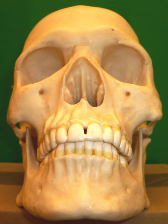
- Glabella: smooth area above root of the nose, between the eyebrows
- Nasal Bones: Where glasses rest. Forms the superior bony wall of the nasal cavity
3
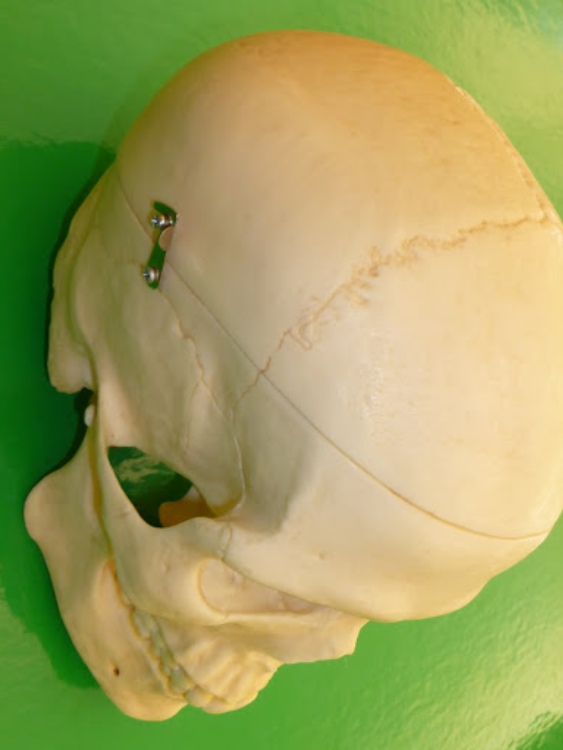
- Coronal Suture: The coronal suture is one of the major fibrous joints (sutures) in the human skull that connects the frontal bone to the two parietal bones.
- Temporal Bone: Side of skull (temple) The earhole is the giveaway.
4
- Supraorbital foramen/notch: a hole through the brow ridge, provides passage for nerve, artery and vein
5
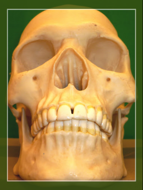
- Maxilla: The maxilla is a paired bone that forms the upper jaw and contributes to the structure of the facial skeleton. It is one of the bones of the skull and plays a crucial role in supporting the structures of the face and forming the upper jaw and parts of the hard palate.
- Alveolar Process: Portion of the maxillary bones that form the support for teeth of the maxillary arch
6

- Mandibular Fossa: depression or concavity in the temporal bone of the skull. Look for piece of rubber on model.
- Styloid Process: : pointy bone sticking downward
- Mastoid Process:large lump behind the ear
7

- External Acoustic Meatus: : the ear hole.
- Zygomatic Bones: Cheek bones, defines the face.
- Zygomatic Process: The zygomatic process is a bony projection that extends from the temporal bone and contributes to the formation of the zygomatic arch, which is a part of the cheekbone.
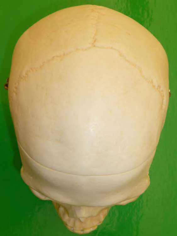
8
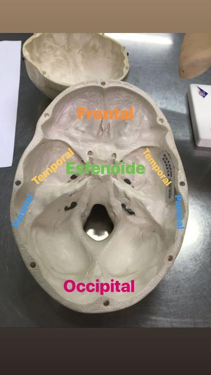
- Occipital bone: The occipital bone is a large, unpaired bone located at the back of the skull. It forms the posterior part and base of the skull, protecting the cerebellum and brainstem.
- Lambdoidal suture:The lambdoid suture is one of the primary sutures in the human skull, joining the parietal bones to the occipital bone. The lambdoid suture runs horizontally across the posterior part of the skull, connecting the parietal bones to the occipital bone.
9
- Ethmoid bone: Located between eyes. Contributes to medial wall of orbit, walls and roof of nasal cavity, and nasal septum.
- Cribriform Plate: The cribriform plates are located on each side of the ethmoid bone, forming part of the skull's anterior cranial fossa.
- Crista galli: situated in the midline of the ethmoid bone, rising vertically from the cribriform plate. It extends superiorly (upward) and separates the left and right olfactory foramina.
- Petrous part of Temporal bone: a pyramid-shaped portion located at the base of the skull, deep within the skull structure. located at the base of the skull, between the sphenoid and occipital bones. It is situated on each side of the skull and is well-protected by dense bone. You can pinch it.
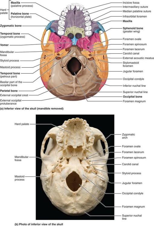
10
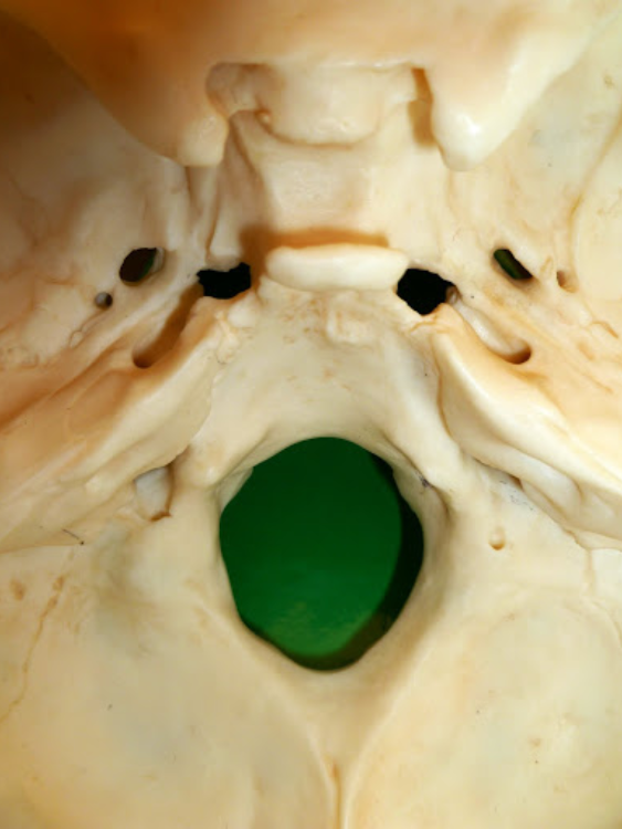
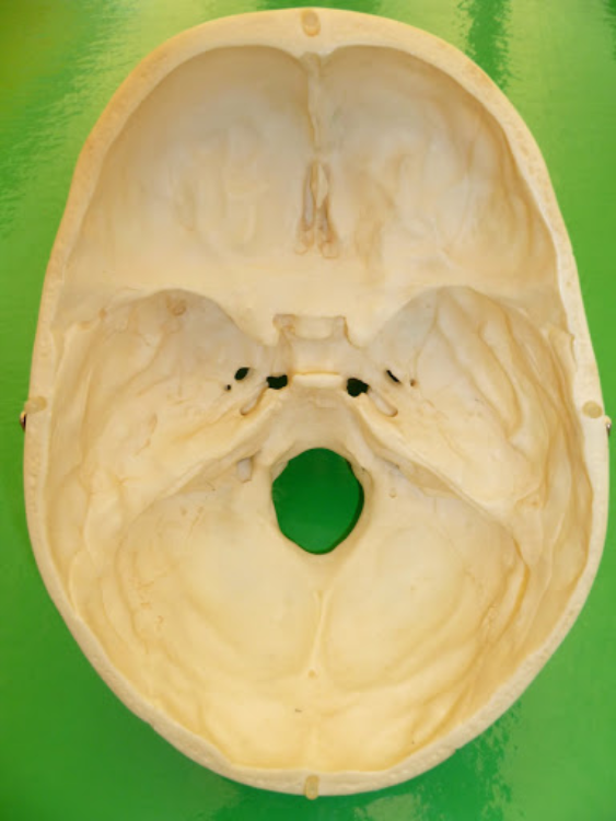
- Occipital Condyle: paired, rounded projections located on the inferior aspect of the occipital bone. situated on either side of the foramen magnum, which is a large opening at the base of the skull. The foramen magnum allows the passage of the spinal cord.
-
Internal Acoustic Meatus: on temporal inner
ear
connected to external acoustic meatus, inside sound canal. physically look inside to see it. - Jugular Foramen: On temporal bone, 2 large holes nearest the magnum foramen. Wide as a pinky. Pipe cleaner can go through it.
- Carotid Canal: No wider than pipe cleaner, in front of jugular foramen.
11
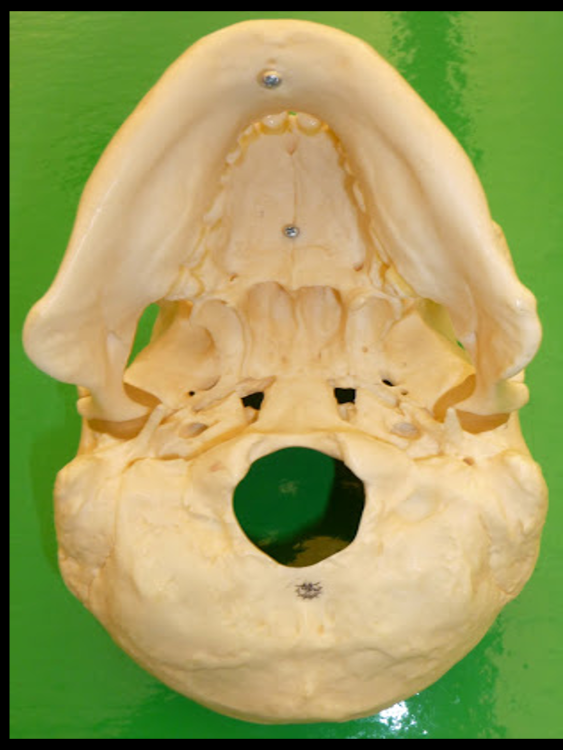
- Foramen Magnum: a large opening located at the base of the skull, specifically in the occipital bone. It is a crucial anatomical structure through which the spinal cord passes, connecting the brain to the spinal canal.
- Pterygold Processes: projects downward from the junction of the body and greater wing of the sphenoid bone. bony projections arising from the sphenoid bone, forming part of the lateral walls of the skull and contributing to the base of the skull.
12
...
13
- external occipital protuberance: a bony prominence located on the external surface of the occipital bone at the back of the skull. It serves as an anatomical landmark and an attachment point for ligaments and muscles.
- Squamous Suture: This is the flat and scale-like portion of the temporal bone that forms the lateral side of the skull. It contributes to the zygomatic process, which, along with the zygomatic bone, forms the zygomatic arch (cheekbone).
14
...
15
...
16
...
17
...
18
...
19
...
20
...
21
...
22
- Sella Turcica- bony saddle-shaped structure located on the superior surface of the body of the sphenoid bone, which is a butterfly-shaped bone at the base of the skull. Indentation in the middle.
- Palatine Process: The palatine process refers to the horizontal, plate-like extensions of the maxillary bones that contribute to the formation of the hard palate—the bony structure at the roof of the mouth. Front 2/3 hole of mouth-right before soft palate.
- Palatine Bones: pair of L-shaped facial bones that contribute to the formation of the hard palate, part of the floor and lateral walls of the nasal cavity, and a small portion of the eye sockets (orbits). Located at the back of the nasal cavity, forming part of the facial skeleton.
23
- Superior orbital fissure: situated at the junction of the greater and lesser wings of the sphenoid bone. It is positioned in the posterior part of the orbit.
- Optic Canal: Smaller hole, circular opening of the eye socket. Pipe cleaner can fit through it.
24
- Greater and Lesser Wings: The greater and lesser wings are two prominent bony structures of the sphenoid bone, a butterfly-shaped bone located at the base of the skull.
- Greater Wings:The greater wings are large, flattened projections that extend laterally from the body of the sphenoid bone.
- Lesser Wings:The lesser wings are smaller bony projections located superiorly and medially to the greater wings.
25
CHEAT SHEET
26
CHEAT SHEET
27
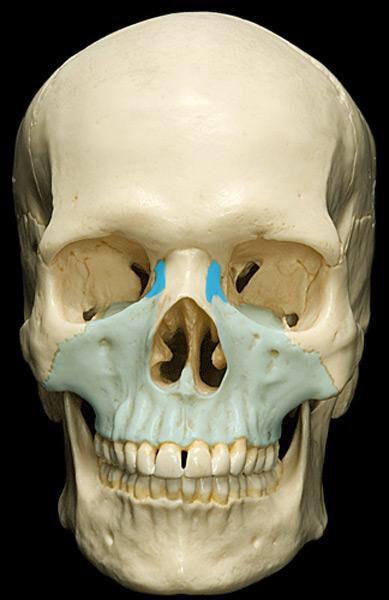
- Maxilla: Forms side of nasal cavity, front teeth.
- Lacrimal Bones: Thin and roughly rectangular in shape. paired, small, flat bones located within the facial skeleton, contributing to the formation of the eye sockets (orbits) and the medial walls of the orbits. Where tears drip out of your face. "LACRIMAL" translates to "CRYING" "TEARS"
28
- Vomer: behind nasal cavity, forming part of the nasal septum. The vomer is typically thin and flat, resembling a plowshare or a blade. Its shape helps to support and stabilize the nasal septum.
- Perpendicular Plate: located in the midline of the ethmoid bone and extends downward from the cribriform plate. a thin, vertical projection that descends from the cribriform plate, forming the upper part of the nasal septum.
29
- Inferior Nasal Conchae: The inferior nasal conchae, also known as the inferior turbinates, are thin, curved bones within the nasal cavity. Situated on the lateral walls of the nasal cavity, projecting inward like shelves.
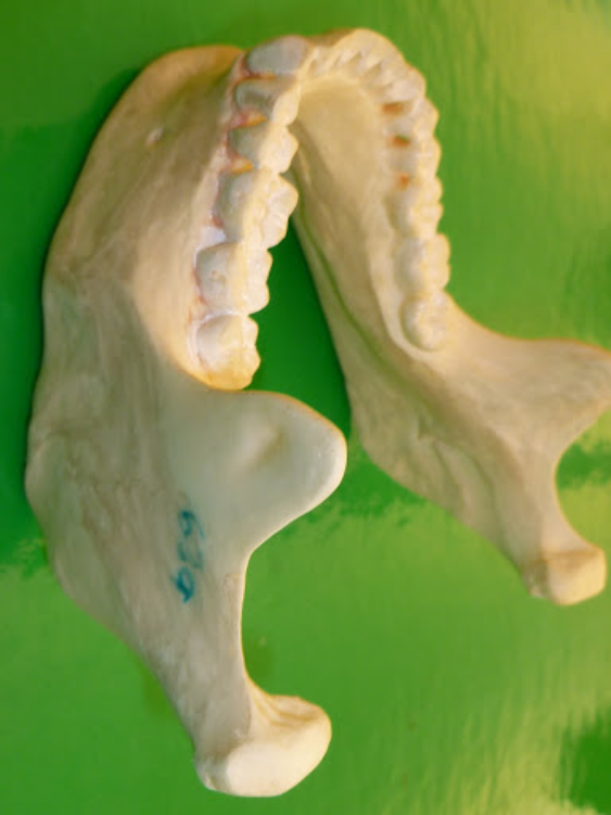
- Mandible: Commonly known as the jawbone, is the largest and strongest bone in the human face.
- The body of the mandible:forms the lower jaw and contains the alveolar processes that support the lower teeth.
30

- Mental Foramen: The mental foramen is an opening or hole located on the external surface of the mandible, which is the lower jawbone. Holes on both side, near chin (on model)
- Mental Eminence (protuberance): Bump near chin (on model)
- Alveolar Process: The alveolar process refers to the bony ridge within the maxillary (upper jaw) and mandibular (lower jaw) bones that contains the sockets, or alveoli, for the teeth. The alveolar processes provide support and housing for the roots of the teeth, securing them in position.
31
- Ramus: The mandibular ramus is a part of the mandible (lower jawbone). The mandible has two rami (plural for ramus), each extending upward from the body of the mandible. The mandibular ramus has two main processes:
- Coronoid Process: The coronoid process is situated at the upper end of each mandibular ramus. The coronoid process is situated at the upper end of each mandibular ramus.
- Mandibular Condyle: a bony prominence located at the posterior (back) end of each mandibular ramus, which is part of the lower jawbone (mandible).
32
- Central and Lateral Incisors: Central and lateral incisors are types of teeth found in both the upper (maxillary) and lower (mandibular) dental arches. The central incisors are the most anterior (front) teeth in both the maxillary and mandibular dental arches. In each dental arch, there are two central incisors—one on the right side and one on the left.
- Canines:The central incisors are the most anterior (front) teeth in both the maxillary and mandibular dental arches.In each dental arch, there are two central incisors—one on the right side and one on the left.
33
- Premolars 1-2: situated between the canines and molars in both the upper and lower dental arches.There are two premolars in each quadrant of the mouth, making a total of eight premolars in the permanent dentition.
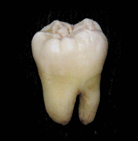
- Molars 1-3: The molars are the large, flat teeth located at the back of the mouth. Third molar, commonly known as wisdom teeth.
34
...
35
...
36
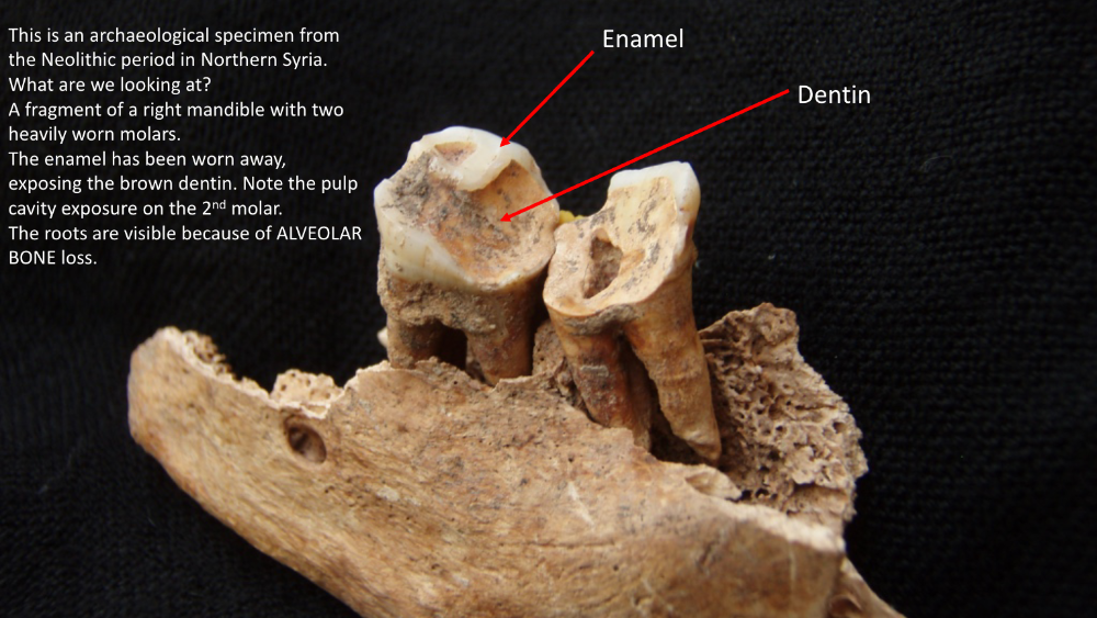
- Crown: the visible, exposed part of the tooth above the gumline. It is the portion of the tooth that is covered by enamel, which is the hardest substance in the human body. Here are key points about the crown of a tooth
- Enamel: the outermost and hardest layer of a tooth, covering the crown. can be worn away, irreplaceable, permanent, does not grow back.
- Dentin: Brown/Yellow. Dentin makes up the majority of the tooth structure and is located between the enamel or cementum and the pulp chamber. It forms the crown and root of the tooth.