The notochord is a structure present in _________.
- All of the listed responses are correct.
- humans
- chickens
- salamanders
- frogs
All of the listed responses are correct.
Ex.
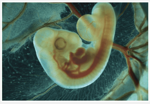
The notochord is a structure present in frogs, salamanders, chickens, and humans during embryonic development.
These animals are all chordates. The notochord, a rod that extends along the dorsal side of the embryo, forms from cells of the dorsal mesoderm during the neurulation stage of chordate embryonic development cells. Neurulation then continues throughout the early steps in the formation of the brain and spinal cord. Neurulation is one component of the larger process of organogenesis, in which regions of the three embryonic germ layers develop into the rudiments of organs.
A critical role of calcium ions (Ca2+) in development immediately following fertilization is that of __________.
- triggering the acrosomal reaction
- triggering the cortical reaction as a slow block to polyspermy
- triggering the fast block to polyspermy
- neutralizing the effect of hydrolytic enzymes in the egg
- depolarizing the egg cell membrane
triggering the cortical reaction as a slow block to polyspermy
Ex.
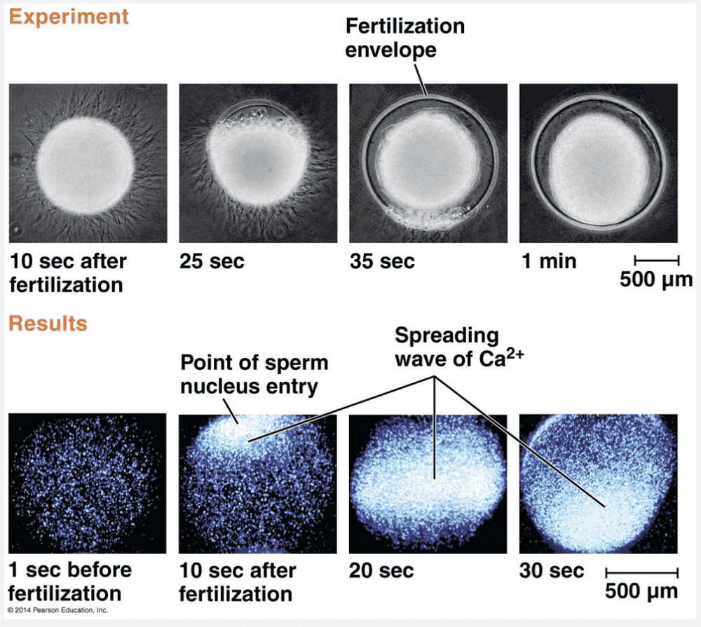
A critical role of calcium ions (Ca2+) in development immediately following fertilization is that of triggering the cortical reaction as a slow block to polyspermy.
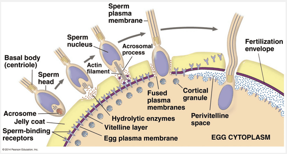
Binding of sperm to the egg activates a signal transduction pathway that triggers the release of Ca2+ from the endoplasmic reticulum into the cytosol. The resulting increase in Ca2+ levels causes cortical granules to fuse with the plasma membrane. This cortical reaction lifts the vitelline layer away from the egg and hardens the layer into a protective fertilization envelope. Additional enzymes clip off and release the external portions of the remaining receptor proteins, along with any attached sperm.
The calcium ions are involved in releasing, not neutralizing, hydrolytic enzymes in the egg associated with the cortical reaction.
The acrosomal reaction precedes fertilization and involves the sperm penetrating the jellylike coat of an egg.
Sodium ions diffusing into the egg cause depolarization of the membrane.
Depolarization of the membrane comprises the fast block to polyspermy.
The role of the cytoskeleton in morphogenesis is ___________.
- to change the shape of cells
- to change the location of cells
- to anchor all cells so that they are not displaced from the developing embryo
- substantial during early stages only
- to change the shape and location of cells
to change the shape and location of cells
Ex.
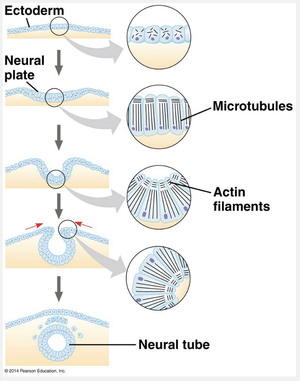
The role of the cytoskeleton in morphogenesis is to change the shape and location of cells.
The cytoskeleton is the set of microtubules, intermediate filaments, and microfilaments that extend throughout the cytoplasm of a cell. In morphogenesis, which is the development of the form and structures of an organism, the cytoskeleton directs various types of movements, one of which is called convergent extension. This is a rearrangement that causes a sheet of cells to become narrower (converge) as it becomes longer (extends). As their shapes change, the cells elongate, with their ends pointing in the direction they will move, and then they wedge between each other to form fewer columns of cells. The cytoskeleton is also responsible for cell migration. During the process of organogenesis in vertebrates, cells from the neural crest and from somites migrate to locations throughout the embryo. In this migration, cells “crawl” within the embryo by using cytoskeletal fibers to extend and retract cellular protrusions.
Reorganization of the cytoskeleton is a major force in changing cell shape and position throughout development.
Rather than simply anchoring cells so that they are not displaced from the developing embryo, changes in the cytoskeleton permit a variety of integrated responses during morphogenesis.
During gastrulation, the primitive __________ forms.
- eye
- nervous system
- None of the listed responses is correct.
- heart
- gut
gut
Ex.
During gastrulation, the primitive gut forms.
During gastrulation, a set of cells at or near the surface of the blastula moves to an interior location, cell layers are established, and a primitive digestive tube is formed. Gastrulation is a dramatic reorganization of the hollow blastula into a two-layered or three-layered embryo called a gastrula. The cell layers produced by gastrulation are collectively called the embryonic germ layers (from the Latin germen, to sprout or germinate). In the late gastrula, ectoderm forms the outer layer and endoderm lines the embryonic digestive compartment or tract.
The “eye” and the “heart” are both incorrect because they form during organogenesis.
“Nervous system” is incorrect because the nervous system forms during neurulation.
A sea urchin sperm penetrates the jelly coat of an egg and adheres to receptor proteins on the egg's surface as a result of __________.
- the acrosomal reaction
- cleavage
- gastrulation
- depolarization
- the cortical reaction
the acrosomal reaction
Ex.
A sea urchin sperm penetrates the jelly coat of an egg and adheres to receptor proteins on the egg's surface as a result of the acrosomal reaction.
When sea urchins release their gametes into the water, the jelly coat that surrounds the egg exudes soluble molecules that attract the sperm, which swim toward the egg. As soon as the head of a sea urchin sperm contacts the jelly coat of a sea urchin egg, molecules in the jelly coat trigger the acrosomal reaction in the sperm.
“Depolarization” is incorrect because this occurs within about one to three seconds after a sperm binds to an egg. By preventing additional sperm from fusing with the egg’s plasma membrane, this depolarization acts as a fast block to polyspermy.
“The cortical reaction” is incorrect because this reaction occurs after depolarization when vesicles called cortical granules fuse with the egg’s plasma membrane.
“Cleavage” is incorrect because this is a step in which a series of cell divisions divide, or cleave, the zygote into a many-celled embryo. This step occurs after fertilization.
“Gastrulation” is incorrect because this step involves the formation of the three embryonic germ layers.
During cleavage the single large zygote is converted into a __________.
- multicellular embryo consisting of smaller cells called blastomeres
- fetus
- multicellular embryo with a blastopore and an archenteron
- three-layered embryo called a gastrula
- multicellular embryo consisting of a zona pellucida and a zygote
multicellular embryo consisting of smaller cells called blastomeres
Ex.
During cleavage the single large zygote is converted into a multicellular embryo consisting of smaller cells called blastomeres.
Once fertilization is complete, many animal species undergo a succession of rapid cell divisions that characterize the cleavage stage of early development. During cleavage, the cell cycle consists primarily of the S (DNA synthesis) and M (mitosis) phases. Cells essentially skip the G1 and G2 (gap) phases, and little or no protein synthesis occurs. Development is initiated by the cleavage stage, during which a series of cell divisions divide (cleave) the zygote into a many-celled embryo. As a result, cleavage partitions the cytoplasm of the large fertilized egg into many smaller cells called blastomeres.
“Three-layered embryo called a gastrula” is incorrect because this occurs after the blastomeres form.
“Multicellular embryo with a blastopore and an archenteron” is incorrect because this is the gastrula stage of the embryo.
“Multicellular embryo consisting of a zona pellucida and a zygote” is incorrect because the zygote diploid cell that forms when the sperm and egg nuclei join.
“Fetus” is incorrect because the fetus has formed tissues, organs, and organ systems.
The functional relationship between the zone of polarizing activity (ZPA) and the apical ectodermal ridge (AER) in vertebrate limb development is that ____________.
- they simultaneously release signaling molecules in order to provide positional information to the cells comprising the limb
- the ZPA organizes skeletal development, whereas the AER organizes external limb features
- signals secreted by the ZPA trigger the secretion of signaling molecules from the AER
- they have antagonistic effects on limb growth
- the ZPA functions early in development to promote overall limb growth, and the AER functions to promote growth only later
they simultaneously release signaling molecules in order to provide positional information to the cells comprising the limb
Ex.
The functional relationship between the zone of polarizing activity (ZPA) and the apical ectodermal ridge (AER) in vertebrate limb development is that they simultaneously release signaling molecules in order to provide positional information to the cells comprising the limb.
The ZPA is a specialized block of mesodermal tissue that regulates development along the anterior-posterior axis of the limb. The signaling molecule that the ZPA secretes is called Sonic hedgehog, named after both a video game character and a similar protein in Drosophila that also regulates development. Cells nearest the ZPA form posterior structures, such as the most posterior of the digits (equivalent to a human little finger); cells farthest from the ZPA form anterior structures, including the most anterior digit (like a human thumb). The AER secretes a protein signal, called fibroblast growth factor (FGF), that promotes limb-bud outgrowth.
It is important that the ZPA and AER function simultaneously, rather than sequentially, so that they provide positional information.
It is important that the ZPA and AER each secrete their own signaling molecule, rather than one molecule being triggered by the other molecule.
Together, the ZPA and AER organize limb development along the anterior-posterior and proximal-distal axes, not primarily along an interior-to-exterior axis.
Instead of having antagonistic effects, both the ZPA and the AER play roles in promoting limb development.
The eggs of humans differ significantly from those of reptiles in that the eggs of humans __________.
- develop only after fertilization
- are amniotic
- lack yolk as a substantial store of energy
- develop extraembryonic membranes
- undergo a cleavage stage
lack yolk as a substantial store of energy
Ex.
The eggs of humans differ significantly from those of reptiles in that the eggs of humans lack yolk as a substantial store of energy.
Unlike the large, yolky eggs of many vertebrates, human eggs are quite small and store little in the way of food reserves. Yolk is most plentiful and has its most pronounced effect on development in the eggs of birds, other reptiles, many fishes, and insects. In these animals, the volume of yolk is so great that cleavage furrows cannot pass through it, and only the region of the egg lacking yolk undergoes cleavage.
Humans and most reptiles have sexual reproduction in which development of an egg proceeds only following fertilization.
Both humans and reptiles have eggs that undergo cleavage, although the cleavage patterns differ in some ways.
Reptiles and mammals have amniotic eggs.
The amniotic egg is characterized by extraembryonic membranes that support embryonic development in terrestrial environments.
What is the embryonic origin of the lining of the digestive tube?
- All three germ layers
- Mesoderm
- Ectoderm
- Both endoderm and mesoderm
- Endoderm

Endoderm
Ex.
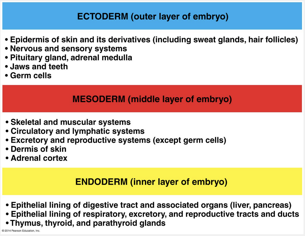
The embryonic origin of the lining of the digestive tube stems from the endoderm.
During gastrulation, a set of cells at or near the surface of the blastula moves to an interior location, cell layers are established, and a primitive digestive tube is formed. Gastrulation is a dramatic reorganization of the hollow blastula into a two-layered or three-layered embryo called a gastrula. The cell layers produced by gastrulation are collectively called the embryonic germ layers (from the Latin germen, to sprout or germinate). In the late gastrula, ectoderm forms the outer layer and endoderm lines the embryonic digestive compartment or tract.
“Ectoderm” is incorrect because this is the outer layer of the embryo and gives rise to the epidermis of the skin and its derivatives, the nervous and sensory systems, the pituitary gland and the adrenal medulla, jaws and teeth, and germ cells.
“Mesoderm” is incorrect because this is the middle layer of the embryo and gives rise to the skeletal and muscular systems, the circulatory and lymphatic systems, the excretory and reproductive systems (except germ cells), the dermis, and the adrenal cortex.
“Both endoderm and mesoderm” is incorrect because only the endoderm gives rise the epithelial lining of the digestive track and associated organs such as the liver and pancreas.
“All three germ layers” is incorrect because only the endoderm gives rise the epithelial lining of the digestive track and associated organs such as the liver and pancreas.
_________ have embryonic development based on a segmented body plan.
- shrimp
- frogs
- humans
- chickens
- All of the listed responses are correct.
All of the listed responses are correct.
Ex.
Humans, chickens, frogs, and shrimp all have embryonic development based on a segmented body plan.
Following the formation of the notochord during the development of chordates, which include mammals, reptiles (including birds), and amphibians, cells located in strips of mesoderm lateral to the notochord separate into blocks called somites. These somites are arranged serially on both sides along the length of the notochord. Somites play a significant role in organizing the segmented structure of the chordate body: Parts of the somites dissociate into mesenchymal cells, which migrate individually to new locations. Some of these cells form the vertebrae. Somite cells that become mesenchymal also form the muscles associated with the vertebral column and the ribs.
By contributing to the formation of vertebrae, ribs, and associated muscles, serially repeating structures of the embryo (somites) form repeated structures in the adult. Chordates, including humans, are thus segmented, although in the adult form, the segmentation is much less obvious than it is in shrimp and many other segmented invertebrates.
How does a zygote differ from an ovum?
- A zygote consists of more than one cell.
- A zygote has more chromosomes.
- A zygote is smaller.
- A zygote divides by meiosis.
- A zygote is much larger.
A zygote has more chromosomes.
Ex.
A zygote has more chromosomes: A zygote is diploid whereas an egg is haploid. Fertilization is the formation of a diploid zygote from a haploid egg and sperm. A major function of fertilization is the combining of haploid sets of chromosomes from two individuals into a single diploid cell, the zygote.
“A zygote is smaller” and “a zygote is much larger” are both incorrect because the egg becomes the diploid zygote at fertilization; therefore, the zygote is the same size as the egg.
“A zygote consists of more than one cell” is incorrect because the zygote is diploid single cell that forms when the haploid sperm fertilizes the haploid egg.
“A zygote divides by meiosis” is incorrect because meiosis is the process that produces haploid gametes such as sperm and eggs.
What is the most likely trigger of egg activation?
- Fertilization
- Formation of the fertilization envelope
- None of the listed responses is correct.
- A rise in Ca2+ concentration
- The onset of embryonic development
A rise in Ca2+ concentration
Ex.
The most likely trigger of egg activation is a rise in Ca2+ concentration.
A rise in cytosolic Ca2+ concentration began at the point of sperm entry and spread in a wave to the other side of the egg. Soon after the wave passed, the fertilization envelope arises. Researchers tracked the release of free Ca2+ in sea urchin eggs after sperm binding to see if it correlated with formation of the fertilization envelope. A fluorescent dye that glows when it binds free Ca2+ was injected into unfertilized eggs. The researchers then added sea urchin sperm and observed the eggs with a fluorescence microscope.
“The onset of embryonic development” is incorrect because egg activation begins about 10 seconds after fertilization.
“Formation of the fertilization envelope” is incorrect because the fertilization envelope forms after a rise in Ca2+ concentration at about 25 seconds after sperm entry.
“Fertilization” is incorrect because fertilization occurs at sperm entry. The rise in Ca2+ concentration is seen at 10 seconds after fertilization.
Which of the following extraembryonic membranes functions in gas exchange and encloses a chamber for the deposition of wastes of a bird embryo?
- Yolk sac
- Allantois
- Amnion
- Fertilization envelope
- Chorion
Allantois
Ex.
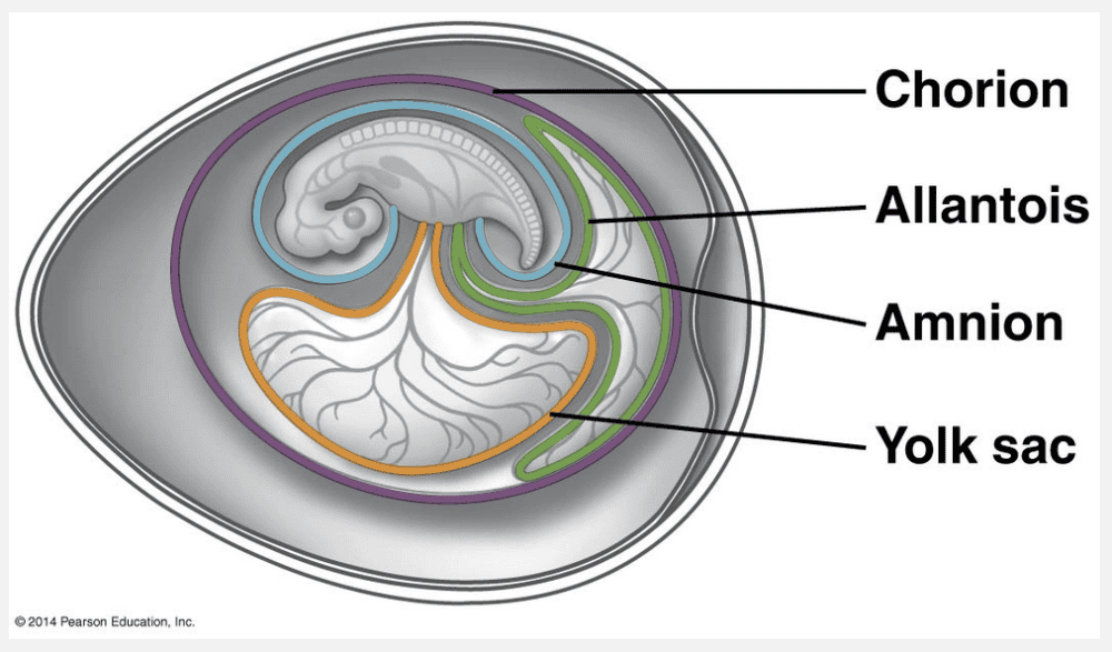
The allantois is the extraembryonic membrane that encloses a chamber involved in the deposition of wastes in the bird embryo.
Mammals and reptiles, including birds, are amniotes. For the most part, the extraembryonic membranes have similar functions in mammals and reptiles, consistent with a common evolutionary origin: this would apply to bird embryos too. The allantois disposes of wastes in the reptilian egg.
“Amnion” is incorrect because this fluid-filled extraembryonic membrane physically protects the developing embryo.
“Chorion” is incorrect because this extraembryonic membrane is the site of gas exchange.
“Fertilization envelope” is incorrect because this forms seconds after fertilization occurs.
“Yolk sac” is incorrect because it encloses a source of nutrition for the developing bird embryo.
The role of cilia in organogenesis in human embryos is to __________.
- serve as the first point of attachment in the development of the cleavage furrow that divides a cell
- increase absorption of the nutrients necessary to support cell growth
- secrete growth factors
- help distribute and receive signaling molecules
- serve as points of attachment between adjacent cells in the process of forming tissues
help distribute and receive signaling molecules
Ex.
The role of cilia in organogenesis in human embryos is to help distribute and receive signaling molecules.
Like other mammals, humans have stationary and motile cilia. Stationary primary cilia, or monocilia, jut from the surface of nearly all cells, one per cell. In contrast, motile cilia are restricted to cells that propel fluid over their surfaces, such as the epithelial cells of airways and the flagella on sperm that propel sperm movement. In development, monocilia act as antennae on the cell surface, receiving signals from multiple signaling proteins, including Sonic hedgehog. When the monocilia are defective, signaling is disrupted. The motion of motile cilia in a particular part of the embryo is also essential for normal development. Evidence indicates that movement of the cilia generates a leftward fluid flow, which breaks the symmetry between the left and right sides in the development of the internal organs.
Cilia function primarily to receive, rather than to secrete, signaling molecules important in organogenesis.
Cilia do not serve as the first point of attachment in the development of the cleavage furrow dividing a cell, a process that occurs in cells in the cleavage stage of early development.
Cilia typically extend from the free side of cells comprising the surface of a tissue and do not serve as a point of attachment between adjacent cells.
Increasing absorption of nutrients in a cell is not a primary function of cilia in organogenesis.
Up to the eight-cell stage, the blastomeres of a mouse embryo can each form a complete embryo if isolated. This indicates that __________.
- cells of mammalian embryos remain totipotent until at least the eight-cell stage
- differentiation does not depend on cytoplasmic determinants
- cytoplasmic determinants are unevenly distributed during the early cleavage divisions
- the mouse embryo is strongly polarized
- mammalian embryos do not experience the progressive restriction of potency characteristic of other classes
cells of mammalian embryos remain totipotent until at least the eight-cell stage
Ex.
Up to the eight-cell stage, the blastomeres of a mouse embryo can each form a complete embryo if isolated. This indicates that cells of mammalian embryos remain totipotent until at least the eight-cell stage.
Spemann found that the fates of embryonic cells are affected by both the distribution of determinants and the pattern of cleavage relative to this distribution. Furthermore, the work of Spemann and others demonstrated that the first two blastomeres of the frog embryo are totipotent, meaning that they can each develop into all the different cell types of that species. In mammals, embryonic cells remain totipotent through the eight-cell stage, much longer than in many other animals.
“Differentiation does not depend on cytoplasmic determinants” is incorrect because the developmental potential of the two blastomeres (of fertilized salamander eggs) normally formed during the first cleavage division depends on their acquisition of cytoplasmic determinants localized in the gray crescent.
“The mouse embryo is strongly polarized” is incorrect because in mammals like the mouse, no polarity is obvious until after cleavage.
“Cytoplasmic determinants are unevenly distributed during the early cleavage divisions” is incorrect because as seen in the nematode C. elegans, cytoplasmic granules called P granules are distributed throughout the newly fertilized egg but move to the posterior end of the zygote before the first cleavage division.
“Mammalian embryos do not experience the progressive restriction of potency characteristic of other classes” is incorrect because regardless of how uniform or varied early embryonic cells are in a particular species, the progressive restriction of developmental potential is a general feature of development in all animals.
In an experiment on eye development, a thin piece of aluminum foil was placed between the bulging wall of the brain and the overlying ectoderm where the eye normally forms. Wherever the foil was placed, the lens failed to develop, because the foil __________.
- changed the gravity in the region
- rearranged the cytoplasmic determinants in the adjacent cells
- prevented the acrosomal reaction
- prevented the inductive contact that normally stimulates the lens to form
- prevented the cortical reaction
prevented the inductive contact that normally stimulates the lens to form
Ex.
In an experiment on eye development, a thin piece of aluminum foil was placed between the bulging wall of the brain and the overlying ectoderm where the eye normally forms. Wherever the foil was placed, the lens failed to develop, because the foil prevented the inductive contact that normally stimulates the lens to form.
As embryonic cells acquire distinct fates, the cells begin to influence each other’s fates by induction. At the molecular level, the response to an inductive signal is usually to switch on a set of genes that make the receiving cells differentiate into a specific tissue. Induction is an essential process in the development of many tissues in most animals.
When Hans Spemann and Hilde Mangold examined the dorsal lip from a pigmented newt gastrula to the ventral side of a nonpigmented newt gastrula they found that the transplanted dorsal lip was able to induce cells in a different region of the recipient to form structure different from the normal fate. The transplanted dorsal lip “organized” the later development of an entire extra embryo. In the experiment were foil was placed between the bulging wall of the brain and the overlying ectoderm, the lens failed to form because the inductive signals from the bulging wall of the brain did not make contact with the overlying ectoderm.
“Changed the gravity in the region” is incorrect because gravity plays a role in establishing the anterior-posterior axis of the chick egg as it travels down the hen’s oviduct and but no role in lens development.
“Rearranged the cytoplasmic determinants in the adjacent cells” is incorrect because cytoplasmic determinants do not rearrange, but in the Spemann and Mangold experiment the dorsal lip from a pigmented newt was transplanted to different region of a nonpigmented newt inducing a structure different from the normal fate.
“Prevented the acrosomal reaction” and “prevented the cortical reaction” are both incorrect because these reactions occur in sequence when a sperm makes contact with an egg followed by cortical granule release.
The ectoderm germ layer gives rise to ___________.
- the dermis and epidermis of the skin
- the skeletal and muscular systems
- the epithelial lining of the respiratory, excretory, and reproductive tracts
- the adrenal, thymus, thyroid, and parathyroid glands
- the jaws, teeth, and epidermis of the skin
the jaws, teeth, and epidermis of the skin
Ex.
The ectoderm germ layer gives rise to the jaws, teeth, and epidermis of the skin.
Following the cleavage stage of early development, a set of cells at or near the surface of the resulting blastula moves to an interior location and establishes a layered embryo called a gastrula. The cell layers produced are collectively called the embryonic germ layers. In the late gastrula, the ectoderm is the outer layer, and the endoderm lines the embryonic digestive compartment or tract. In cnidarians and a few other radially symmetric animals, only these two germ layers form during gastrulation. In contrast, vertebrates and other animals with bilateral symmetry are triploblasts and have a third germ layer, the mesoderm, which forms between the ectoderm and the endoderm.
Holoblastic cleavage of an egg differs from meroblastic cleavage in that ____________.
- holoblastic cleavage results in an asymmetric cleavage pattern
- holoblastic cleavage occurs only in insects
- holoblastic cleavage occurs only in reptiles
- in holoblastic cleavage, the cleavage furrow goes all the way through the egg
- in holoblastic cleavage, all the cleavage furrows are along the same axis
in holoblastic cleavage, the cleavage furrow goes all the way through the egg
Ex.
Holoblastic cleavage of an egg differs from meroblastic cleavage in that in holoblastic cleavage, the cleavage furrow goes all the way through the egg.
Holoblastic cleavage is seen in many groups of animals—including echinoderms, annelids, amphibians, and mammals—whose eggs contain a relatively small amount of yolk. Yolk is most plentiful and has its most pronounced effect on cleavage in the eggs of birds, other reptiles, many fishes, and insects. In these animals, the volume of yolk is so great that cleavage furrows cannot pass through it, and only the region of the egg lacking yolk undergoes cleavage. This incomplete cleavage of a yolk-rich egg is said to be meroblastic (from the Greek meros, partial).
In both holoblastic and meroblastic cleavage, the cleavage pattern can be asymmetric.
In all cleavage, the pattern involves perpendicular cleavage furrows.
Cleavage in reptiles is characteristically meroblastic, not holoblastic.
Insect cleavage is meroblastic, rather than holoblastic.
The slow block to polyspermy __________.
- produces a fertilization envelope that prevents sperm from reaching the egg cytoplasm
- is less effective than the fast block to polyspermy
- leads to the formation of the vitelline layer
- occurs only in invertebrates
- occurs minutes after the fast block to polyspermy
produces a fertilization envelope that prevents sperm from reaching the egg cytoplasm
Ex.
The slow block to polyspermy produces a fertilization envelope that prevents sperm from reaching the egg cytoplasm. This helps prevent the fatally disruptive circumstance in which multiple sperm enter the egg.
After one sperm binds to the egg, vesicles that lie just beneath the egg plasma membrane, called cortical granules, fuse with the egg plasma membrane. Contents of the cortical granules are released into the space between the plasma membrane and the surrounding vitelline layer, a structure formed by the egg’s extracellular matrix. Enzymes and other macromolecules from the granules then trigger a cortical reaction, which lifts the vitelline layer away from the egg and hardens the layer into the protective fertilization envelope. Additional enzymes clip off and release the external portions of the remaining receptor proteins, along with any attached sperm.
The vitelline layer is present before the slow block to polyspermy occurs.
The slow block to polyspermy occurs after the fast block to polyspermy but still within seconds after a sperm binds to the egg.
The slow block to polyspermy, by removing all receptors and attached sperm, is a more effective obstacle to polyspermy than the fast block because of the depolarization of the egg cell membrane.
The slow block to polyspermy occurs in invertebrates and vertebrates.
How is the animal hemisphere of a frog blastula different from the vegetal hemisphere?
- The vegetal hemisphere is where the blastocoel is located.
- The cells of the vegetal hemisphere are pigmented more darkly than the cells of the animal hemisphere.
- The cells of the vegetal hemisphere have more yolk than the cells of the animal hemisphere.
- The blastomeres in the animal hemisphere are larger than the cells in the vegetal hemisphere.
- The cells of the vegetal hemisphere do not participate in gastrulation.
The cells of the vegetal hemisphere have more yolk than the cells of the animal hemisphere.
Ex.
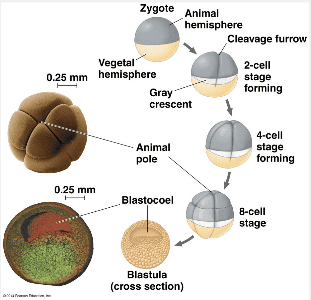
The animal hemisphere of a frog blastula differs from the vegetal hemisphere in that the cells of the vegetal hemisphere have more yolk than the cells of the animal hemisphere.
In frog development, the asymmetric distribution of yolk in the embryo affects the relative size of cells produced in the two hemispheres. This division is equatorial (perpendicular to the line connecting the poles) and produces an eight-celled embryo. However, as each of the four blastomeres begin this division, the high concentration of yolk around the vegetal pole displaces the mitotic apparatus toward the animal pole. Consequently, the cleavage furrow is also displaced from the egg equator toward the animal pole, yielding smaller blastomeres in the animal hemisphere than in the vegetal hemisphere. The displacing effect of the yolk persists in the subsequent divisions that produce a blastula. In frogs, these unequal cell divisions cause the blastocoel to form entirely in the animal hemisphere.
“The cells of the vegetal hemisphere do not participate in gastrulation” is incorrect because the cleavage furrow passes entirely through the egg.
“The cells of the vegetal hemisphere are pigmented more darkly than the cells of the animal hemisphere” is incorrect because the darker pigmented cells are found in the animal hemisphere.
“The blastomeres in the animal hemisphere are larger than the cells in the vegetal hemisphere” is incorrect because the cleavage furrow is displaced from the egg equator yielding smaller blastomeres in the animal hemisphere.
“The vegetal hemisphere is where the blastocoel is located” is incorrect because of unequal cell divisions, the blastocoel is located in the animal hemisphere.
Which of the following is the correct sequence of stages in embryogenesis?
- gastrula, blastula, cleavage, and zygote
- zygote, cleavage, blastula, and gastrula
- zygote, cleavage, gastrula, and blastula
- zygote, blastula, cleavage, and gastrula
- cleavage, blastula, zygote, and gastrula
zygote, cleavage, blastula, and gastrula
Ex.
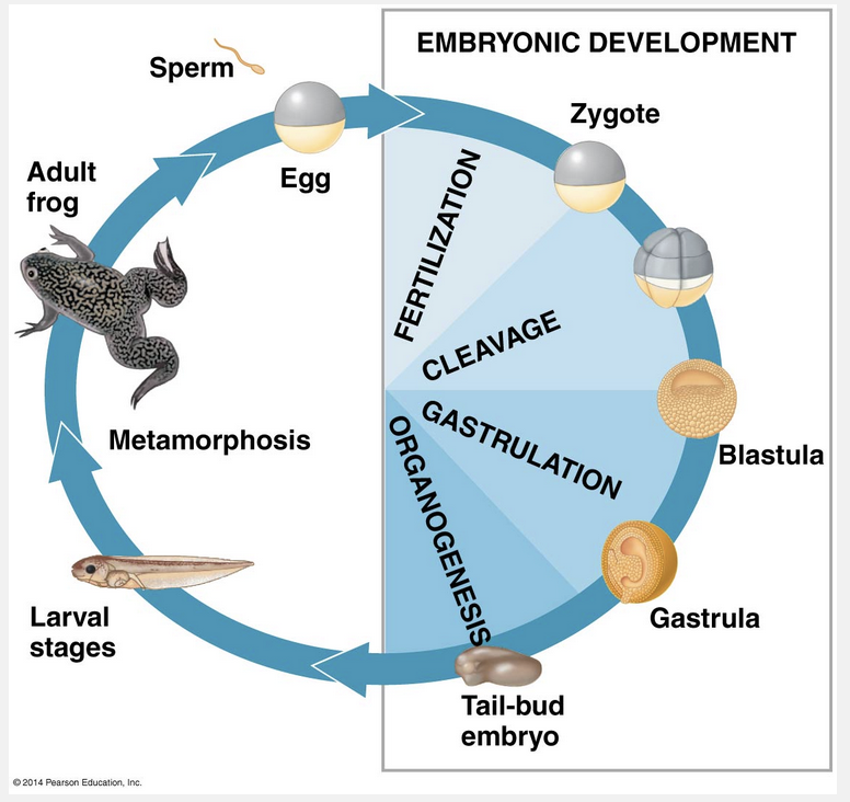
Zygote, cleavage, blastula, and gastrula is the correct sequence of stages during embryogenesis.
As shown in the figure below, the first is fertilization, the fusion of sperm and egg, which forms a zygote. Development proceeds with the cleavage stage, during which a series of cell divisions divide, or cleave, the zygote into a many-celled embryo. These cleavage divisions, which typically are rapid and lack accompanying cell growth, convert the embryo into a hollow ball of cells called a blastula. Next, the blastula folds in on itself, rearranging into a three-layered embryo, the gastrula, in a process called gastrulation. During organogenesis, the last major stage of embryonic development, local changes in cell shape and large-scale changes in cell location generate the rudimentary organs from which adult structures grow.
“Zygote, cleavage, gastrula, and blastula” is incorrect because gastrula forms from the blastula.
“Zygote, blastula, cleavage, and gastrula” is incorrect because the process called cleavage leads to the creation of a many-celled embryo called the blastula.
“Gastrula, blastula, cleavage, and zygote” is incorrect because this is in the reverse order of events of embryogenesis.
“Cleavage, blastula, zygote, and gastrula” is incorrect because the zygote is formed during the fertilization of the sperm and egg.
Which of the following statements is an accurate description of apoptosis?
- Apoptosis is a disorder in vertebrates that often results in the failure of an embryo to complete development.
- Apoptosis is an important general component of proper embryonic development.
- Apoptosis takes place only before the cleavage stage of embryonic development.
- Apoptosis is the primary means by which cell differentiation occurs.
- Apoptosis normally occurs only in the metamorphosis of amphibians.
Apoptosis is an important general component of proper embryonic development.
Ex.
Apoptosis is an important general component of proper embryonic development.
Apoptosis is a type of programmed cell death. At various times in embryonic development, individual cells, sets of cells, or whole tissues cease to develop, die, and are engulfed by neighboring cells. In the development of both nervous and immune systems, large numbers of cells undergo apoptosis. In the vertebrate nervous system, for instance, many more neurons are produced during development than exist in the adult. In general, neurons survive if they make functional connections with other neurons but die if they do not make such connections. In the adaptive immune system, cells that are self-reactive are often eliminated by apoptosis, as are effector cells that arise after encountering a pathogen but are no longer required after the infection has been eliminated.
Apoptosis is not a disorder in vertebrates that often results in the failure of an embryo to complete development.
Apoptosis would terminate development if it took place before the cleavage stage of embryonic development.
Apoptosis, by eliminating a cell, could not be a means by which differentiation occurs in that cell.
Apoptosis does normally occur in amphibian metamorphosis—for example, in cells in the tail of a tadpole—but it also occurs during the development of animals that do not go through a metamorphosis.
Reptiles possess which of the following extraembryonic membranes that mammals possess?
- Only the amnion
- Chorion, allantois, amnion, and yolk sac
- Only the allantois and amnion
- Only the chorion, allantois, and amnion
- Only the chorion and allantois
Chorion, allantois, amnion, and yolk sac
Ex.
Reptiles possess all of the extraembryonic membranes that mammals possess: the chorion, allantois, amnion, and yolk sac. After forming, these membranes provide a “life-support system” for further embryonic development.
All vertebrate embryos require an aqueous environment for their development, whereas the embryos of fishes and amphibians usually develop in the surrounding sea or pond and need no specialized, water-filled enclosure. Thus, the extensive colonization of land by vertebrates was possible only after the evolution of structures that would allow reproduction in dry environments; such structures include the shelled egg of reptiles and monotremes and the uterus of marsupials and placental mammals. Inside the shell or uterus, the embryos of these animals are surrounded by fluid within a sac that is formed by one of the extraembryonic membranes, the amnion. Mammals and reptiles, including birds, are therefore called amniotes. The three other extraembryonic membranes common to reptiles and mammals function to transport or store oxygen, nutrients, and waste products.
A major difference between reproduction in most marine invertebrates and reproduction in most terrestrial vertebrates is that __________.
- most terrestrial vertebrates produce eggs with no yolk
- most terrestrial vertebrates have asexual reproduction
- most terrestrial vertebrates have internal fertilization
- only terrestrial vertebrates have zygotes that go through a process of cleavage following fertilization
- most terrestrial vertebrates do not lay eggs
most terrestrial vertebrates have internal fertilization
Ex.
Unlike sea urchins and most other marine invertebrates, most terrestrial vertebrates have internal fertilization.
The sperm must cross several obstacles to reach the egg in the female reproductive system and fuse with it in fertilization. In mammals, for example, support cells of the developing follicle surround the egg before and after ovulation. A sperm must travel through this layer of follicle cells before it can reach the zona pellucida, the extracellular matrix of the egg. There, the binding of the sperm to a sperm receptor induces an acrosomal reaction, which facilitates sperm entry. Overall, the process of fertilization is much slower in mammals than it is in marine invertebrates such as sea urchins.
Internal fertilization implies sexual reproduction.
Most terrestrial vertebrates lay eggs following internal fertilization.
Most terrestrial vertebrate eggs have substantial yolk as stored nutrients for the developing embryo.
Once fertilization is complete, the zygotes of many animals, both marine and terrestrial and invertebrate and vertebrate, go through a cleavage stage in which the cell is rapidly divided into many smaller cells.
Which of the following statements is an accurate description of the cleavage stage of early development?
- The yolk is mostly consumed.
- The pattern of cleavage is similar in all species.
- Although the rate of cleavage differs, the number of divisions that occur in the cleavage stage is the same for all species.
- There is no increase in mass.
- Embryonic germ layers are formed during cleavage.
There is no increase in mass.
Ex.
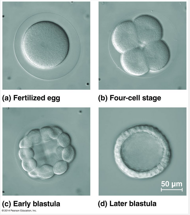
There is no increase in mass in the cleavage stage of early development. This is because during cleavage, the cell cycle consists primarily of the S (DNA synthesis) and M (mitosis) phases. Since the G1 and G2 (gap) phases are essentially skipped, little or no protein synthesis occurs. Instead, cleavage partitions the cytoplasm of the large fertilized egg into many smaller cells called blastomeres. The first five to seven cleavage divisions produce a hollow ball of cells, the blastula, that surrounds a fluid-filled cavity called the blastocoel.
The pattern of cleavage divisions differs among species; in some species it is uniform across the embryo, and in other species it is asymmetric.
The number of cleavage divisions differs among species.
The yolk persists throughout the cleavage stage, and differences in the amount of yolk possessed by different species account for some of the differences in cleavage patterns.
Embryonic germ layers do not form until after the cleavage stage.
One difference between the blastula and gastrula stages of development is that __________.
- the blastula consists of more cell layers
- the blastula is a solid ball of cells, but the gastrula is hollow
- blastula cells are more differentiated than gastrula cells
- there are many more cells in a blastula
- there is an opening from the cavity inside the gastrula to the outside
there is an opening from the cavity inside the gastrula to the outside
Ex.
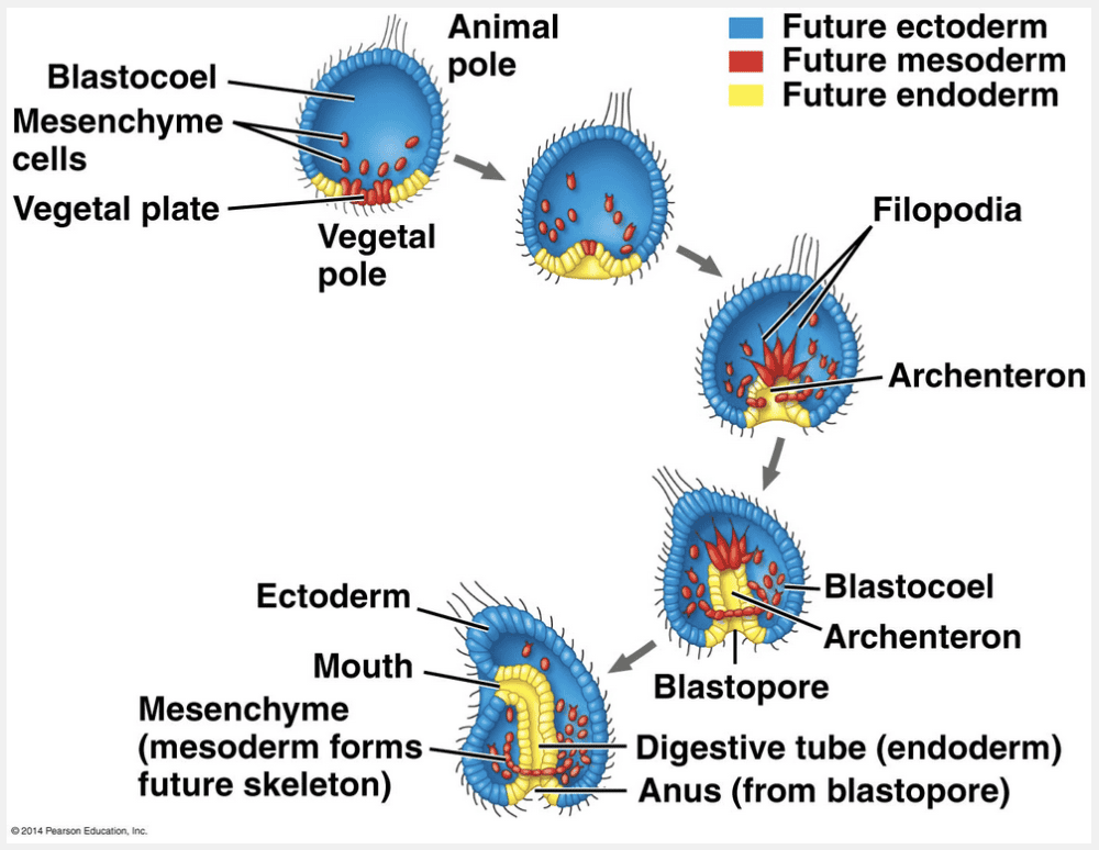
One difference between the blastula and gastrula stages of development is that there is an opening from the cavity inside the gastrula to the outside.
Gastrulation in the sea urchin, as shown below, begins at the vegetal pole of the blastula. There, cells called mesenchyme cells individually detach from the blastocoel wall and enter the blastocoel. The remaining cells near the vegetal pole flatten slightly and cause that end of the embryo to buckle inward as a result of cell shape changes. This process—the infolding of a sheet of cells into the embryo—is called invagination. Extensive rearrangement of cells transforms the shallow depression into a deeper, narrower, blind-ended tube called the archenteron. The open end of the archenteron, which will become the anus, is called the blastopore.
“Blastula cells are more differentiated than gastrula cells” is incorrect because during gastrulation specialized cells such as mesenchyme cells from.
“There are many more cells in a blastula” is incorrect because gastrulation occurs after the blastula forms and cells are now organizing into germ layers.
“The blastula consists of more cell layers” is incorrect because at the end of gastrulation two embryonic germ layers have formed in diploblastic organisms such as the cnidarians, and three embryonic germ layers have formed in triploblastic organisms such as sea urchins, chicks, and humans.
“The blastula is a solid ball of cells, but the gastrula is hollow” is incorrect because the blastula is a hollow ball of cells.
Which stage of development is common in amphibians but is not seen in mammals?
- Organogenesis
- Gastrulation
- Metamorphosis
- None of the listed responses is correct.
- Cleavage
Metamorphosis
Ex.
The stage of development that is common in amphibians but not seen in mammals is metamorphosis.
In a frog, for example, a major developmental period is metamorphosis, when the larva (tadpole) is transformed into an adult. In some cases, a structure functions in a larval or other immature form of the organism and then is eliminated during later development. One familiar example is provided by the cells in the tail of a tadpole, which undergo apoptosis during frog metamorphosis.
“Gastrulation” is incorrect because is incorrect because this step involves the formation of the three embryonic germ layers.
“Cleavage” is incorrect because this is a step in which a series of cell divisions divide, or cleave, the zygote into a many-celled embryo.
“Organogenesis” is incorrect because during this step the three embryonic germ layers begin to develop into the rudiments of organs.
Which of the following statements is true?
- Cell adhesion molecules are located inside of cells.
- The cytoskeleton inhibits cell migration.
- Cell adhesion molecules are secreted glycoproteins found in the extracellular matrix.
- Cells reach specific destinations through the action of cell adhesion molecules and the extracellular matrix.
- The extracellular matrix is not involved in the migration of cells.
Cells reach specific destinations through the action of cell adhesion molecules and the extracellular matrix.
Ex.
The following statement is true: Cells reach specific destinations through the action of cell adhesion molecules and the extracellular matrix.
The cytoskeleton is also involved in cell migration, which relies on cell adhesion molecules and the extracellular matrix to help cells reach specific destinations. The cytoskeleton is responsible not only for cell shape changes but also for cell migration. During organogenesis invertebrates, cells from the neural crest and from somites migrate to locations throughout the embryo. Cells “crawl” within the embryo by using cytoskeletal fibers to extend and retract cellular protrusions.
Transmembrane glycoproteins called cell adhesion molecules play a key role in cell migration by promoting interaction between pairs of cells. Cell migration also involves the extracellular matrix (ECM), the meshwork of secreted glycoproteins and other macromolecules lying outside the plasma membranes of cell. The ECM helps to guide cells in many types of movements, such as migration of individual cells and shape changes of cell sheets. Cells that line migration pathways regulate movement of migrating cells by secreting specific molecules into the ECM.
“The cytoskeleton inhibits cell migration” and “the extracellular matrix is not involved in the migration of cells” are both incorrect because the cytoskeleton is responsible not only for cell shape changes but also for cell migration.
“Cell adhesion molecules are secreted glycoproteins found in the extracellular matrix” is incorrect because these proteins are transmembrane glycoproteins called cell adhesion molecules play a key role in cell migration by promoting interaction between pairs of cells.
“Cell adhesion molecules are located inside of cells” is incorrect because these proteins are transmembrane glycoproteins called cell adhesion molecules that play a key role in cell migration by promoting interaction between pairs of cells.
In mammals, the __________ facilitates implantation in the uterine wall.
- blastocyst
- trophoblast
- amnion
- hypoblast
- epiblast
trophoblast
Ex.
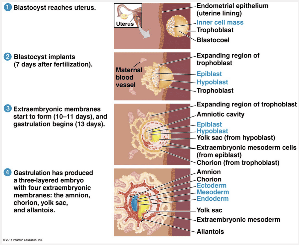
In mammals, the trophoblast facilitates implantation in the uterine wall.
The trophoblast, the outer epithelium of the blastocyst, does not contribute to the embryo itself but instead supports embryo growth in a number of ways. It initiates implantation by secreting enzymes that break down molecules of the endometrium, the lining of the uterus. This allows the blastocyst to invade the endometrium. As the trophoblast thickens through cell division, it extends finger-like projections into the surrounding maternal tissue. Invasion by the trophoblast leads to erosion of capillaries in the endometrium, causing blood to spill out and bathe trophoblast tissues. Around the time of implantation, the inner cell mass of the blastocyst forms a flat disk with an upper layer of cells, the epiblast, and a lower layer, the hypoblast. As in birds, the human embryo develops almost entirely from epiblast cells.
“Blastocyst” is incorrect because this is the mammalian equivalent of the blastula. A group of cells called the inner cell mass will develop into the embryo proper.
“Amnion” is incorrect because this is one of the four embryonic membranes formed by the embryo and functions in protecting the embryo.
“Epiblast” is incorrect because this is the upper layer of the inner cell mass of the blastocyst. The embryo develops almost entirely form epiblast cells.
“Hypoblast” is incorrect because this is the lower layer of the blastocyst. The hypoblast cells are pushed aside during gastrulation.
A fate map produced by a developmental biologist would show __________.
- the process of differentiation in a single cell
- the structures arising from each region of an embryo
- the relative developmental rates of embryos with differing genotypes
- the success of different treatments for developmental disorders
- macroevolutionary changes across populations comprising a lineage
the structures arising from each region of an embryo
Ex.
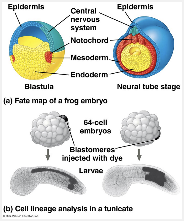
A fate map produced by a developmental biologist would show the structures arising from each region of an embryo.
This type of diagram can be produced by tracing the ancestry of embryonic cells through direct observation with a microscope. In the 1920s, German embryologist Walther Vogt used this approach to determine where groups of cells from the blastula end up in the gastrula.
Later researchers developed techniques that allowed them to mark an individual blastomere during cleavage and then follow the marker as it was distributed to all the mitotic descendants of that cell. Coupled with experiments in which particular cells or groups of cells are destroyed by a laser beam or through mutations, careful microscopic observations have produced a much more comprehensive fate mapping of the soil-dwelling nematode Caenorhabditis elegans.
Fate maps show changes across cell divisions, rather than within single cells.
Although a single fate map wouldn’t show relative developmental rates of embryos with different genotypes, several fate maps could be the basis for a comparative approach.
Fate maps show the ancestry of cells within an individual rather than changes across generations.
Fate maps do not tabulate the success of different treatments for developmental disorders.