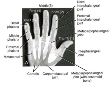
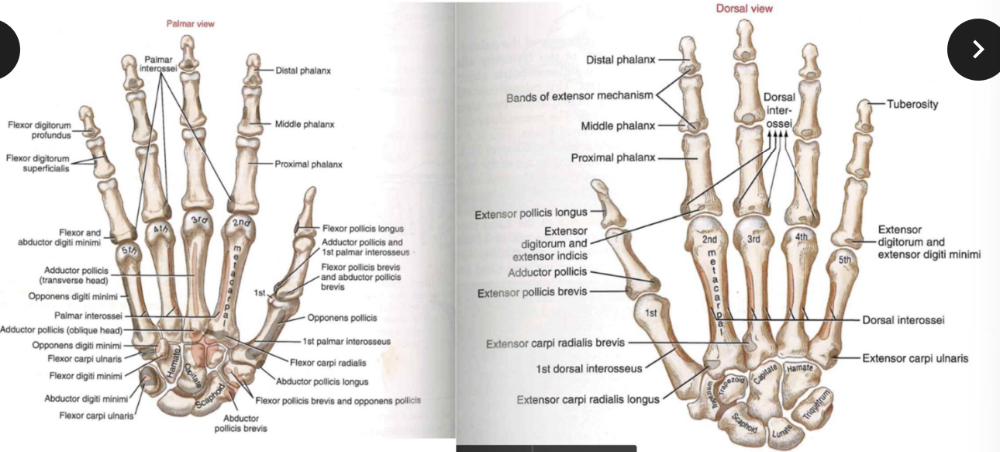
Palmar view: major bones + joints
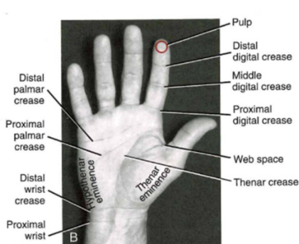
Palmar view: external landmarks
Osteology: metacarpals
Radial view of bones in third ray
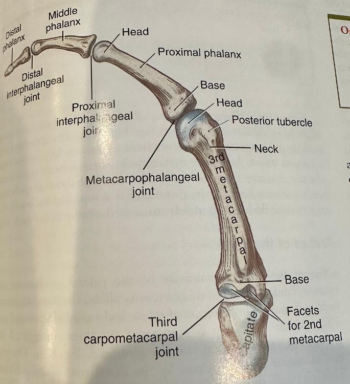
- metacarpals
- phalanges
- capitate bone of wrist
Osteology: Phalanges
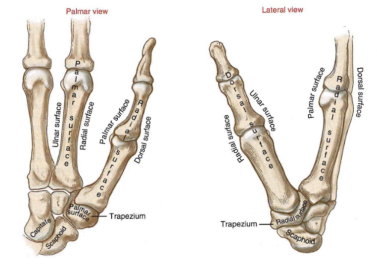
- palmar and lateral view
Osteology: The Arches of the Hand
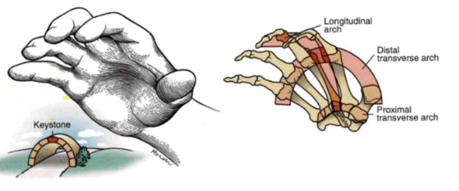
- natural concavity of palm
- longitudinal arch system
- transverse arch system (x2)
AROM of hand
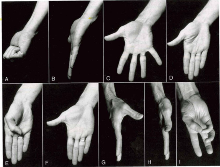
- finger motion (A-D)
- thumb motion (E-I)
- finger adduction // thumb flexion // thumb exension
- Thumb abduction
- Thumb adduction
- thumb opposition
Arthrology: CMC joints (moblity)
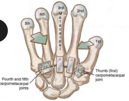
- mobility across 5 carpometacarpal joints
- peripheral joints (1,4,5) MORE MOBILE than central 2 joints
Arthrology: CMC joints (articular surfaces)
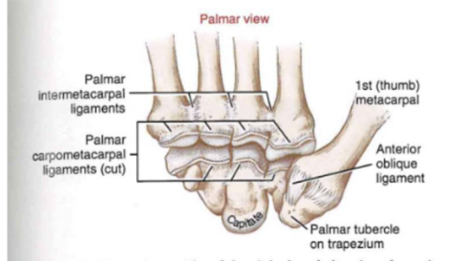
- A. surfaces of 2nd-> 5th carpometacarpal joints (ligaments = cut)
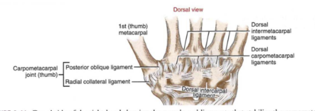
Arthrology: CMC joints and ligaments
Arthrology: The 1st carpometacarpal
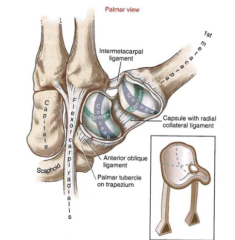
- exposed to show saddle-shaped appearance
- right thumb
Arthrology: The 1st carpometacarpal (ABD/ADD)
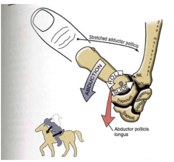
- stretched: intermetacarpal ligamet (IML), radial collateral ligament (RCL), and adductor pollicis muscle
- CONCAVE+CONVAX = ROL + SLide in same direction
Arthrokinematics of the 1st CMC: Flex/Ext
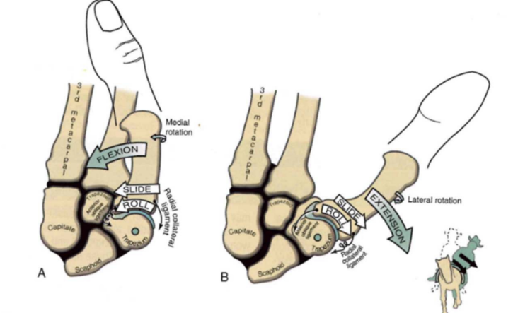
- flexion: slight medial roation + RCL elongation + slack oblique anterior ligament
- extension: slight lateral rotation + oblique anterior ligament elongation
Arthrokinematics of the 1st CMC: Opposition
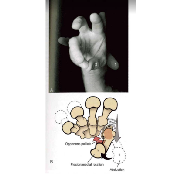
- 2 Phases of opposition
- abduction and flexion with medial rotation
- tight posterior oblique ligament + contraction opponents pollicis
Metacarpophalangeal joints: radial
- radial collateral ligament and connective tissues
- (metacarpophalangeal, proximal interphalangeal, distal interphalangeal points)
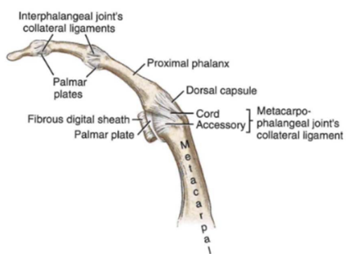
Metacarpophalangeal joints: periarticular connective tissues
- periarticular connective tissues at metacarpophalangeal joints
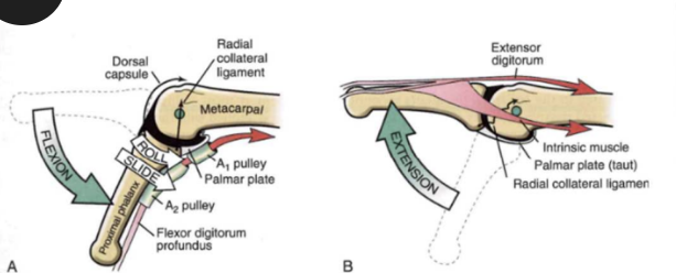
Arthrokinematics: MCPs (FLEX/EXTEN)
-
flexion: activation of flexor digitorum profundus
muscle
- tendon at A1 and A2 pulleys
- tight=> dorsal capsule and R.ColLig.
- ROLL SLIDE = SAME DIRE
- Extension: extensor digitorum+ intrinsic
muscle activate
- tight= palmar plate
- slack= RCL cord
Arthrokinematics: MCPs (ABDUCTION)
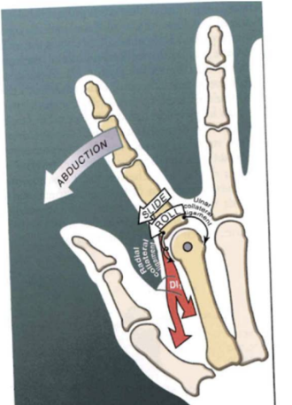
- first dorsal interosseus muscle (DI1)
- tight= U.Col.Lig
- slack= R.Col.Lig
- ANTERIOR POSTERIOR DIRECTION
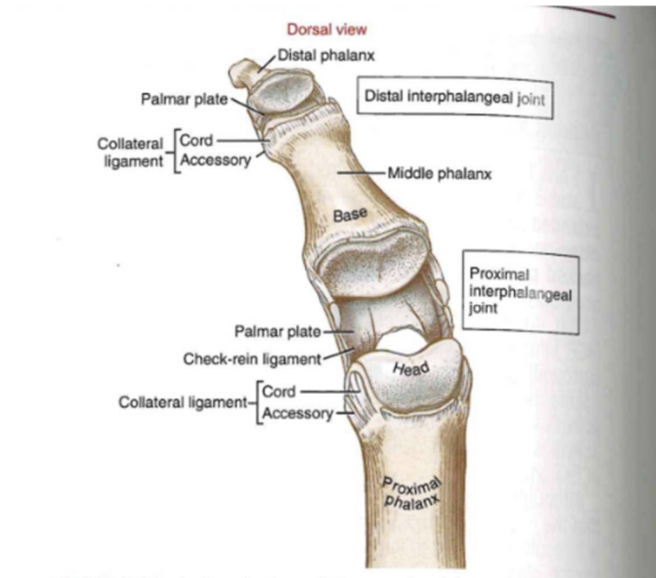
General features of proximal interphalangeal ligaments (IPs)
IPs: Arthrokinematics
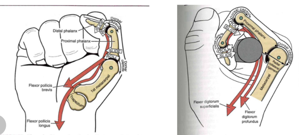
- active flexion
- metacarpophalangeal and interphalangeal joints (thumb)
- Powered by flexor pollicis longus +PLong. brevis
- CONCAVE+CONCAVE = same direction
Muscular Function of the hand: intrinsic
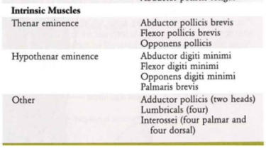
- all attachments within the hand
- themear eminence // hypothenar eminence // other
Muscular Function of the hand: Extrinsic muscles
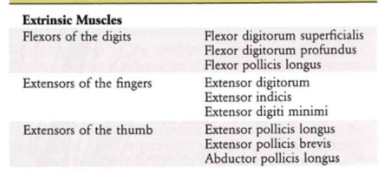
- proximal attachments outside of the hand
- flexor of the digits
- extensors of fingers
- exntensors of thumb
Extrinsic flexors of the digits: muscles
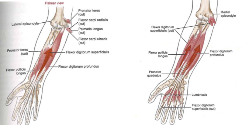
- Flexor digitorum superficialis
- FDP
- Flexor pollicis longus
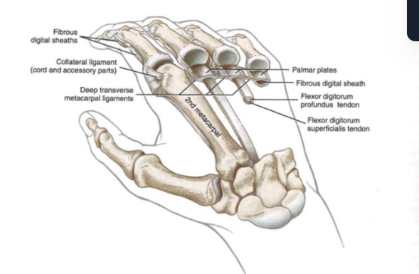
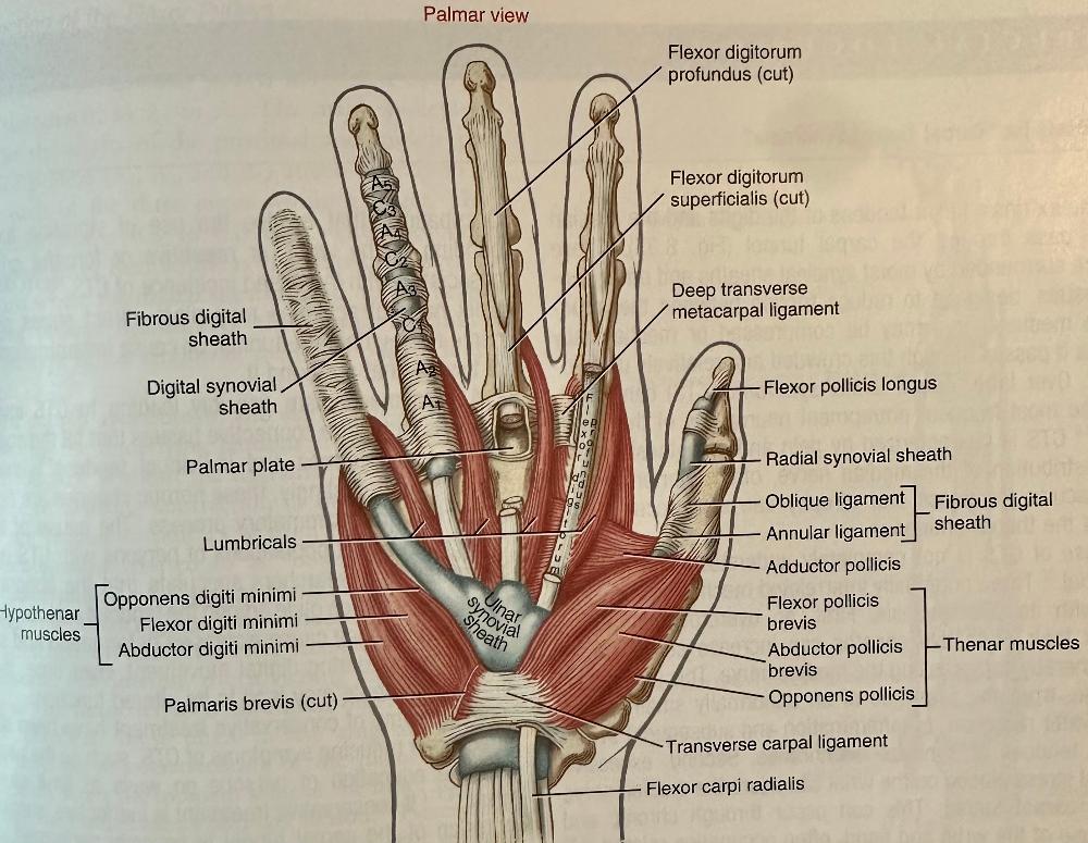
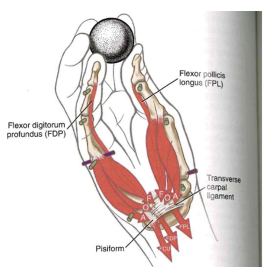
Palmar view of the hand (sheath/ligaments/muscles/bones)
Muscle activation for proximal interphalangeal joint flexion (pulling finger down/in)
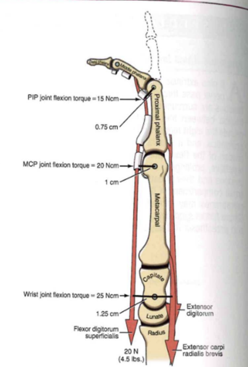
- force by the flexor digitorum superficialis = flexion torque on
all joint it crosses
- flexor torques progressively increase in the proximal direction
- Isoloate only flexion => extensor digitorum+ extensor carpi radialis brevis RESIST flexion effect (on wrist and metacarpal joints)
Tenodesis
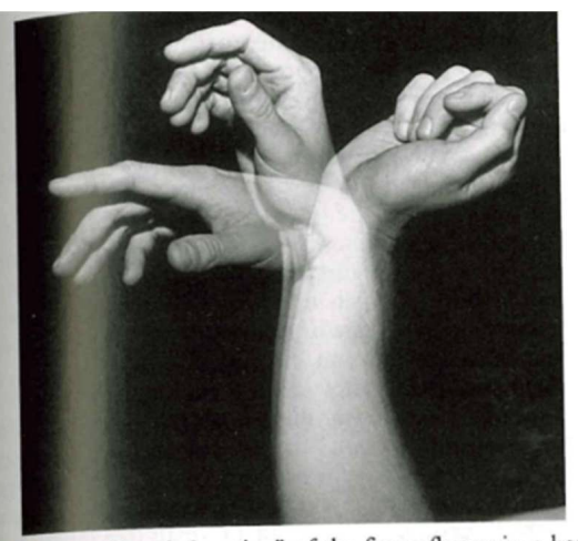
- wriust extends/moves back (palm up) => thumb and fingers automatically flex (b/c extrinsic digital flexors stretch)
Extrinsic extensors of the digits Muscles
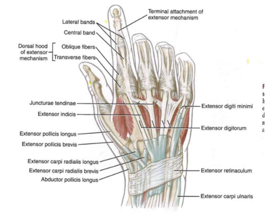
- Extensor digitorum (communis)
- Extensor indicis
- Extensor digit minimi
Extrinsic extensors of the digits Muscles (with arm)
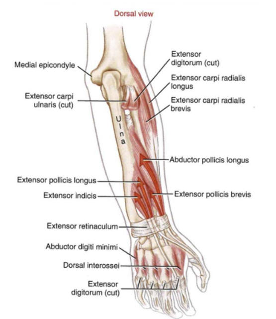
Extrinsic extensors of the thumb (muscles)
- Extensor pollicis longus
- Extensor pollicis brevis
- Abductor pollicis longus
Intrinsic muscles of the hand
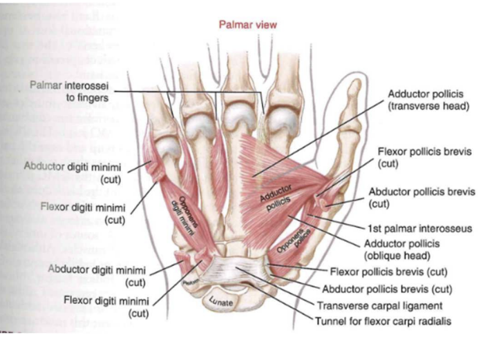
Thenar eminence (muscles)
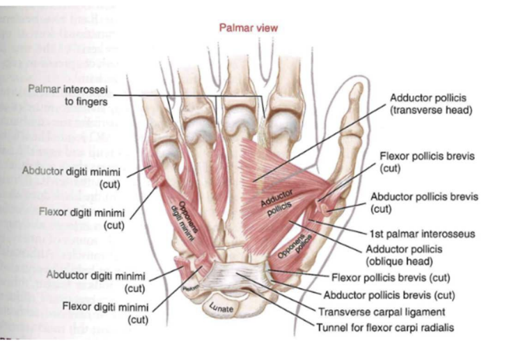
- Abductor pollicis brevis
- Flexor pollicis breview
- Opponens pollicis
Hypothenar eminence (muscles)
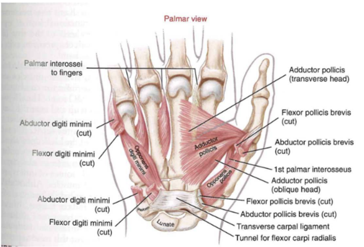
- Flexor digiti minimi
- Abductor digiti minimi
- Opponens digiti minimi
- Palmaris brevis
Thenar & Hypothenar
- used during the opposition of thumb and small finger
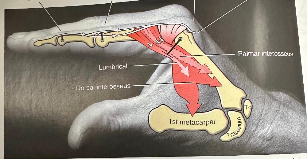
Adductor Pollicis
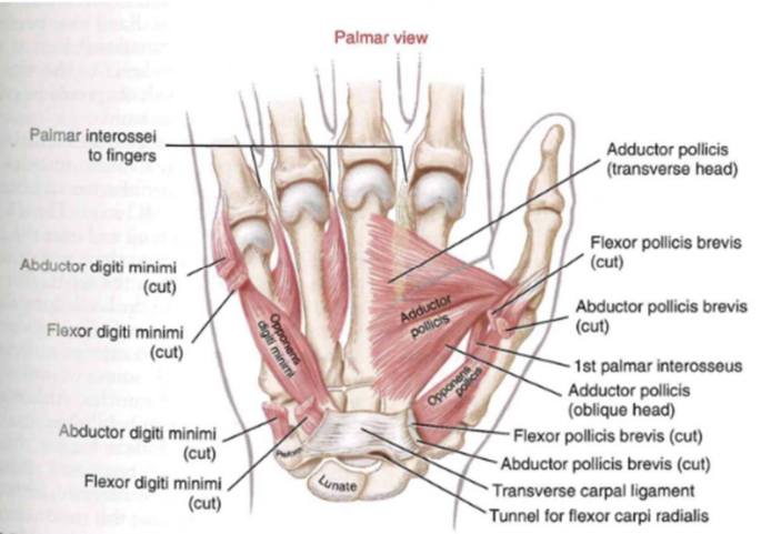
- combo of flexion and adduction torque at the base of the thumb
- oblique and transverse head
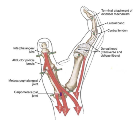
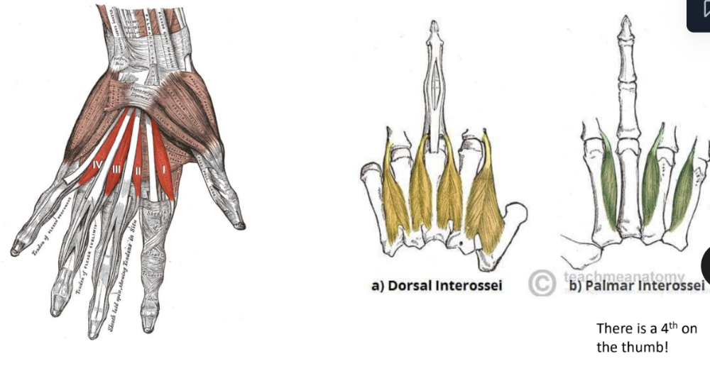
Lumbricals & Interossei
Lumbricals
- innervated: median nerve (laterally) + ulnar nerve (medially)
- distal attachments: oblique fibers of dorsal hood (externsor mechanism on lateral band)
- Contractile: generate small force over long distance
- primary actions: MCP joint flexion and PIP and DIP joint extension
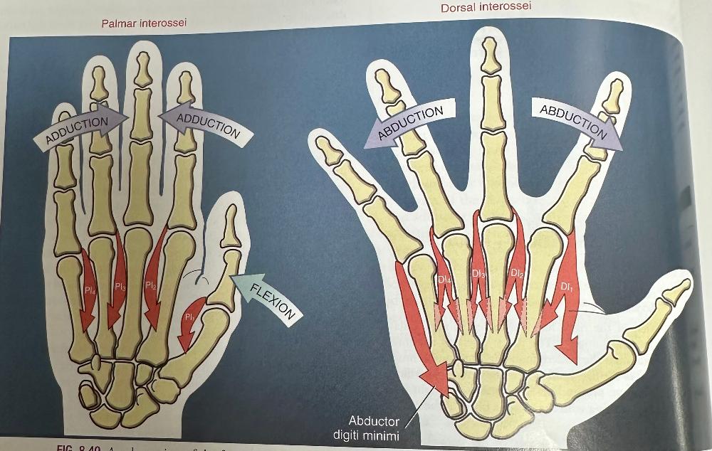
Dorsal Interossei
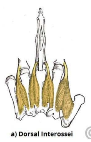
- innervated: ulnar nerve
- distal attachments: oblique fibers of dorsal hood (side base of proximal phalanx)
- Contractile: small force over shortdistance
- primary actions: Abduction of MCP joint flexion and PIP and DIP joint extension
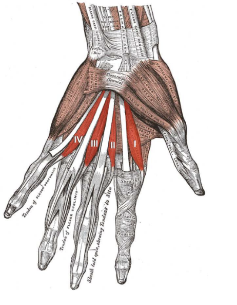
Palmar Interossei
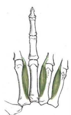
- innervated: ulnar nerve
- distal attachments: oblique fibers of dorsal hood (side base of proximal phalanx)
- Contractile: nondistnct
- primary actions: Adduction of MCP joint flexion and PIP and DIP joint extension
Adduction and Abduction of hand
Combined actions of lumbricals and interossei
- shown as flexors at the metacarpophalangeal joint + extensors at the interphalangeal joints
Extensor Mechanism & Interaction
between extrinsics and intrinsics
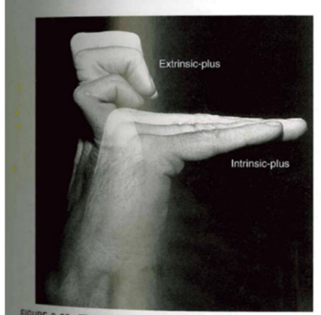
- Intrinsic plus: fingers flat across
- MCP joint flexion and IP joint extension
- Extrinsic plus:
fingers curled back
- extrinsic muscles => MCP joint hyperextension and IP joint flexion
- Opening and closing the hand
Full extension of finger
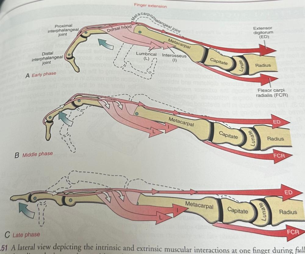
- Early phase: extensor digitorum extends metacarpophalangeal joint
-
Middle phase: intrinsic muscles+ extensors dig. to
extend proximal+distal interphalangeal joints
- flexor torque prevents E.dig from hyperextending the metacarpal joint
- Late Phase: muscle activation continues thru full finger extension
Full flexion of finger
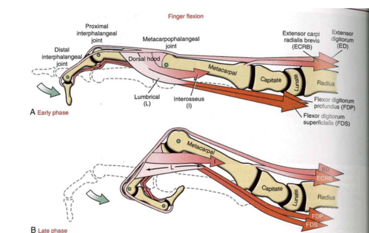
-
Early phase: flexor dig. superficial and
interosseus muscles flex joints
- inactive lumbrical
- Late Phase: muscle action = unchanged, lumbricals stretched across both ends,
hand grips
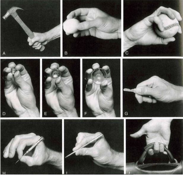
- power grip
- precision grip (egg)
- precision grip (baseball)
- Precision grip (with alternating concavity of distal transverse arch)
- power key pinch
- tip-to-tip prehension pinch
- pad-to-pad prehension pinch
- hook grip