Arrange the following structures in the correct sequence in which urine passes through them to the external environment: (1) ureter, (2) renal pelvis, (3) calyx, (4) urinary bladder, (5) urethra.
- 1, 2, 3, 4, 5
- 2, 4, 1, 3, 5
- 3, 4, 1, 5, 2
- 3, 2, 1, 4, 5
3, 2, 1, 4, 5
Ex.
The correct sequence in which urine passes to the external environment is (3) calyx, (2) renal pelvis, (1) ureter, (4) urinary bladder, (5) urethra.
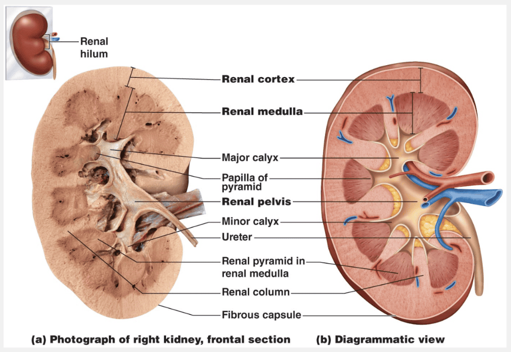
A frontal section through a kidney reveals three distinct regions: cortex, medulla, and pelvis. The most superficial region, the renal cortex, is light-colored and has a granular appearance. Deep to the cortex is the darker, reddish-brown renal medulla, which exhibits cone-shaped tissue masses called medullary or renal pyramids. The broad base of each pyramid faces toward the cortex, and its apex, or papilla (“nipple”), points internally. The pyramids appear striped because they are formed almost entirely of parallel bundles of microscopic urine-collecting tubules and capillaries. The renal columns, inward extensions of cortical tissue, separate the pyramids. Each pyramid and its surrounding cortical tissue constitutes one of approximately eight lobes of a kidney.
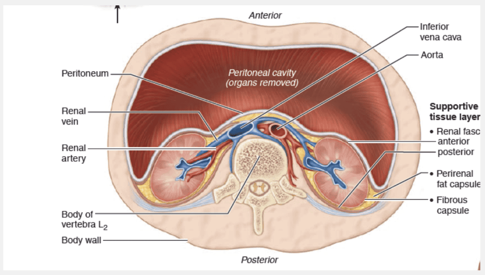
The renal pelvis, a funnel-shaped tube, is continuous with the ureter leaving the hilum. Branching extensions of the pelvis form two or three major calyces (ka′lih-sēz; singular: calyx). Each major calyx subdivides to form several minor calyces, cup-shaped areas that enclose the papillae. The calyces collect urine, which drains continuously from the papillae, and empty it into the renal pelvis. The urine then flows through the renal pelvis and into the ureter, which moves it to the bladder to be stored. The walls of the calyces, pelvis, and ureter contain smooth muscle that contracts rhythmically to propel urine by peristalsis.
Every day the kidneys filter nearly __________ of fluid from the bloodstream.
- 200 liters
- 100 liters
- 500 liters
- 50 liters
200 liters
Ex.
Every day the kidneys filter nearly 200 liters of fluid from our bloodstream, allowing toxins, metabolic wastes, and excess ions to leave the body in urine while returning needed substances to the blood. Much like a water purification plant that keeps a city’s water drinkable and disposes of its wastes, the kidneys are usually unappreciated until they malfunction and body fluids become contaminated.
Which two structures make up each nephron?
- renal corpuscle and renal tubule
- renal tubule and peritubular capillary
- nephron loop and collecting duct
- glomerulus and glomerular capsule
- renal cortex and renal medulla
renal corpuscle and renal tubule
Ex.
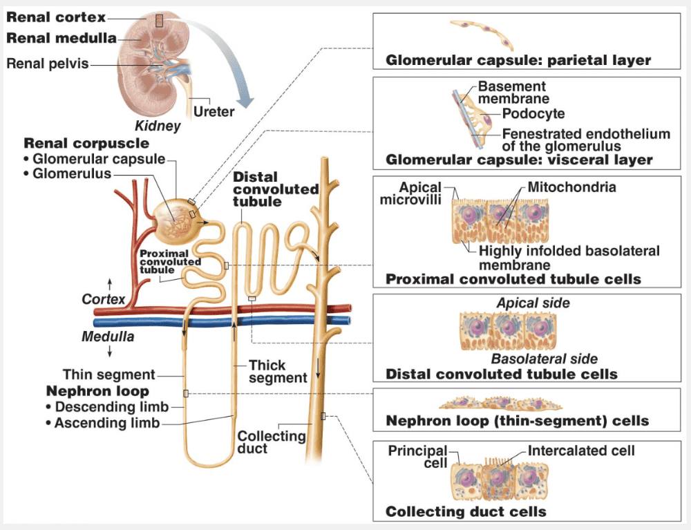
Each nephron consists of a renal corpuscle and a renal tubule.
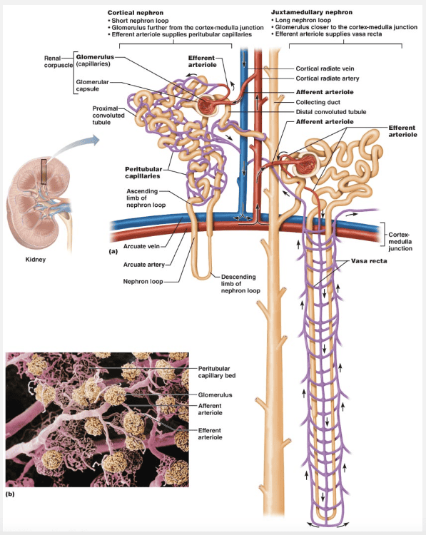
Each renal corpuscle consists of a tuft of capillaries called a glomerulus and a cup-shaped hollow structure called the glomerular capsule. The renal tubule has three major parts. It leaves the glomerular capsule as the proximal convoluted tubule, drops into a hairpin loop called the nephron loop, and then winds and twists again as the distal convoluted tubule before emptying into a collecting duct. The peritubular capillaries cling closely to adjacent renal tubules and empty into nearby venules. The most superficial region of the kidney is the the renal cortex. Deep to the cortex is the darker, reddish-brown renal medulla.
Match the following vessel found in the kidney with its location: Blood vessel leading directly into the glomerulus.
- Peritubular capillary
- Efferent arteriole
- Afferent arteriole
- Segmental artery
Afferent arteriole
Ex.
The afferent arteriole is the blood vessel leading directly into the glomerulus.
The vascular pathway through a kidney is as follows: renal artery → segmental arteries → interlobar arteries → arcuate arteries → cortical radiate arteries → afferent arterioles → glomeruli → efferent arterioles → peritubular capillary beds → cortical radiate veins → arcuate veins → interlobar veins → renal vein.
Into what part of the nephron is plasma filtered?
- Collecting duct
- Proximal convoluted tubule
- Glomerular capsule
- Distal convoluted tubule
Glomerular capsule
Ex.
Plasma is filtered into the glomerular capsule.
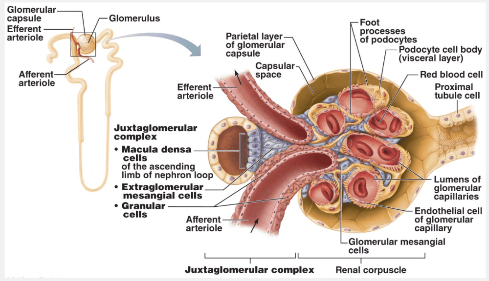
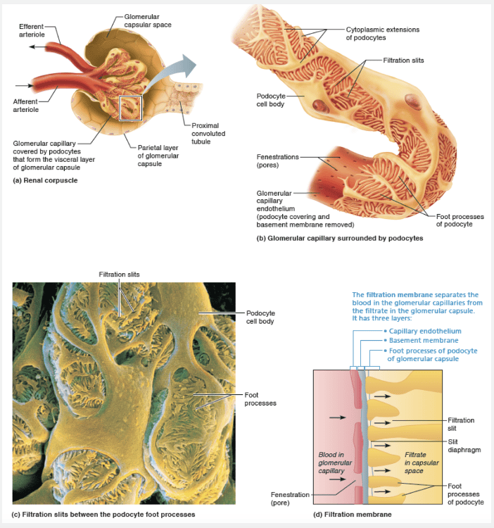
Each renal corpuscle consists of a tuft of capillaries called a glomerulus (glo-mer′u-lus; glom = ball of yarn) and a cup-shaped hollow structure called the glomerular capsule (or Bowman’s capsule). The glomerular capsule is continuous with its renal tubule and completely surrounds the glomerulus, much as a well-worn baseball glove encloses a ball.
The endothelium of the glomerular capillaries is fenestrated (penetrated by many pores), which makes these capillaries exceptionally porous. This property allows large amounts of solute-rich but virtually protein-free fluid to pass from the blood into the glomerular capsule. This plasma-derived fluid or filtrate is the raw material that the renal tubules process to form urine.
Which of the following is incorrect with regards to pyelonephritis?
- Pyelonephritis is more common in females than males.
- Pyelonephritis is an infection or inflammation of the entire kidney.
- In untreated pyelonephritis, the kidney may be severely damaged and antibiotic therapy often fails to treat the disorder.
- In severe cases of pyelonephritis, the kidney swells, abscesses form, and the pelvis fills with pus.
In untreated pyelonephritis, the kidney may be severely damaged and antibiotic therapy often fails to treat the disorder.
Ex.
It is incorrect that untreated pyelonephritis often fails to treat the disorder because antibiotic therapy can usually treat the infection successfully.
Infections or inflammations that affect the kidney are called pyelonephritis. Kidney infections in females are usually caused by fecal bacteria that spread from the anal region to the urinary tract. Less often they result from blood borne bacteria (traveling from other infected sites) that lodge and multiply in a kidney.In severe cases of pyelonephritis, the kidney swells, abscesses form, and the pelvis fills with pus. Untreated, the kidney may be severely damaged, but antibiotic therapy can usually treat the infection successfully.
The __________ cushions the kidney and helps attach it to the posterior body wall.
- perirenal fat capsule
- renal fascia
- ureter
- fibrous capsule
perirenal fat capsule
Ex.
The perirenal fat capsule cushions the kidney and helps attach it to the posterior body wall.
Three layers of supportive tissue surround each kidney. From superficial to deep, these are:
- The renal fascia, an outer layer of dense fibrous connective tissue that anchors the kidney and the adrenal gland to surrounding structures
- The perirenal fat capsule, a fatty mass that surrounds the kidney and cushions it against blows. The fibrous capsule, a transparent capsule that prevents infections in surrounding regions from spreading to the kidney.
Match the following part of the nephron with its correct function: Primary site of glucose and amino acid reabsorption.
- Nephron loop, descending limb
- Nephron loop, ascending limb
- Proximal convoluted tubule
- Distal convoluted tubule
Proximal convoluted tubule
Ex.
The proximal convoluted tubule is the primary site of glucose and amino acid reabsorption.
The entire renal tubule is involved in reabsorption to some degree, but the PCT cells are by far the most active reabsorbers and the events just described occur mainly in this tubular segment. Normally, the PCT reabsorbs all of the glucose and amino acids in the filtrate and 65% of the Na+ and water. The bulk of the electrolytes are reabsorbed by the time the filtrate reaches the nephron loop. Nearly all of the uric acid and about half of the urea are reabsorbed in the proximal tubule, but both are later secreted back into the filtrate.
The major calyces are the __________.
- basic functional units of the kidneys
- large branches of the renal pelvis
- cone-shaped structures located in the renal medulla
- expanded ends of renal pyramids
- expanded ends of nephrons
large branches of the renal pelvis
Ex.
The major calyces are the large branches of the renal pelvis.
The renal pelvis, a funnel-shaped tube, is continuous with the ureter leaving the hilum. Branching extensions of the pelvis form two or three major calyces (ka′lih-sēz; singular: calyx). Each major calyx subdivides to form several minor calyces, cup-shaped areas that enclose the papillae. The calyces collect urine, which drains continuously from the papillae, and empty it into the renal pelvis. The urine then flows through the renal pelvis and into the ureter, which moves it to the bladder to be stored. The walls of the calyces, pelvis, and ureter contain smooth muscle that contracts rhythmically to propel urine by peristalsis.
Select the blood vessel that directly drains the glomerulus.
- Peritubular capillary
- Segmental artery
- Efferent arteriole
- Afferent arteriole
Efferent arteriole
Ex.
The efferent arteriole directly drains the glomerulus.
The vascular pathway through a kidney is as follows: renal artery → segmental arteries → interlobar arteries → arcuate arteries → cortical radiate arteries → afferent arterioles → glomeruli → efferent arterioles → peritubular capillary beds → cortical radiate veins → arcuate veins → interlobar veins → renal vein.
__________ are the structural and functional units of the kidneys.
- Renal pyramids
- Nephrons
- Major calyces
- Glomerular capsules
Nephrons
Ex.
Nephrons (nef′ronz) are the structural and functional units of the kidneys. Each kidney contains over 1 million of these tiny blood-processing units, which carry out the processes that form urine. In addition, there are thousands of collecting ducts, each of which collects fluid from several nephrons and conveys it to the renal pelvis.
Select the deepest layer of supportive tissue that surrounds each kidney.
- Perirenal fat capsule
- Renal hilum
- Fibrous capsule
- Renal fascia
Fibrous capsule
Ex.
The fibrous capsule is the deepest layer of supportive tissue that surrounds each kidney.
Three layers of supportive tissue surround each kidney. From superficial to deep, these are:
- The renal fascia, an outer layer of dense fibrous connective tissue that anchors the kidney and the adrenal gland to surrounding structures
- The perirenal fat capsule, a fatty mass that surrounds the kidney and cushions it against blows
- The fibrous capsule, a transparent capsule that prevents infections in surrounding regions from spreading to the kidney.
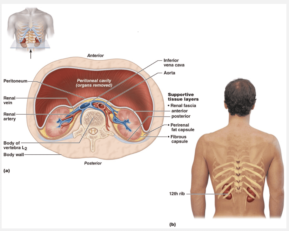
The renal hilum is a vertical cleft in the concave medial surface of the kidney.
The blood supply to the nephron is the __________.
- segmental artery
- efferent arteriole
- afferent arteriole
- interlobular artery
- renal artery
afferent arteriole
Ex.
The blood supply to the nephron is the afferent arteriole.
The vascular pathway through a kidney is as follows: renal artery → segmental arteries → interlobar arteries → arcuate arteries → cortical radiate arteries → afferent arterioles → glomeruli → efferent arterioles → peritubular capillary beds → cortical radiate veins → arcuate veins → interlobar veins → renal vein.
The renal hilum lies on the __________ surface of the kidney.
- inferior
- medial
- lateral
- superior
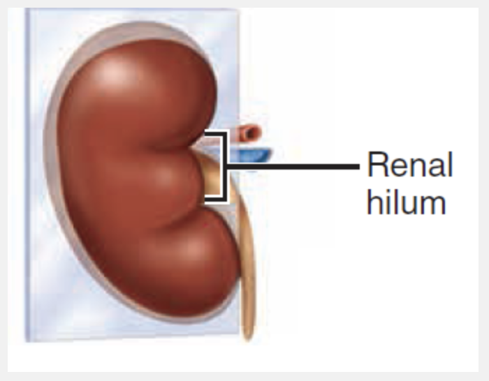
medial
Ex.
The renal hilum lies on the medial surface of the kidney.
An adult’s kidney has a mass of about 150 g (5 ounces) and its average dimensions are 11 cm long, 6 cm wide, and 3 cm thick—about the size of a large bar of soap. The lateral surface is convex. The medial surface is concave and has a vertical cleft called the renal hilum that leads into an internal space called the renal sinus. The ureter, renal blood vessels, lymphatics, and nerves all join each kidney at the hilum and occupy the sinus. Atop each kidney is an adrenal (or suprarenal) gland, an endocrine gland that is functionally unrelated to the kidney.
The renal __________ is continuous with the ureter.
- medulla
- cortex
- pelvis
- glomerulus
pelvis
Ex.
The renal pelvis, a funnel-shaped tube, is continuous with the ureter leaving the hilum.
Branching extensions of the pelvis form two or three major calyces (ka′lih-sēz; singular: calyx). Each major calyx subdivides to form several minor calyces, cup-shaped areas that enclose the papillae. The calyces collect urine, which drains continuously from the papillae, and empty it into the renal pelvis. The urine then flows through the renal pelvis and into the ureter, which moves it to the bladder to be stored. The walls of the calyces, pelvis, and ureter contain smooth muscle that contracts rhythmically to propel urine by peristalsis.
Which of the following is true with regards to an important sign of renal trauma?
- May cause a ureter to become kinked
- May cause backup of urine
- May cause excess glucose to be present in the urine
- May result in hematuria
May result in hematuria
Ex.
The presence of hematuria (blood in the urine) is an important sign of renal trauma.
The upper parts of both kidneys are protected by the thoracic cage, and the perirenal fat provides cushioning. However, the lower parts of the kidneys, especially the right kidney, are susceptible to blunt trauma such as from falls, motor vehicle accidents, or contact sports injuries. The renal artery is especially vulnerable to injury from rapid deceleration during car crashes, and may be lacerated (torn) or develop a thrombosis (blood clot).
Which of the following phrases describes the granular cells of the juxtaglomerular complex?
- Prevent infection from other areas spreading to the kidney
- Anchor kidneys to surrounding structures
- Specialized chemoreceptors
- Specialized mechanoreceptors
- Long nephrons that deeply invade the medulla
Specialized mechanoreceptors
Ex.
Granular cells are specialized mechanoreceptors.
Granular cells [also called juxtaglomerular (JG) cells] are in the arteriolar walls. They are enlarged smooth muscle cells with prominent secretory granules containing the enzyme renin. Granular cells act as mechanoreceptors that sense the blood pressure in the afferent arteriole.
Match the urinary term with its characteristic: Juxtamedullary nephrons.
- Specialized chemoreceptors
- Anchor kidneys to surrounding structures
- Prevent infection from other areas spreading to the kidney
- Specialized mechanoreceptors
- Long nephrons that deeply invade the medulla
Long nephrons that deeply invade the medulla
Ex.
Juxtamedullary nephrons are long nephrons that deeply invade the medulla.
Nephrons are generally divided into two major groups, cortical and juxtamedullary.
- Cortical nephrons account for 85% of the nephrons in the kidneys. Except for small parts of their nephron loops that dip into the outer medulla, they are located entirely in the cortex.
- Juxtamedullary nephrons (juks″tah-mĕ′dul-ah-re) originate close to (juxta = near to) the cortex-medulla junction, and they play an important role in the kidneys’ ability to produce concentrated urine. They have long nephron loops that deeply invade the medulla, and their ascending limbs have both thin and thick segments.
Which of the following statements about the urinary system is incorrect?
- It carries out gluconeogenesis in the body.
- It produces erythropoietin, which stimulates red blood cell formation.
- It produces renin, which helps regulate blood pressure.
- It metabolizes vitamin C to its active form.
It metabolizes vitamin C to its active form.
Ex.
The kidney metabolizes vitamin D, not vitamin C, to its active form.
The kidneys perform a chemical balancing act that would be tricky even for the best chemical engineer. They maintain the body’s internal environment by:
- Regulating the total volume of water in the body and the total concentration of solutes in that water (osmolality).
- Regulating the concentrations of the various ions in the extracellular fluids. (Even relatively small changes in some ion concentrations such as K+ can be fatal.)
- Ensuring long-term acid-base balance.
- Excreting metabolic wastes and foreign substances such as drugs or toxins
- Producing erythropoietin and renin (re′nin; ren = kidney), important molecules for regulating red blood cell production and blood pressure, respectively.
- Converting vitamin D to its active form.
- Carrying out gluconeogenesis during prolonged fasting
Under normal resting conditions, the __________ arteries deliver one-fourth of the total cardiac output (about 1200 ml) to the kidneys each minute.
- segmental
- cortical radiate
- renal
- interlobar
renal
Ex.
Under normal resting conditions, the renal arteries deliver one-fourth of the total cardiac output (about 1200 ml) to the kidneys each minute.
The kidneys continuously cleanse the blood and adjust its composition, so it is not surprising that they have a rich blood supply. Under normal resting conditions, the large renal arteries deliver one-fourth of the total cardiac output to the kidneys—about 1200 ml each minute
Which vessels closely surround renal tubules?
- Efferent arterioles
- Afferent arterioles
- Peritubular capillaries
- Segmental arteries
Peritubular capillaries
Ex.
The peritubular capillaries cling closely to adjacent renal tubules and empty into nearby venules. Because they arise from the efferent arterioles (which have high resistance), they only experience low pressure. As a result, these low-pressure, porous capillaries readily absorb solutes and water from the tubule cells as these substances are reclaimed from the filtrate. Renal tubules are closely packed together, so the peritubular capillaries of each nephron absorb substances from several adjacent nephrons.